Introduction
The parapharyngeal space is a very complex region of the head and neck which is difficult to access because of its surrounding boundaries. The space itself contains several important structures, from which many differing pathologies arise. Neoplastic pathology of the parapharyngeal space can mainly be divided into salivary, neurogenic and vascular tumours. It is sometimes very difficult to access these tumours, let alone diagnose them.
In the presented case, it was not possible to perform a needle biopsy of the patient's parapharyngeal mass using an external approach. Therefore, a transoral approach was adopted using an endo-cavitary ultrasound (US) probe with a core biopsy needle attached, in order to confirm the nature of the parapharyngeal mass. The current paper represents the first report of the use of such methodology and instrumentation to obtain a diagnostic tissue sample.
Case report
A 42-year-old woman was admitted with a history suggestive of a cerebrovascular accident, and resulting left-sided hemiplegia.
She subsequently underwent a head and neck magnetic resonance imaging (MRI) scan (Figure 1). This revealed a right-sided parapharyngeal mass with associated compression of the right internal carotid artery (ICA). Angiography revealed complete thrombus occlusion of the right ICA. This was presumed to have arisen from a right ICA dissection secondary to long-standing compression.
Axial magnetic resonance imaging scan of parapharyngeal space mass.

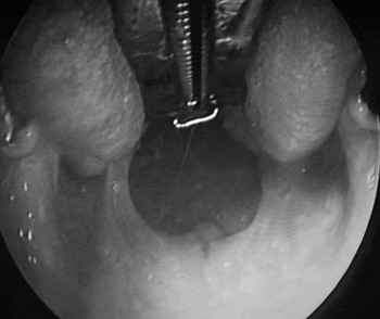
The parapharyngeal lesion was not palpable or visible, externally or intra-orally (Figure 2). Obtaining a tissue biopsy was technically challenging and required image guidance. Ultrasound-guided percutaneous biopsy was attempted but proved unsuccessful due to the deep location of the parapharyngeal mass.
Intra-oral examination.

Therefore, an US-guided core biopsy was performed transorally under general anaesthesia, utilising a prostatic US probe (Figure 3). The procedure was uneventful, with a good sample being obtained under accurate guidance.
Endo-cavitary ultrasound probe.

The histopathology report confirmed pleomorphic adenoma. The patient subsequently underwent resection of the parapharyngeal mass via a trans-cervical approach, without the need for mandibulotomy.
Discussion
The parapharyngeal space is a well defined region of loose connective tissue which lies adjacent to the nasopharynx and the oropharynx, between the base of the skull and the hyoid bone.Reference Oliai, Sheth, Burroughs and Ali1, Reference Pang, Goh and Tan2 The shape of the parapharyngeal space may be likened to an inverted pyramid, with the cranial base forming its base and the greater cornu of the hyoid bone forming its apex. The styloid process divides this space into the pre- and post-styloid compartments.Reference Pang, Goh and Tan2 The pre-styloid compartment contains the parotid gland, fat and lymph nodes. The post-styloid compartment contains the carotid sheath with the internal carotid artery, internal jugular vein, cranial nerves IX to XII, and lymph nodes.
Tumours of the parapharyngeal space are uncommon and account for only 0.5 per cent of all head and neck neoplasms. They are usually one of three types: salivary, neurogenic or paraganglioma.Reference Pang, Goh and Tan2 Parapharyngeal space tumours may grow very large, yet they often remain relatively asymptomatic and are frequently discovered incidentally.Reference Oliai, Sheth, Burroughs and Ali1 The most common parapharyngeal space tumour is pleomorphic adenoma.Reference Farag, Lin, Koch, Caifano, Cummings and Farinola3 Approximately 50 per cent of parapharyngeal space neoplasms originate from the deep lobe of the parotid or from the minor salivary glands.Reference Rebol, Takae and Bumber4
Imaging of parapharyngeal space tumours is extremely important, as identification of the tumour's location relative to its adjacent structures aids diagnosis. Obtaining a tissue sample of the lesion is a necessary diagnostic tool.Reference Rebol, Takae and Bumber4 However, this can be technically challenging, due to the inaccessible nature of the parapharyngeal space and the multiple adjacent structures which could be damaged in the process. Percutaneous US-guided biopsy may be possible and is usually successful. Alternatively, if the tumour is palpable intra-orally, it is easily amenable to intra-oral fine needle aspiration.Reference Rebol, Takae and Bumber4
We report a challenging case in which the parapharyngeal space mass was located higher than usual and a percutaneous US-guided biopsy was unsuccessful, despite being performed by a dedicated head and neck radiologist. The mass was not palpable intra-orally or externally. Therefore, an image-guided, intra-oral technique was used, following modification of a Siemens EC-10C5 endo-cavitary US probe (Siemens, Iowa, USA) with a CIVCO needle guide attachment (Civco medical solutions, Iowa, USA). An 18-gauge core biopsy cannula was used to obtain the sample. The procedure was successfully carried out under general anaesthesia for patient comfort. This paper represents the first report of this technique, which allows biopsy of the parapharyngeal space lesion in an accurate and well controlled fashion under general anaesthesia. The technique also permits access to lesions located high in the parapharyngeal space, including retropharyngeal lymph nodes that are difficult to reach percutaneously.
Acknowledgements
We would like to thank Dr S Morley for his radiological expertise in dealing with this case, and also for providing us with the MRI scans.