Introduction
Prebiotics are defined as feed supplements that beneficially affect the host by selectively stimulating the growth or activity of the limited number of bacterial species inhabiting the digestive tract, without being digested (Patterson and Burkholder, Reference PATTERSON and BURKHOLDER2003). Natural and synthetic oligosaccharides are known types of prebiotics. Among the oligosaccharides are mannan-oligosaccharides (MOS), fructose-oligosaccharides (FOS), galacto-oligosaccharides, isomalto-oligosaccharides, xylo-oligosaccharides, and chito-oligosaccharides (Iji and Tivey, Reference IJI and TIVEY1998). MOS is used abundantly in poultry as an alternative to antibiotic growth promoters (AGPs).
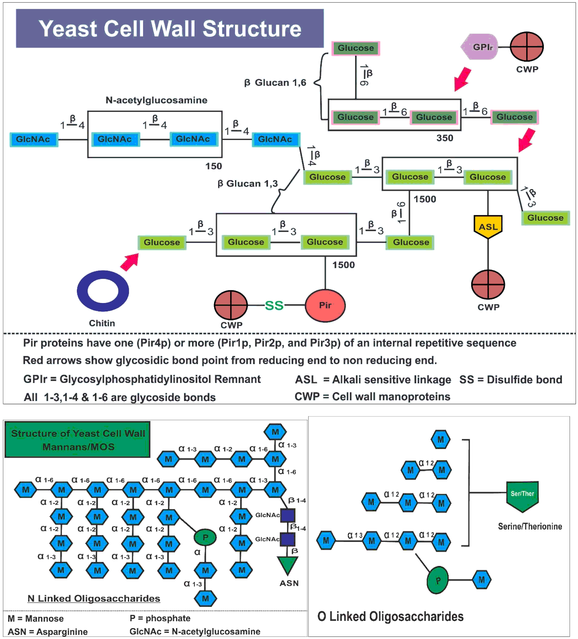
MOS is a yeast cell wall fragment derived from Saccharomyces cerevisiae. In order to extract MOS, the cell wall of yeast is first cracked open by physical, chemical and /or enzymatic procedures. After that, centrifugation, spray drying and alkaline extraction produce α-mannoproteins. In the last step, α-mannoproteins are concentrated by membrane ultrafiltration and spray drying, resulting in a product of light brown colour will be MOS (Kwiatkowski and Kwiatkowski, Reference KWIATKOWSKI and KWIATKOWSKI2012). The cell wall of Saccharomyces cerevisiae yeast accounts for 25% of the total cell with a complex structure. It consists of mainly four components including mannoproteins or cell wall proteins (CWP), glucans, chitin, and N-acetylglucosamine (GlcNAc). CWP are comprised of 50-90% of carbohydrate by weight, of which are characterised as mannans. These consist of D- mannose subunits which are covalently bonded with proteins by two linkages (Lipke and Ovalle, Reference LIPKE and OVALLE1998). First are the O-linked oligosaccharides which consist of five mannose residues 0-glycosylated with threonine/serine. Second are N-linked oligosaccharides which are N glycosylated (Lesage and Bussey, Reference LESAGE and BUSSEY2006; Lipke and Ovalle, Reference LIPKE and OVALLE1998; Cawley and Ballou, Reference CAWLEY and BALLOU1972; Mrsa et al., Reference MRSA, KLEBL and TANNER1993; Klis et al., Reference KLIS, BOORSMA and DE GROOT2006) ( Figure 1 ).
Structure of yeast cell wall and yeast cell wall mannans (MOS).

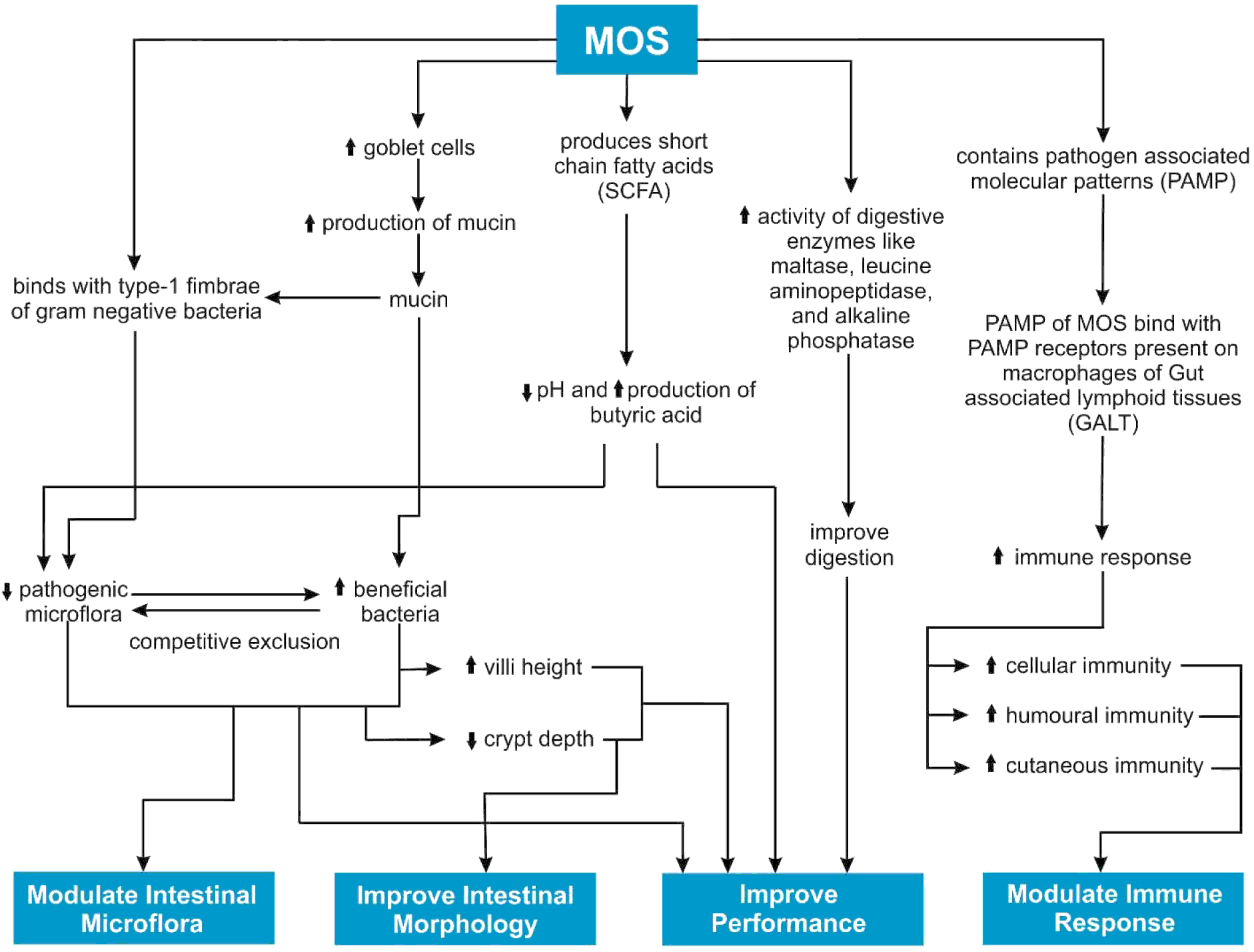
MOS can tolerate temperatures of 120°C for 20 minutes without losing its capability to bind pathogenic bacteria. (Shane, Reference SHANE2001). Therefore, feed milling operations do not affect its activity. This indigestible sugar is involved in a wide variety of functions including reduction in pathogenic bacteria (Spring et al., Reference SPRING, WENK, DAWSON and NEWMAN2000), enhancing beneficial bacteria (Baurhoo et al., Reference BAURHOO, LETELLIER, ZHAO and RUIZ-FERIA2007a), increasing villus height and decreasing crypt depth (Baurhoo et al., Reference BAURHOO, PHILLIP and RUIZ-FERIA2007b; Yang et al., Reference YANG, IJI and CHOCT2009), modulating immune response (Shashidhara and Devegowda, Reference SHASHIDHARA and DEVEGOWDA2003; Khalaji et al., Reference KHALAJI, ZAGHARI and NEZAFATI2011), and improving performance of broilers (Rosen, Reference ROSEN2007). The purpose of this article is to review the different mechanisms of MOS and their subsequent effect on intestinal microflora, intestinal morphology, immunity and performance of broilers ( Figure 2 ).
Mechanisms of MOS.

Mechanisms of MOS
mechanisms involved in modulating intestinal microflora
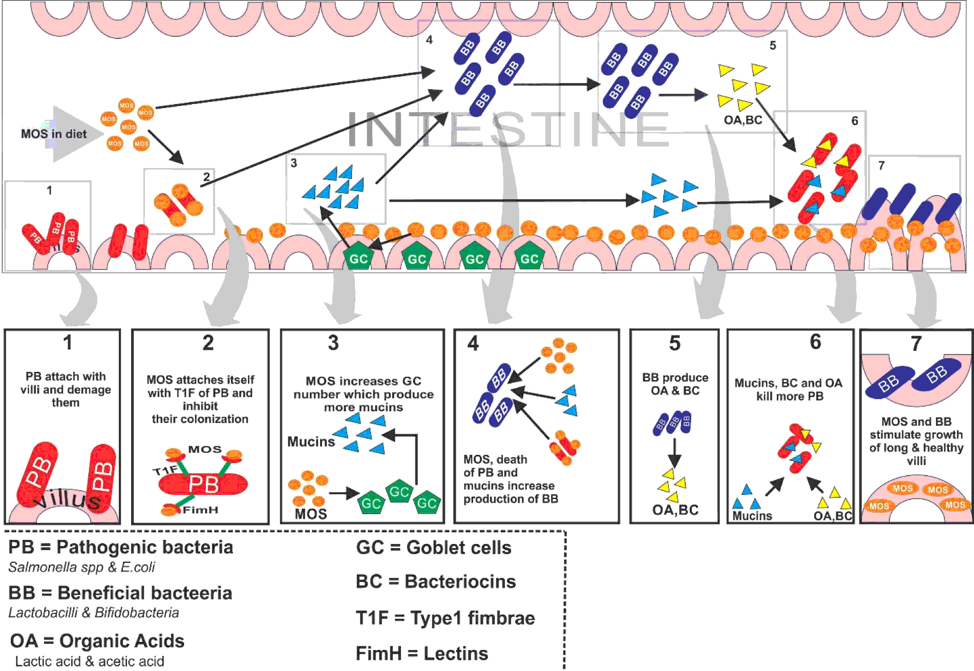
A well-balanced microflora within the digestive tract is important for normal functioning (Anderson et al., Reference ANDERSON, MCCRACKEN, AMINOC, SIMPSON, MACKIE, VERSTEGEN and GASKINS2000) and MOS maintains this balance by reducing the load of pathogenic bacteria and enhancing the growth of beneficial bacteria through different mechanisms ( Figure 3 ).
Bacteria require attachment for colonisation in intestinal mucosa enabling their harmful effects. In Gram-negative bacteria like Salmonella and E. coli, the attachment is carried out with type-1 fimbriae, which contain mannose-specific lectins named FimH. These lectins have a high affinity for mannose residues on intestinal mucosa. MOS reduce the attachment of Gram-negative bacteria with intestinal mucosa by binding with bacterial FimH of type-1 fimbriae. In this way, the bacteria move through the intestine without allowing colonisation (Fernandez et al., Reference FERNANDEZ, HINTON and VAN GILS2002; Hooge et al., Reference HOOGE, SIMS, SEFTON, CONNOLLY and SPRING2003; Baurhoo et al., Reference BAURHOO, GOLDFLUS and ZHAO2009b).
Mechanisms of MOS involved in modulating microflora and bacterial load from intestine.

The structure of MOS clearly defines its function in gastrointestinal tract of birds. Its brush-like structure which consists of a 1-5 ringed alpha 1,2 and alpha 1,3 mannose linked side chain of which is attached to a 120 ringed alpha 1,6 mannose backbone that resemble the ‘brush handle’. This unique structure makes MOS functional with in the bird's digestive tract by attaching itself with receptors present on wall of digestive tract and membranes of pathogens (Vinogradov et al., Reference VINOGRADOV, PETERSEN and BOCK1998; Kwiatkowski and Kwiatkowski, Reference KWIATKOWSKI and KWIATKOWSKI2012).
Fernandez et al. (Reference FERNANDEZ, HINTON and VAN GILS2002) observed the reduction of Salmonella spp. in the caeca of broilers by supplementing with MOS. In another study, MOS significantly lowered S. typhimurium and S. dublin infections in broilers (Newman, Reference NEWMAN1994). Studies showed that E. coli populations were reduced in the digestive tracts of broilers when they were fed MOS supplemented diets (Spring, Reference SPRING1996). Furthermore, MOS agglutinated five out of seven strains of E. coli when they were subjected to an agglutination test in vitro (Spring et al., Reference SPRING, WENK, DAWSON and NEWMAN2000). MOS tends to reduce E. coli populations of the caecum and ileum of the broiler birds when fed at 0.2% of feed (Koc et al., Reference KOC, SAMLI, OKUR, OZDUVEN and SENKOYLU2010).
When comparing with AGPs (Avilamycin 0.001%) MOS (0.2%/0.1%/0.05% in starter/grower/finisher periods, respectively) showed greater reduction in E. coli populations in the caecum and jejunum (Jamroz et al., Reference JAMROZ, WILICZKIEWICZ, ORDA, WERTELECKI and SKORUPINSKA2004). However, Denev et al. (Reference DENEV, DINEV, NIKIFOROV and KOINARSKI2006) didn't find any significant difference of feeding MOS (0.2% 0-21d and 0.1% 21-42d) on caecal coliforms as compared to both negative and positive (Avilamycin 0.001%) control diets. These contraindicatory results might have occurred because of different dose regimes used in the studies.
MOS increases the synthesis of goblet cells (Baurhoo et al., Reference BAURHOO, LETELLIER, ZHAO and RUIZ-FERIA2007a; Reference BAURHOO, GOLDFLUS and ZHAO2009b) which are present in the villi membrane of the intestine. These cells secrete glycoproteins, namely mucins, which act as the first line of host defence against pathogenic bacteria (Belley et al., Reference BELLEY, KELLER, GOTTKLE and CHADEE1999). Mucins contain mannosyl receptors which competitively bind to the type-1 fimbriae of Gram-negative bacteria and thus assist in the clearance of pathogens from the intestine. Feeding MOS (0.2% and 0.5%) to broilers has been shown to cause a significant increase in goblet cell number at 34 days of age, which in turn diminishes E. coli counts in caecum as compared to basal diet (Baurhoo et al., Reference BAURHOO, FERKET and ZHAO2009a). Baurhoo et al. (Reference BAURHOO, GOLDFLUS and ZHAO2009b) found that feeding MOS (0.15%/0.1% in starter/grower diets, respectively) increased goblet cells and significantly diminished the E. coli populations in caeca of broiler chickens. However, at day 14, MOS didn't enhance goblet cells and thus E. coli numbers in intestine remained unaffected. As with age, the number and length of villi increase along with the number of goblet cells. By producing mucins, goblet cells control E. coli in intestine of broiler. So, the age of the birds, goblet cells and E. coli have some correlation between them (Baurhoo et al., Reference BAURHOO, FERKET and ZHAO2009a).
Reports have shown that MOS enhances the numbers of beneficial bacteria, mainly Lactobacillus and Bifidobacteria spp., in the intestines of broilers (Baurhoo et al., Reference BAURHOO, FERKET and ZHAO2009a; Reference BAURHOO, GOLDFLUS and ZHAO2009b). A decrease in pathogenic bacteria through aforementioned mechanisms may provide a favourable environment for proliferation of beneficial bacteria, mainly Lactobacillus spp. (Van der Wielen et al., Reference VAN DER WIELEN, LIPMAN, VAN KNAPEN and BIESTERVELD2002; Baurhoo et al., Reference BAURHOO, FERKET and ZHAO2009a; Reference BAURHOO, GOLDFLUS and ZHAO2009b). At day 34, a reduction in E. coli counts through feeding MOS (0.2% and 0.5%) may allow Lactobacilli spp. to proliferate in a pathogenic bacteria free environment, leading to increased populations as compared to negative control and bacitracin (0.006%) fed birds (Baurhoo et al., Reference BAURHOO, FERKET and ZHAO2009a). Baurhoo et al. (Reference BAURHOO, GOLDFLUS and ZHAO2009b) reported that Lactobacillus spp. count was significantly enhanced in caeca of broilers only at day 34 of age in response to the dietary inclusion of MOS (0.15%/0.1% in starter/grower periods, respectively) when compared to both negative and positive control (Virginiamycin 0.002%) This occurred due to decreased E. coli counts at the same time of trial, as this reduction produced a favourable environment for the growth of Lactobacillus spp.
Other studies have shown that MOS is involved in increasing the intestinal Lactobacillus spp. content of the broilers when given at various dietary inclusion levels (Fernandez et al., Reference FERNANDEZ, HINTON and VAN GILS2002; Denev et al., Reference DENEV, DINEV, NIKIFOROV and KOINARSKI2006; Koc et al., Reference KOC, SAMLI, OKUR, OZDUVEN and SENKOYLU2010). However, the findings of Spring et al. (Reference SPRING, WENK, DAWSON and NEWMAN2000) suggested that supplementation of MOS in broiler diets (0.4%) did not affect intestinal Lactobacillus spp. concentration. Similarly, Khalaji et al. (Reference KHALAJI, ZAGHARI and NEZAFATI2011) did not find any significant difference in Lactobacillus spp. counts among all the treatment groups including MOS (0.05, 0.1 and 0.15% in the diet). During this period, E. coli populations remained unaffected by inclusion of MOS, which could be the reason behind this contraindicatory result.
Bifidobacteria spp. feed on mucin glycoprotein secreted from goblet cells by secreting the unique enzymes 1,2-α-L-fucosidase and endo-α-N-acetyl galactosaminidase (Katayama et al., Reference KATAYAMA, FUJITA and YAMAMOTO2005; Ruas-Madiedo et al., Reference RUAS-MADIEDO, GUEIMONDE, FERNANDEZ-GARCIA, DE LOSREYES-GAVILAN and MARGOLLES2008). Feeding MOS (0.2% and 0.5%) to broiler chickens significantly increased goblet cells and mucin production, and Bifidobacteria spp. counts were enhanced when compared with diets containing virginiamycin (0.002%) and zinc bacitracin (0.006%). Inclusion of MOS increased Bifidobacteria spp. to a greater extent than the control (Baurhoo et al., Reference BAURHOO, FERKET and ZHAO2009a). Baurhoo et al. (Reference BAURHOO, LETELLIER, ZHAO and RUIZ-FERIA2007a; Reference BAURHOO, PHILLIP and RUIZ-FERIA2007b) reported that dietary MOS enhanced Bifidobacteria spp. in the intestines of broiler chickens by increasing goblet cells and mucin production as compared to positive control (Virginiamycin 0.0011%) diet. Similarly, other studies have shown increased Bifidobacteria spp. numbers in intestines when compared to control or AGP supplemented groups (Fernandez et al., Reference FERNANDEZ, HINTON and VAN GILS2002; Kocher et al., Reference KOCHER, GELIOT and TUCKER2005a; Denev et al., Reference DENEV, DINEV, NIKIFOROV and KOINARSKI2006). These beneficial bacteria compete against pathogens for space and nutrients and decrease their load by the mechanism of competitive exclusion (Rolfe, Reference ROLFE2000). Lactobacilli spp. produce bacteriocins (Joerger, Reference JOERGER2003) whereas, Bifidobacteria spp. secrete organic acids and bactericidal substances (Gibson and Wang, Reference GIBSON and WANG1994), which decrease the populations of pathogenic bacteria.
Lactobacilli spp. carry out the fermentation of glucose, converting it into lactic acid and acetic acid within the digestive tract of birds (Audisio et al., Reference AUDISIO, OLIVER and APELLA2000). These mechanisms of control may be proposed for all types of pathogenic bacteria even if they do not contain type-1 fimbriae, such as Clostridium and Campylobacter spp. It has been reported that the intestinal population of C. perfringens in broilers was lowered following the supplementation of MOS (0.2% and 0.1%) in diets (Jamroz et al., Reference JAMROZ, WILICZKIEWICZ, ORDA, WERTELECKI and SKORUPINSKA2004). Similarly, Kocher et al. (Reference KOCHER, GELIOT and TUCKER2005a) observed a decrease in C. perfringens count in the caeca of broiler chickens fed MOS (0.2%/0.1% kg/ton in starter/finisher periods, respectively) supplemented diets. The incidence of Campylobacter jejuni infection was limited when broiler birds were fed diets containing MOS as compared to control, unchallenged group (Schoeni and Wong, Reference SCHOENI and WONG1994). Similarly, Campylobacter spp. counts were reported to be reduced at day 34 due to the increased Lactobacillus and Bifidobacteria spp. counts through feeding of a diet supplemented with MOS as compared to basal diet. But this effect of MOS was not significant compared to a positive control (virginiamycin 0.00165%) diet, which showed that MOS produced a similar effect to antibiotic used in the study (Baurhoo et al., Reference BAURHOO, FERKET and ZHAO2009a; Reference BAURHOO, GOLDFLUS and ZHAO2009b).
mechanisms involved in improving intestinal morphology
A characteristic feature of a healthy bird's digestive tract is a long, intact villus having shallow crypts and low renewal rate (Ferket et al., Reference FERKET, PARKS and GRIMES2002). Long villi and shallow crypts provide a larger surface area for the absorption of nutrients and low renewal rate allows efficient enzyme production and maturation of the intestinal cells (Yang et al., Reference YANG, IJI and CHOCT2009). Improvements in gut morphology are largely associated with the change in the diet and modification in intestinal microflora. (Baurhoo et al., Reference BAURHOO, PHILLIP and RUIZ-FERIA2007b). Beneficial bacteria stimulate the development of a healthy intestinal environment resulting in an increase in villi length through competitive exclusion (Baurhoo et al., Reference BAURHOO, PHILLIP and RUIZ-FERIA2007b).
A study conducted by Baurhoo et al. (Reference BAURHOO, FERKET and ZHAO2009a) revealed that dietary MOS (0.2 and 0.5% in feed) significantly increased villus height in all intestinal segments on day 24 and 34 as compared to virginiamycin (0.002%) and bacitracin (0.006%) whereas, 0.5% MOS improved villus height in duodenum of broilers as compared to the control diet on day 14 of age. Sinovec et al. (Reference SINOVEC, MARKOVIC and GLEDIC2005) reported that MOS (0.2%) significantly improved broiler villus height in all parts of the small intestine on day 21 of age when compared to birds from both negative and positive (Flavomycin 0.002%) control diets. Similarly, the inclusion of MOS (0.2% up to day 21 and 0.1% thereafter) improved the villus height in the jejunum when compared with the positive control diet containing 0.001% virginiamycin (Baurhoo et al., Reference BAURHOO, PHILLIP and RUIZ-FERIA2007b). Supplementation of 0.1% MOS with or without enzymes raised broiler duodenal and jejunal villi height (Oliveira et al., Reference OLIVEIRA, RODRIGUES, MARQUES, GRAVENA, GUANDOLINI and MORAES2008).
MOS may increase the fermentation of ingesta by improving gut health in the distal intestine, resulting in the production of short-chain fatty acids (SCFA). The SCFA are involved in cell proliferation in the intestinal mucosa. These reduce the pH of the brush border microenvironment and block the adhesion of pathogens (Lan et al., Reference LAN, XUN, TAMMINGA, WILLIAMS, VERSTEGEN and ERIDI2004; Oliveira et al., Reference OLIVEIRA, RODRIGUES, MARQUES, GRAVENA, GUANDOLINI and MORAES2008). Studies in a group of broilers fed diets containing 0.5 and 0.3% MOS showed that RNA content of the ileal mucosa, which represents efficiency of various cells in performing functions, and protein/DNA ratio of the jejunal mucosa, which is the measure of cells size (0.1% MOS in feed) were higher in contrast to other treatments without MOS. This demonstrated that improved intestinal structure and function occurred after eliminating pathogenic bacterial load from intestine by MOS (Iji et al., Reference IJI, SAKI and TIVEY2001). Similarly, MOS (0.2% in feed) significantly increased villus height in all parts of the intestine (Marković et al., Reference MARKOVIĆ, ŠEFER, KRSTIĆ and PETRUJKIĆ2009) whereas; Loddi et al. (Reference LODDI, MORAES, NAKAGHI, TUCCI, HANNAS, ARIKI and BRUNO2002) observed that MOS affected villus height in the duodenum only. Yang et al. (Reference YANG, IJI and CHOCT2007b) reported that adding 0.1% and 0.2% MOS in broiler diet resulted in an increased villus height of the small intestine up to day 35 of age, which then decreased gradually in all groups except MOS. In another study, MOS did not affect the villus height in the intestine of broilers (Baurhoo et al., Reference BAURHOO, GOLDFLUS and ZHAO2009b). Similarly, Garces-Narro et al. (Reference GARCES-NARRO, BARRAGAN, SOLER, ROS, ORTEGA, MOCE, MATEOS, GÓMEZ-MUÑOZ, LOPEZ-MENDOZA and RODILLA2006) and Oliveira et al. (Reference OLIVEIRA, CANCHERINI, MARQUES, GRAVENA and MORAES2009b) reported no difference in the villus height of broilers fed MOS and other dietary treatments.
The continuous proliferation and migration of the enterocyte cells from the crypt to the villi is a normal function of the intestine. However, this process speeds up in the presence of infection or toxins from pathogenic bacteria. A healthy gut has low renewal rate which indicates shallow crypts (Shane, Reference SHANE2001). The higher the cell renewal rate, the deeper crypt depth, indicating colonisation of the pathogenic bacteria in the intestine, and it is well established that villi length increases due to reduced enterocyte cell migration or renewal from the crypt to the villus (Baurhoo et al., Reference BAURHOO, LETELLIER, ZHAO and RUIZ-FERIA2007a; Reference BAURHOO, PHILLIP and RUIZ-FERIA2007b). Since MOS reduces the load of pathogenic bacteria in the broilers intestine (Baurhoo et al., Reference BAURHOO, LETELLIER, ZHAO and RUIZ-FERIA2007a; Reference BAURHOO, PHILLIP and RUIZ-FERIA2007b); the cell renewal rate will be less, leading to shallower crypts. Yang et al. (Reference YANG, IJI and CHOCT2009) reported that MOS decreased crypt depth in the small intestine of broilers favouring more rapid growth of chickens. Reduction in crypt depth of all parts of the small intestine of broilers was observed in response to 0.2% MOS in the diet as compared to both positive (Flavomycin 0.002%) and negative control diets (Sinovec et al., Reference SINOVEC, MARKOVIC and GLEDIC2005). Sarica et al. (Reference SARICA, ERDOGAN, KOC and ERDOGAN2005) suggested that MOS caused a significant decrease in crypt depth in all segments of the small intestine except duodenum. Whereas, some studies have shown that supplementation of MOS did not affect the crypt depth to a significant level (Garces-Narro et al., Reference GARCES-NARRO, BARRAGAN, SOLER, ROS, ORTEGA, MOCE, MATEOS, GÓMEZ-MUÑOZ, LOPEZ-MENDOZA and RODILLA2006; Baurhoo et al., Reference BAURHOO, FERKET and ZHAO2009a; Reference BAURHOO, GOLDFLUS and ZHAO2009b). On the other hand, other studies on intestinal morphology revealed that birds fed MOS (0.1 and 0.5% of diet) had deeper crypts as compared to control group (Iji et al., Reference IJI, SAKI and TIVEY2001; Oliveira et al., Reference OLIVEIRA, RODRIGUES, MARQUES, GRAVENA, GUANDOLINI and MORAES2008).
mechanisms involved in improving immune responses
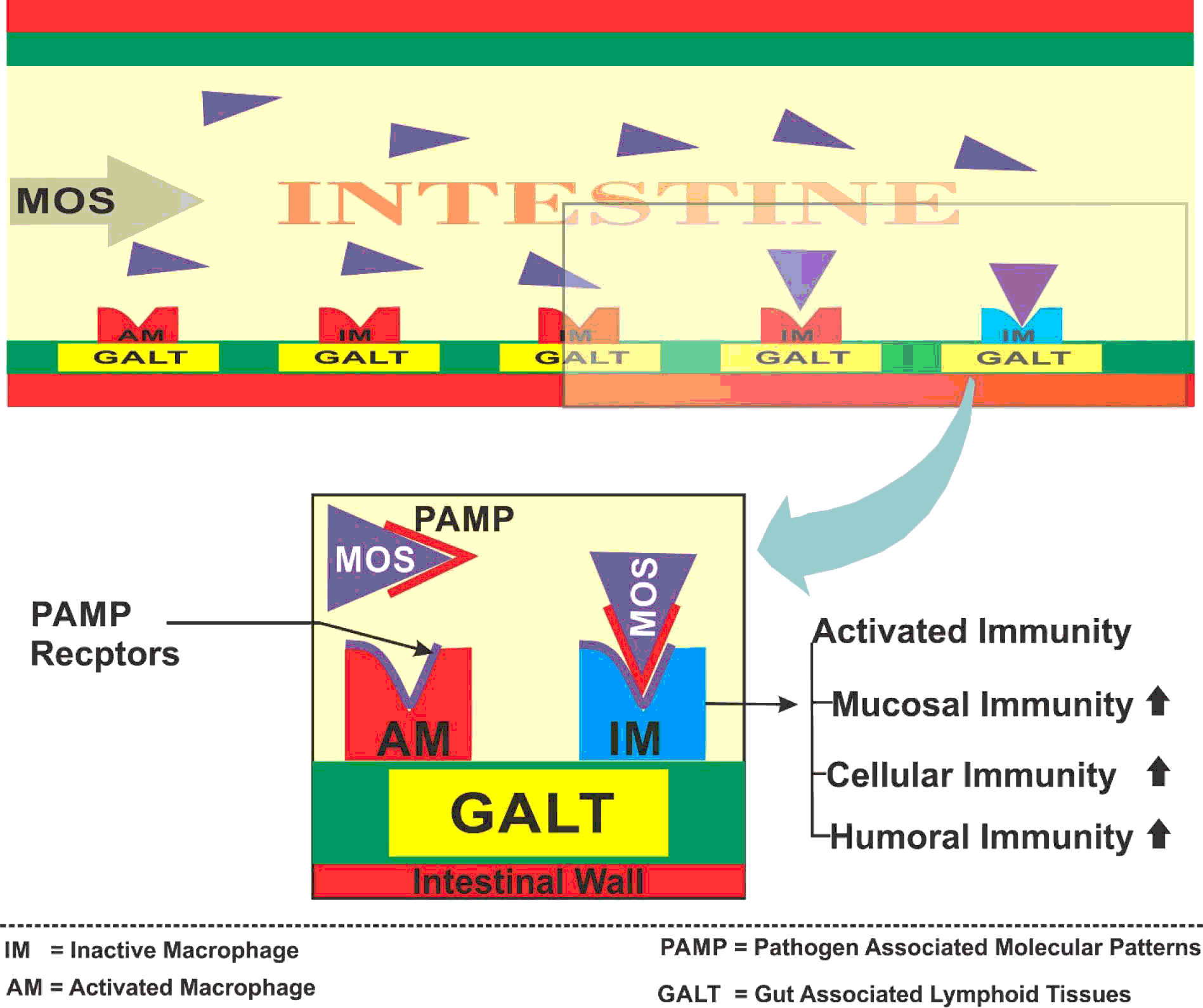
Birds with improved immune status have the ability to resist various pathogens and show better responses to vaccination programmes (Vesna et al., Reference VESNA, LAZAREVIĆ, SINOVEC and TOKIĆ2007). MOS is used to increase the immune potential of broiler chickens by improving cellular, humoral and mucosal immunities (Gómez-Verduzco et al., Reference GÓMEZ-VERDUZCO, CORTES-CUEVAS, LÓPEZ-COELLO, ÁVILA-GONZÁLEZ and NAVA2009; Ozpinar et al., Reference OZPINAR, ERHARD, AHRENS, KUTAY and ESECELI2010; Zakeri and Kashefi, Reference ZAKERI and KASHEFI2011; Figure 4 ).
In the bird's intestine, a variety of macrophages are present, as components of gut-associated lymphoid tissue (GALT). These defence cells recognise pathogens by unique type of molecules that are only on microbes and not the host cells, called ‘pathogens associated molecular patterns’ (PAMP). Yeast cell wall components, like mannan and glucan, and other microbial molecules, like peptidoglycan and lipopolysaccharide, contain PAMP. Mannans bind with the PAMP receptors present on defence cells of GALT and then trigger the immune system of the bird (Shashidhara and Devegowda, Reference SHASHIDHARA and DEVEGOWDA2003). When more receptors of these defence cells are occupied, these macrophages become activated, leading to enhanced cellular immune response (Zakeri and Kashefi, Reference ZAKERI and KASHEFI2011). Khalaji et al. (Reference KHALAJI, ZAGHARI and NEZAFATI2011) reported that supplementation of 0.05, 0.1 and 0.15% MOS in broiler diets resulted in a significantly increased immune response seen in a cutaneous basophil hypersensitivity test (CBHR) to phytohaemagglutinin-P (PHA) whereas, the immune response against sheep red blood cells (SRBC) did not show any significance. Vesna et al. (Reference VESNA, LAZAREVIĆ, SINOVEC and TOKIĆ2007) evaluated the effect of MOS (0.2% in feed) by performing the CBHR to PHA test. They observed that birds fed MOS showed significantly higher results as compared to a control fed group. Similarly, 0.05% MOS in broiler diets increased the degree of the immune response when analysed by CBHR (Gómez-Verduzco et al., Reference GÓMEZ-VERDUZCO, CORTES-CUEVAS, LÓPEZ-COELLO, ÁVILA-GONZÁLEZ and NAVA2009). In contrast, Cotter (Reference COTTER1997) reported a decreased degree of CBHR to PHA with the inclusion of MOS in the diet. Liangping et al. (Reference LIANGPING, LUNJIANG, GUOPING and FANPING1999) assessed the cellular immune response of broiler birds by determining macrophage activity and PHA lymphoblast transforming rate. They concluded that MOS (0.2 and 0.4% in feed) significantly enhanced macrophage activity and PHA lymphoblast transforming rate as compared to basal diet.
Mechanism involving MOS in improving immune responses.

The increase in antibody titre after vaccination is due to an increase in the immune response of birds (Oliveira et al., Reference OLIVEIRA, FIGUEIREDO-LIMA, FARIA FILHO, MARQUES and MORAES2009a). Feeding MOS can lead to the activation of macrophages, and thus improved the immune status of birds. Humoral immunity of broiler chickens given Newcastle B1 vaccine (NDVB1), when calculated by haemagglutination inhibition test (HI), was significantly improved by including 0.1% MOS in the diet (Zakeri and Kashefi, Reference ZAKERI and KASHEFI2011). Gómez-Verduzco et al. (Reference GÓMEZ-VERDUZCO, CORTES-CUEVAS, LÓPEZ-COELLO, ÁVILA-GONZÁLEZ and NAVA2009) showed similar results in broilers fed 0.05% MOS. Similarly, inclusion of MOS (0.1% in breeder and 0.15% in starter diets) showed the highest Newcastle disease (NCD) antibody titre (De Lange, Reference DE LANGE2007). Ozpinar et al. (Reference OZPINAR, ERHARD, AHRENS, KUTAY and ESECELI2010) reported that birds fed 0.15% MOS significantly improved the plasma IgG level as compared to other dietary treatments. Oliveira et al. (Reference OLIVEIRA, FIGUEIREDO-LIMA, FARIA FILHO, MARQUES and MORAES2009a) observed that the antibody titres of infectious bursal disease virus (IBDV) and Newcastle disease virus (NDV) increased in subsequent weeks after vaccination in broiler breeders following the consumption of MOS in feed (0.1% up to 21 days and 0.05% in 22 to 42 days) in comparison to other feed additives. As discussed earlier, MOS increases Lactobacilli and Bifidobacteria spp. populations in the intestine; which may lead to improved immune responses. Bifidobacteria spp. have been reported to stimulate antibody production (Bornet et al., 2002) while Lactobacillus spp. can modulate the overall immune system (Lee et al., Reference LEE, DRONGOWSKI, CORAN and HARMON2000). However, the exact mechanism behind this is still unclear (Oliveira et al., Reference OLIVEIRA, FIGUEIREDO-LIMA, FARIA FILHO, MARQUES and MORAES2009a).
The antibody titre of NCD vaccine at days 10 and 17 of age in broilers was significantly improved in response to 0.2% dietary MOS (Vesna et al., Reference VESNA, LAZAREVIĆ, SINOVEC and TOKIĆ2007). Awaad et al. (Reference AWAAD, ATTA, EL-GHANY, ELMENAWEY, AHMED, HASSAN and ABDELALEEM2011) observed that, even in ochratoxicosis challenged birds, MOS (0.2% in feed) tended to improve humoral immune response against NDV vaccine. Shashidhara and Devegowda (Reference SHASHIDHARA and DEVEGOWDA2003) suggested that feeding MOS (0.05% in trial 1 and 0.2% in trial 2) significantly increased antibody titre against IBDV as compared to other dietary treatments. Furthermore, MOS improved maternal antibody titre in progeny. However, Shafey et al. (Reference SHAFEY, AL-MUFAREJ, SHALABY and JARELNABI2001) reported no significant effects of MOS supplementation on humoral immunity against NCD and IBDV in chickens. Gómez-Verduzco et al. (Reference GÓMEZ-VERDUZCO, CORTES-CUEVAS, LÓPEZ-COELLO, ÁVILA-GONZÁLEZ and NAVA2009) evaluated the mucosal immune response (IgA) and coccidial replication in the intestinal tract of broiler chickens fed 0.05% MOS diet. They observed a significant increase in the mucosal concentration of IgA and a reduction in the coccidial oocyte excretion in faeces with MOS supplementation. The increase in intestinal IgA reduced coccidial infection by attaching to the parasite surface and preventing its binding to the intestinal epithelium. Furthermore, IgA decreased the development of sporozoites or merozoites leading to diminished coccidial shedding in faeces.
mechanisms involved in improving performance
It has been reported in many kinds of research, meta-analysis and holo-analysis studies that addition of MOS in broiler diets as a growth promoter significantly improves the performance of birds than that of negative control (basal) diets. In some studies, MOS showed that the effect on performance was equal or superior to that shown by positive control diet (AGPs) (Hooge, Reference HOOGE2004; Rosen, Reference ROSEN2007; Denev et al., Reference DENEV, DINEV, NIKIFOROV and KOINARSKI2006). There are multiple mechanisms which MOS can exploit to produce a positive impact on the performance of broilers. The increased population of the pathogenic microflora of the gut negatively affects the growth and performance of birds. As the number of pathogenic microflora increases, the production of toxic metabolites rises, leading to increased mucosal turnover rate. Consequently, the absorption of nutrients is greatly reduced (Anderson et al., Reference ANDERSON, MCCRACKEN, AMINOC, SIMPSON, MACKIE, VERSTEGEN and GASKINS2000). The first mechanism of MOS proposed is a reduction in a load of pathogenic bacteria and increase production of beneficial bacteria leading to the establishment of a healthy intestinal environment, which has already been discussed. This, in turn, enhances absorption of nutrients in the intestine and result in improved performance. Benites et al. (Reference BENITES, GILHARRY, GERNAT and MURILLO2008) suggested that birds fed MOS (0.1/0.05/0.05% of starter/grower/finisher diet, respectively) had significantly improved body weight at day 42 of the experiment than those of fed basal diet. Similarly, administration of MOS (0.1/0.075/0.05% in starter/grower/finisher diet, respectively) in the diet led to superior weight gain in broilers when compared to other dietary treatments, with a numerical improvement in feed conversion ratio (FCR) (Žikić et al., Reference ŽIKIĆ, PERIĆ, UŠĆEBRKA, STOJANOVIĆ, MILIĆ and NOLLET2011). Shendare et al. (Reference SHENDARE, GONGLE, RAJPUT, WANJARI and MANDLEKAR2008) assessed the effect of MOS (0.1%) on weight gain and FCR of broiler chickens. They concluded that addition of MOS significantly improved the weight gain (1712.22 vs. 1821.11kg) and FCR (2.15 vs. 1.950) than that of control diet.
Long villi and shallow crypts provide a larger surface area for the absorption of nutrients and low renewal rate allows efficient enzyme production and maturation of the intestinal cells (Yang et al., Reference YANG, IJI and CHOCT2009). The response of the bird's intestine to dietary changes may result in either shortening or lengthening of villi, which further affects digestion and absorption of nutrients or intestinal functioning (Yang et al., Reference YANG, IJI, KOCHER, MIKKELSEN and CHOCT2007a). Baurhoo et al. (Reference BAURHOO, FERKET and ZHAO2009a) observed that improvements in growth and FCR occurred because MOS produce longer villi, increasing their ability for nutrient absorption. In a meta-analysis study on research trials from 1990-2003, Hooge (Reference HOOGE2004) reported that MOS supplementation produced higher weight gains compared to broilers fed the negative control diet. Rosen (Reference ROSEN2007) performed a holo-analysis study to examine the efficacy of MOS and found a positive effect of MOS on body weight gain of broilers. Ozduven et al. (Reference OZDUVEN, SAMLI, OKUR, KOC, AKYUREK and SENKOYLU2009) reported that FCR was significantly improved with the addition of MOS in broiler chickens diet. Marković et al. (Reference MARKOVIĆ, ŠEFER, KRSTIĆ and PETRUJKIĆ2009) compared the effect of MOS on body weight gain and FCR on day 42 of the experiment, and suggested that supplementation with MOS (0.2%) significantly increased body weight and lowered FCR in comparison to negative control diets.
Furthermore, as crypt depth is indicative of cell replacement rate, more replacement requires more energy and protein which can negatively affect the development of the bird by diverting energy and nutrients away from growth. Therefore, shallow crypts are desired to enhance the growth rate of chicken (Savage et al., Reference SAVAGE, ZAKRZEWSKA and ANDREASAN1997; Miles et al., Reference MILES, BUTCHER, HENRY and LITTELL2006). Kumprecht and Zobac (Reference KUMPRECHT and ZOBAC1997) used MOS (0.2%) in finisher diets and reported higher body weights than seen for birds fed the control group. In a study, MOS produced varying results, such that from day 1-21, MOS (0.2%) showed highest weight gain among all dietary treatment including AGPs (Flavomycin) whereas, from day 22-42, no significant effect of MOS was seen on gain (Ashayerizadeh et al., Reference ASHAYERIZADEH, DABIRI, MIRZADEH and GHORBANI2011). In the first experiment of a study conducted by Hooge et al. (Reference HOOGE, SIMS, SEFTON, CONNOLLY and SPRING2003), MOS (0.1% in the starter, and 0.05% ppm in grower and finisher) improved body weight of broiler chickens when compared to negative (basal) control group. Similarly, in the second experiment, MOS showed significantly higher body weight than the control diet.
Improvements in performance was correlated with the better feed digestion and digestive enzyme activity. The latter includes maltase, leucine aminopeptidase, and alkaline phosphatase, which increased in the presence of MOS (Iji et al., Reference IJI, SAKI and TIVEY2001). Mohammed et al. (Reference MOHAMMED, HASSAN and EL-BARKOUKY2008) evaluated MOS performance benefits effect by comparing it with AGPs (Enramycin). The inclusion of MOS (0.1% in feed during 1-28d and 0.05% 29-42d) significantly enhanced body weight of broilers compared to the negative control group. Addition of 0.15% MOS in starter diets effectively improved the body weight (Nollet et al., Reference NOLLET, HUYGHEBAERT and SPRING2007), and Blake et al. (Reference BLAKE, HESS, MAKLIN, BILGILI, SEFTON and KOCHER2006) fed broiler diets supplemented with MOS (0.2/0.15/0.05% during starter/grower/withdrawal, respectively) and reported a significant improvement in body weight at day 14 of the trial. These mechanisms for MOS increased production of intestinal short chain fatty acids (SCFA) like propionic and butyric acid (Yang et al., Reference YANG, IJI and CHOCT2007b). Further, MOS decreased the pH in the gut through enhanced production of these acids and lactic acid from the promotion of Lactobacillus spp. (Iji and Tivey, Reference IJI and TIVEY1999). Butyric acid and the corresponding decrease in pH play a vital role in growth promotion (Ahsan et al., Reference AHSAN, CENGİZ, RAZA, KUTER, CHACHER, IQBAL, UMAR and CAKIR2016).
In a further broiler trial, up to day 42 of age, dietary MOS (0.2% in feed) gave more body weight and better FCR than both the negative (basal) and the positive control (AGP) fed birds (Sinovec et al., Reference SINOVEC, MARKOVIC and GLEDIC2005). Denev et al. (Reference DENEV, DINEV, NIKIFOROV and KOINARSKI2006) showed that MOS produced significantly higher body weight and improved FCR than both positive (Avilamycin) and negative control diets when added in feed (0.2% 0-21d and 0.1% 21-42d). The addition of MOS at 0.1% in the starter and 0.05% onwards significantly improved body weight and FCR ins broiler compared to both basal and antibiotic supplemented diets including those containing zinc bacitracin (ZnB). Also, the combination of MOS and ZnB gave better weight gain when compared to other dietary treatments (Mateo, Reference MATEO1999). In subsequent trials, similar results were reported (Mateo and Billena, Reference MATEO and BILLENA2000; Mateo et al., Reference MATEO, BILLENA and CARANDANG2000; Bozkurt et al., Reference BOZKURT, KÜÇÜKYILMAZ, ÇATLI and ÇINAR2008; Reference BOZKURT, KÜÇÜKYILMAZ, ÇATLI and ÇINAR2009; Kocher et al. (Reference KOCHER, DENEV, DINEV, NIKIFOROV and SCHEIDEMANN2005b).
Hooge (Reference HOOGE2004) conducted a meta-analysis study to assess the overall benefits of MOS supplementation on broilers. He concluded that MOS induced similar effects on weight gain as antibiotics. No difference was observed by Mohammed et al. (Reference MOHAMMED, HASSAN and EL-BARKOUKY2008) between the birds fed MOS and AGPs diets. Similarly, Jamroz et al. (Reference JAMROZ, WILICZKIEWICZ, ORDA, WERTELECKI and SKORUPINSKA2004) found that supplementation of MOS (0.1 and 0.2%) resulted in similar weight gain as those of AGPs fed groups. Hooge et al. (Reference HOOGE, SIMS, SEFTON, CONNOLLY and SPRING2003) concluded that MOS and an AGP shuttle program (MOS + bacitracin methylene disalicylate and MOS + virginiamycin) gave a better result than other dietary treatments, because of the synergistic effect of MOS and AGPs.
Conclusions
MOS shows a strong inhibiting effect against type-1 fimbriae bacteria and enhances the proliferation of beneficial bacteria. Its exact mechanism of action in pathogenic bacteria other than type 1 fimbriae is not particularly clear, although different mechanism has been proposed, including improving immune status, thereby reducing nutrient and energy losses in supporting immune responses to pathogens. MOS can improve intestinal morphology and, combined with its immunomodulatory effect, enhances disease resistance capabilities in birds. In feeding trials, MOS showed equal or superior growth promoting effect to AGPs, and it has a great potential to replace AGPs in broiler nutrition.