INTRODUCTION
In recent years, the study of biodiversity has become one of the hottest topics in the field of environmental sciences and ecology, primarily because of the growing concern as to what will be the effects on ecosystem processes and functioning due to the observed loss of diversity in all of its biotic scales (genetic to biome variation). Interest has nowadays shifted towards investigating biodiversity patterns at large spatial and temporal scales and revealing ecosystem key mechanisms. Nevertheless, species inventories still remain very important as they provide the basis to evaluate what, in what numbers and in what ways they might affect the ecosystems and shape biodiversity patterns. This is especially true for the deep-sea sediments, the largest planetary habitat, for which only a small fraction of the benthic organisms living there have been described (Snelgrove et al., Reference Snelgrove, Blackburn, Hutchings, Alongi, Grassle, Hummel, King, Koike, Lambshead, Ramsing and Solis-Weiss1997).
Harpacticoida are primarily free-living meiobenthic organisms that have successfully inhabited most marine habitats. They are the second most abundant meiobenthic taxon after the numerically dominant Nematoda (Coull & Bell, Reference Coull, Bell and Livingston1979; Hicks & Coull, Reference Hicks and Coull1983; Higgins & Thiel, Reference Higgins and Thiel1988); they are ubiquitous in the deep sea, where they have proportionally increasing abundance and diversity compared to macrobenthos (Thistle, Reference Thistle2001; Baguley et al., Reference Baguley, Montagna, Lee, Hyde and Rowe2006); they appear to have high functional diversity (Baguley et al., Reference Baguley, Montagna, Lee, Hyde and Rowe2006) and they exhibit morphological adaptations (Montagna, Reference Montagna1982).
In the course of the MTP–MATER project, meiofauna samples were collected in June 1999 at different depths in the Mediterranean Sea along a west–east transect (5 to 35° E) with a view to understanding spatial patterns of deep-sea biodiversity and their controlling mechanisms. As expected, harpacticoid copepods were the second most abundant meiofaunal metazoans after nematodes (4–21%) (Danovaro et al., Reference Danovaro, Gambi, Lampadariou and Tselepides2008). Within this set of samples, a specimen of a new harpacticoid species of the genus Dahmsopottekina Özdikmen, 2009 (formerly Talpina Dahms & Pottek, Reference Dahms and Pottek1992) was discovered. During the DYFAMED–BENTHOS survey, established to investigate the possible coupling of benthic to pelagic processes, two more specimens were also collected in sediment traps set in the benthic boundary layer of the DYFAMED permanent station in the deep NW Mediterranean Sea (Guidi-Guilvard, Reference Guidi-Guilvard2002).
Dahmsopottekina species were initially assigned to the genus Metahuntemannia Smirnov, 1946, which was subdivided by Becker (Reference Becker1979) in the spinosa- and talpa- group and later allocated to Huntemanniidae Por, Reference Por, Schriever, Schminke and Shih1986 (Por, Reference Por, Schriever, Schminke and Shih1986). In 1992, Dahms & Potteck (1992) upgraded the two sister groups to generic level (Metahuntemania s. str. and Talpina) based on differences in body form, rostrum, antennule, mandible, P1 and caudal rami. So far, 11 Dahmsopottekina species have been described, all of which have a deep-sea (>400 m) distribution. The fact that only a small number of individuals (so far a total of 21) have been found in the oceans indicates that Dahmsopottekina species are sparsely distributed. Among them, seven species are described on the basis of one specimen only, while as for the rest, specimens of the same species were either found in the same sample or the same region. In fact, none of the Dahmsopottekina species has been reported from any other area outside its type locality suggesting a remarkable degree of endemism at species level (Dahms & Pottek, Reference Dahms and Pottek1992).
In the present study, we describe a new species of Dahmsopottekina from the Mediterranean Sea. Similar to its congeners, the new species has a deep-sea sparse distribution and is characterized by a modification in the first locomotor thoracopod that enables digging, and pronounced sexual dimorphism (Dahms & Pottek, Reference Dahms and Pottek1992).
MATERIALS AND METHODS
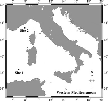
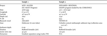
Three specimens, two females and one male, were found in two meiobenthic samples collected from the Mediterranean deep sea during two expeditions. One female specimen, the holotype, was collected in June 1999 during the ‘TransMediterranean’ expedition across the Mediterranean under the MTP–MATER project (Site 1, Figure 1); two more specimens, one male (allotype) and one female (paratype) were found in a sample that was collected in the course of DYFAMED–BENTHOS survey in 1997 (Site 2, Figure 1). Information regarding the sites, sampling and processing of the samples is provided in Table 1.
Location of Dahmsopottekina guilvardi sp. nov. collection sites.

Information on the sites, sampling and processing of the two samples where Dahmsopottekina guilvardi sp. nov. specimens were found.

N/A, not applicable.
The specimens were processed and dissected under an MZ 125 Leica stereomicroscope and were subsequently mounted on slides using glycerol as embedding medium. Examination of the specimens and drawings were made using a camera lucida on a DMR Leica interference contrast microscope.
Abbreviations used in the text: aes, aesthetasc; benp, basendopodite; cpth, cephalothorax; CR, caudal rami; enp, endopodite; exp, exopodite; GDS, genital double somite; P1–P6, pereiopods 1–6.
The type material is deposited in the collection of the Senckenberg Museum in Frankfurt am Main, Germany (SMF).
TYPE MATERIAL
Holotype: adult female 932 µm long, 180 µm wide, dissected and mounted on 13 slides (SMF 32138); paratypes: one male (allotype) 593 µm long, 110 µm wide, dissected and mounted on 7 slides (SMF 32139); one female 689 µm long, mounted on 1 slide (SMF 32140).
TYPE LOCALITY
The new species was found in the western Mediterranean (Figure 1). Details of the habitat are presented in Table 1.
ETYMOLOGY
The new species is named after Dr Laurence Guidi-Guilvard who provided the authors with the paratypes.
DESCRIPTION
Female
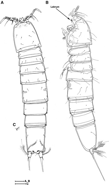
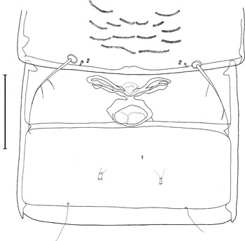
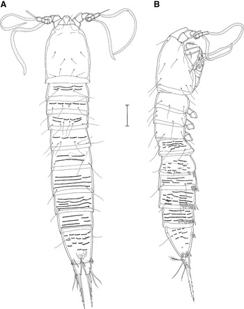
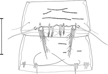
Body vermiform with no distinction between prosome and urosome (Figure 2); urosome longer than prosome (540 versus 392 µm); anal somite elongated, almost one-third of urosome length; with the exception of the penultimate somite, all somites bear dorsally and/or laterally pairs of sensilla; tube pores present on the GDS, the somites anterior and posterior to the GDS, the anal somite and the CR; details on sensillum and tube pore number and pattern are shown in Figure 2; inner thickening of the cuticle crossing the GDS midlength indicates the ancestral articulation between second and third urosomites (Figures 2A & 3); anal operculum convex.
Dahmsopottekina guilvardi sp. nov. Female: habitus in dorsal (A) and lateral (B) view with details of a tube pore (C). Scale bars: (A, B) 50 µm; (C) 20 µm.

Dahmsopottekina guilvardi sp. nov. Female: genital double somite with genital field and P5. Scale bar: 50 µm.

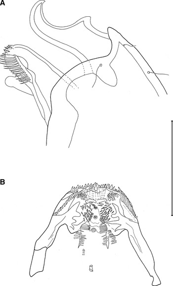
Rostrum prominent, plough-like in shape (Figure 4A); bifid and articulated with the cpth, bearing a pair of short bare setae midlength, near the dorsal margin (Figure 5A); 1 pair of sensilla ventrally inserted, near the articulation with cpth and 1 sensillum at the distal edge of the left side of the bifid projection (Figure 5A).
Dahmsopottekina guilvardi sp. nov. Female: (A) rostrum and labrum, lateral view; (B) labrum, ventral view. Scale bar: 50 µm.

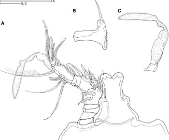
Dahmsopottekina guilvardi sp. nov. Female: antennule (A) with details of first (B) and fourth (C) segment. Scale bars: (A) 50 µm; (B, C) 20 µm.

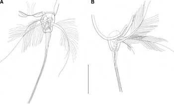
Caudal rami twice longer than wide with 6 setae (Figure 6); setae II and III slender, plumose and of equal size; terminal seta (V) very long and robust accompanied by an outer slender and bare seta (IV) and by a minute spine (VI) slightly exceeding the insertion point; seta VII is slender and plumose, almost half as long as setae II and III and close to a small inner dorsal lobe; 2 tube pores dorsally located on either side of seta II, a third one is located close to the margin with the anal somite; ornamentation appears from midlength downwards on both rami due to surface irregularities.
Dahmsopottekina guilvardi sp. nov. Female: caudal rami in dorsal (A) and lateral (B) view. Scale bar: 50 µm.

Genital field with one copulatory pore located midventrally, anterior to the articulation of the two fused somites (Figure 3); gonopore as a transversal slit located midventrally; seminal receptacle not discernible.
Antennule 6-segmented (Figure 5A); first segment with row of spinules at the inner margin (Figure 5B); segments 4 (Figure 5C) and 5 of equal, very small size; last segment elongated about one-third of the antennule length; armature formula: 1, 6, 5, aes, 1, 11 + aes; 1 inner bipinnate seta at segments 1–3; 3 transformed, aes-like distal setae accompany the aes of last segment; proximal and distal outer setae of last segment biarticulated and plumose.
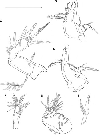
Antenna 3-segmented (Figure 7A); coxa with no armature; allobasis with 1 plumose abexopodal seta; enp with 2 inner and 2 distal spines and with 2 distal bipinnate setae (outermost is shorter); enp ornamented with 5, 3 and 2 long spinules located proximally, midlength and distally respectively; exp very small, 1-segmented with 1 small bipinnate spiniform seta.
Dahmsopottekina guilvardi sp. nov. Female: (A) antenna; (B) mandible; (C) mandibular palp; (D) maxilla; (E) maxilla claw; (F) maxilliped. Scale bar: 50 µm.

Labrum very prominent, extending from cpth as much as the rostrum (Figure 4A); ventrally, of trapezoid shape with very rich and symmetrical ornamentation (Figure 4B); one long semi-circular row of spinules at the distal edge accompanied by a much narrower inner one; a row of spiniform setules associated with cuticular ridges laterally at each side, distal of which spinules are arranged orbicularly; more spinules are associated with the cuticular ridges centrally on a comb-like form; 3 large pores arranged in a line in the middle of the labrum; 1 tube pore is located midwidth at the proximal end of labrum.
Mandible with 5 bidentate teeth at gnathobase which decrease in size from distal to inner margin (Figure 7B); 1 robust pinnate spine and 1 spiniform bipinnate seta stemming midlength at the inner side of gnathobase; 2 rows of spinules located underneath the teeth and seta and 1 small row of spinules present laterally near the outer margin; mandibular palp 2-segmented (Figure 7C); basis with 1 terminal seta at the inner side; enp with 1 inner seta and 1 terminal spiniform seta accompanied by a small outer spine.
Maxillule almost identical to that of D. fodens, the only difference being the lack of one short spine at the outer margin of basis that follows the plumose spine (drawing not provided due to mishandling of the relevant preparation which caused deformation of both maxillules).
Maxilla robust (Figure 7D); syncoxa with 2 endites, each armed with 3 terminal, spiniform, slightly bent inwards setae; distal endite outer seta spinulose; a row of subterminal spinules on each endite; several rows of spinules at the syncoxa located anteriorly and laterally; allobasis claw-like in shape (Figure 7E) with many spinules at the inner distal margin, 2 transformed aes-like setae and 2 spines posteriorly.
Maxilliped 2-segmented; syncoxa with 1 distal bipinnate seta surrounded anteriorly by long spinules (Figure 7F); a row of spinules found posteriorly at midlength; basis with 1 robust, spiniform bipinnate seta accompanied by long setules in a shrubby arrangement (outer distal part of segment); a row of setules covers laterally half the length of basis (inner distal part).
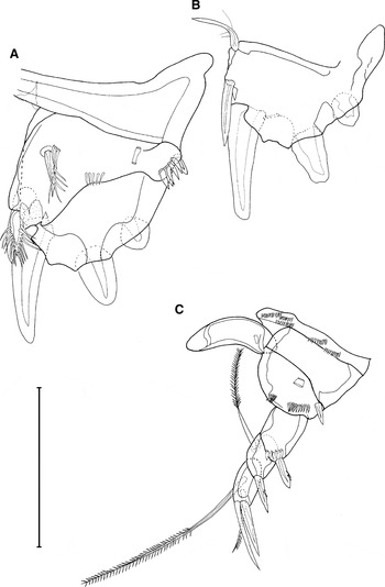
P1 very robust and modified, directed upwards (Figure 8A); coxae of both counterparts fused; basis fused with enp; outer spine stout, accompanied ventrally by long spinules; inner seta spiniform and bipinnate accompanied by 3 long spinules; a row of smaller spinules is located midwidth, near the edge of the basis; distal margin of benp with a pointed tip and a stout bipinate spine inserted at the inner side of the pointed tip; exp 1-segmented, inwardly bent and distally widened, with 3 outer strong spines and 2 distal setae, one spiniform and one plumose (Figure 8B).
Dahmsopottekina guilvardi sp. nov. Female: (A) P1; (B) exopod of P1; (C) P2. Scale bar 50 µm.

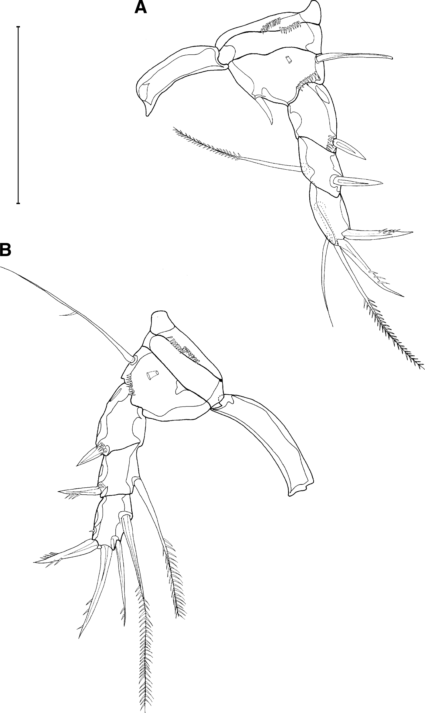
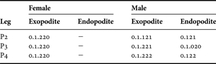
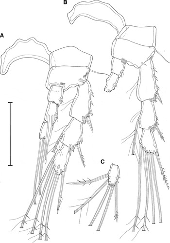
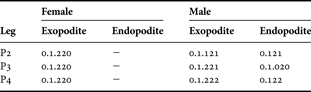
P2–P4 uniramus (Figures 8C & 9); intercoxal sclerites arched; praecoxae with several rows of spinules; coxae plain, with no ornamentation; bases of all swimming legs with a tube pore near the margin with coxae and a row of spinules above insertion of exp; basis of P2 with 1 outer spine and a row of spinules at the inner lateral margin, where it should be the insertion of enp; P3 basis with an inner spine and an outer seta; P4 basis with an outer seta; enp absent; exp 3-segmented, with segments of almost equal size; 1 inner bipinnate seta found at the second and third segment of all swimming legs; most of spines pinnate; spinules found at the first segment, near the insertion of the outer seta; armature formula as in Table 2.
Dahmsopottekina guilvardi sp. nov. Female: (A) P3; (B) P4. Scale bar: 50 µm.
Swimming leg armature formula for Dahmsopottekina guilvardi sp. nov.
P5 represented at each side by a long seta, resembling the outer seta of benp (Figure 3); 1 inner spinule is located close to the seta; 1 tube pore is found above the spinule.
P6 absent.
Male
Male body of same form but smaller than the female (holotype/allotype = 1.6); sexual dimorphism appears in many characters. Differences as follows:
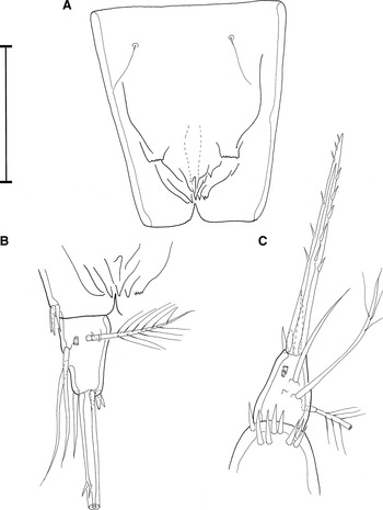
Body: urosome is only slightly longer than prosome (313 versus 280 µm); anal somite less elongated (a quarter of urosome length); with the exception of chpth and CR, body covered with rows of spinules, more markedly at the abdominal somites (Figure 10); ventrally and laterally, all abdominal somites bear a row of spicules above articulation with next somite, which is longer at the intermediate somites; anal somite with 2 rows of spicules posteriorly and 1 anteriorly; tube pores present at segments 3 and 4 of urosome, at anal somite and at CR; sensilla similar with the female in number and pattern but longer; anal operculum not present; integumental folds appear along the anal somite, above the area where operculum should appear (Figure 11A, B).
Dahmsopottekina guilvardi sp. nov. Male: habitus in dorsal (A) and lateral (B) view. Scale bar: 50 µm.

Dahmsopottekina guilvardi sp. nov. Male: (A) anal somite; (B) caudal ramus in dorsal view; (C) caudal ramus in lateral view. Scale bar: 50 µm.

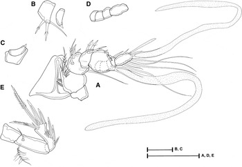
Rostrum of rather trapezoid shape and very much recurved (Figures 10A & 12A); 1 sensillum located dorsally, almost midlength, on the right side (Figure 10A).
Dahmsopottekina guilvardi sp. nov. Male: (A) antennule in ventral view; (B) details of the third and fourth antennule segments (dorsal view); (C) antennule third and fourth segment junction (dorsal view); (D) segments 6–9 of antennule; (E) antenna. Scale bars: (A, D, E) 50 µm; (B, C) 20 µm.

Caudal rami surface without ornamentation (Figure 11B, C); terminal seta shorter, with strong spinules from mid-length downwards; inner distal spine much bigger, exceeding CR; outer distal seta spiniform; seta VII is articulated and the only plumose one; setae II and III are coming out from surface processes; 1 tube pore is located dorsally near seta VII and 2 tube pores are located laterally near seta III; a set of spicules surrounds the inner distal spine.
Antennule 9-segmented, haplocer (Figure 12A); geniculation occurs between the fourth and the fifth segment; third segment with 3 inner spines (Figure 12B), fourth segment very small with 1 very small spine at its inner corner (Figure 12B, C); fifth segment swollen, with 2 small inner spines and 1 distal aes; sixth and seventh segment very small (Figure 12D) and bare; eighth segment with 1 hyaline seta; last segment with all outer elements as bi-articulated setae; 2 setae that accompany the aes of the last segment instead of 3 are aes-like transformed; a third accompanying element in the form of a small spine is located ventrally on the basis of the aes; armature formula: 1, 6, 3, 1, 2 + aes, 0, 0, 1, 9 + aes.
Antenna differs only slightly in spinules arrangement (Figure 12E).
Labrum although with same structure and ornamentation as the female is flat, not exceeding the cpth.
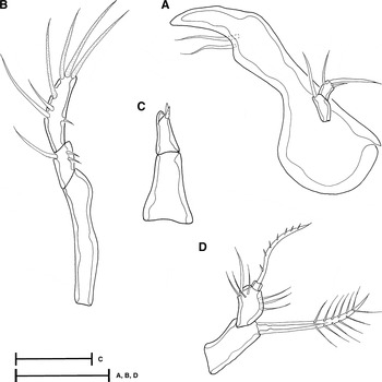
Mandible without a typical gnathobase (Figure 13A); distal end beak-shaped, with no tooth, slightly curved inwards; inner margin with 2 setae distally; mandibular palp as in the female.
Dahmsopottekina guilvardi sp. nov. Male: mouthparts. (A) mandible; (B) maxillule; (C) maxilla (ventral view); (D) maxilliped. Scale bars: (A, B, D) 20 µm; (C) 50 µm.

Maxillule 3-segmented (Figure 13B); praecoxa elongated, unarmed; coxa quite smaller (about 1/5 of praecoxa) with 1 outer distal seta and 2 inner spinules; basis with 3 distal aes-like setae and 3 outer setae, of which the distal one is aes-like; 1 spinule at the inner side of basis.
Maxilla 2-segmented, very much reduced, cone-like, with no endites (Figure 13C); syncoxa unarmed; allobasis with 2 terminal spines, one behind the other on a ventral view.
Maxilliped 3-segmented (Figure 13D); syncoxa with no spinules; basis with spinules in the same arrangement with the female but fewer; enp a tiny distal segment carrying a slender spiniform pinnate seta.
P1 with slightly longer coxa than the female (Figure 14A); traces of the former articulation of basis and enp evident below inner seta of benp; outer spine of basis and accompanying spinules longer and more slender; benp with an extra distal seta protruding from the pointed tip; exp and its outer spines more slender; inner distal seta plumose; distal spiniform seta longer and evidently more robust compared to the female; set of spinules at the insertion point of all spines.
Dahmsopottekina guilvardi sp. nov. Male: (A) P1; (B) P2; (C) endopod of P2. Scale bar: 50 µm.

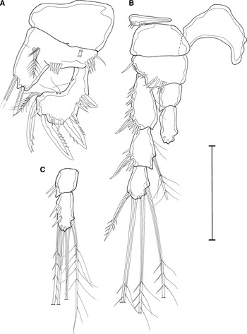
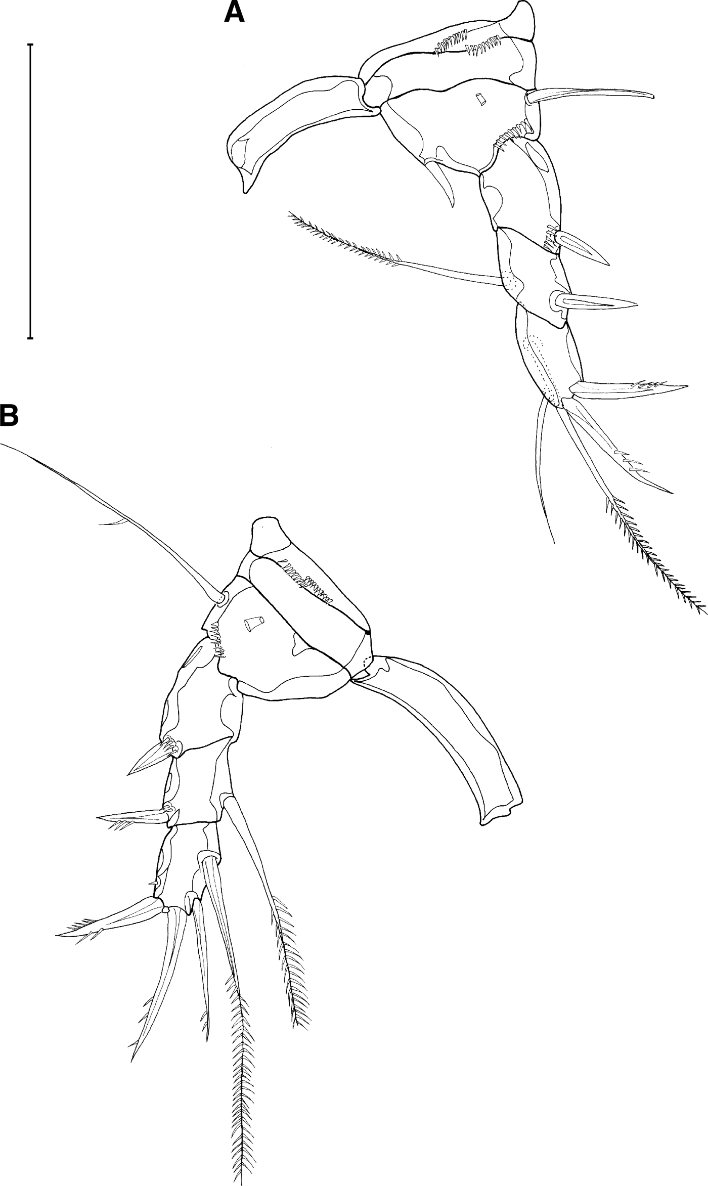
P2–P4 biramus and noticeably longer than the female's one (Figures 14B, C & 15); intercoxal sclerites wider and more arched; praecoxae smaller and less ornamented (Figure 14B); coxae quite wider; bases without tube pores and in general fewer but longer spinules; enp of P2 and P4 2-segmented (Figures 14B, C & 15B, C) with the last segment longer; enp of P3 3-segmented (Figure 15A); first and second segment of all enp with outer row of spinules; first and last segment of P3 enp of almost equal size, middle segment longer with an inner strong modified seta that exceeds the distal edge of the third segment; segments of all exp decrease in length from proximal to distal one; first and second segments with spinules along the outer and distal margin; last segment of P3 exp with 1 tube pore at the distal margin; outer spine of last segment of all exp pinnate; second segment of P3–P4 exp also with an outer pinnate seta; last segment of all exp with distal setae which are pinnate at the outer side and plumose at the inner side; all the other setae in both exp and enp plumose; all setae markedly longer than the female's one; armature formula presented in Table 2.
Dahmsopottekina guilvardi sp. nov. Male: (A) P3; (B) P4; (C) endopod of P4. Scale bar: 50 µm.

P5 well developed and plate-like (Figure 16); basis and enp fused; basis with 1 plain seta; enp with 2 spiniform bipinnate setae, the inner one of which is slightly bigger; exp 1-segmented with 1 spine and 2 setae; inner seta plumose, outer seta longer and spiniform; 2 tube pores appear at each side, one above the spine of exp and one above the outer seta of enp.
Dahmsopottekina guilvardi sp. nov. Male: ventral view of urosome showing P5–P6. Scale bar: 50 µm.

P6 plate-like, asymmetrical, distinctly separated only on the left side (Figure 16); it consists of 3 spines, the innermost of which is longer (about double in length).
DISCUSSION
Harpacticoid fauna of the Mediterranean deep sea is poorly known. To date, only three species have been described, all of which have been collected from Anaximenes Seamount (eastern Mediterranean) and belong to the family Ancorabolidae (Gheerardyn & George, Reference Gheerardyn and George2010; Schulz & George, Reference Schulz and George2010). The new species constitutes the first record of the genus Dahmsopottekina from the Mediterranean Sea. Similar to its congeners, Dahmsopottekina guilvardi sp. nov. was collected from deep-sea habitats, appears to be sparsely distributed and is highly sexually dimorphic.
Dahmsopottekina guilvardi sp. nov. presents most of the diagnostic characters of the genus as described by Dahms & Pottek (Reference Dahms and Pottek1992), namely: habitus vermiform; somites with a peculiar pattern of distinct and symmetrical denticle rows on surface (present in the male of Dahmsopottekina guilvardi sp. nov.); anal somite elongated; rostrum plough-like (in the female); third segment of antennule without denticulated spiniform seta; mandible enp with 1–3 setae; gnathobase with 2 stout and curved elements; maxilliped with a long spine at basis, spinulated along the tapering tip; coxae of P1 fused; P1 highly transformed and sexually dimorphic, with exp 1- or 3-segmented bending inwardly in the females and lobe-like enp bearing a plumose seta; P5 either reduced to a single lobe bearing only the outer seta of benp or with a separate exp; CR small, longer than wide, with a small dorsal lobe next to dorsal seta and pore with bag-like tube on dorsal surface.
Morphological similarities and differences with congeners
As has already been pointed out and confirmed by the present study, Dahmsopottekina demonstrates a high degree of sexual dimorphism (Becker, Reference Becker1979; Dahms & Pottek, Reference Dahms and Pottek1992). Furthermore, the genus is characterized by a female biased sex-ratio (Dahms & Pottek, Reference Dahms and Pottek1992) which along with its sparse distribution has resulted in the description of species based primarily on female specimens. Therefore, it was necessary to place the new species among its congeners using female characters only.
Dahms & Pottek (Reference Dahms and Pottek1992) defined two morphological groups within Dahmsopottekina; one characterized by the presence of a 1-segmented P1 exp in the females and mandibular enp with 3 setae (which consists of the bulk of Dahmsopottekina species) and a second one to which belong the species with a 3-segmented P1 exp and up to 2 setae in the mandibular enp. Dahmsopottekina guilvardi sp. nov. is assigned to the first group, within which it exhibits striking similarities with D. fodens and to a lesser degree with D. bathyalis and D. noodti, all of which lack an enp at P2–P4. Further similarities with those species from the Antarctic are the bifid rostrum, which becomes plough-like in D. fodens and D. noodti, the overall appearance and armature of the maxilliped and the fusion of P1 coxae. The new species shares some more characters with D. fodens as they appear to have identical antenna, labrum, maxillule and maxilla. Furthermore, although the maxilliped of D. fodens is not fully described, there is significant indication that the maxillipeds are also alike. Despite the difference in setal formula between D. guilvardi sp. nov. and D. fodens, there is a great resemblance in P2–P4's overall appearance, ornamentation and seta type. Dahmsopottekina guilvardi sp. nov. has the same number of setae in P2–P4 as D. bathyalis; however, they differ with regard not only to setal formula but also with regard to form and length of setae. The aforementioned differences are more pronounced when the new species is compared to the rest of its congeners, with the exception of D. fodens. Although D. guilvardi sp. nov. appears quite different from D. peruana, a deep-sea species from the Pacific Ocean, the most important difference being the presence of a 3-segmented enp in P2–P4 in the latter, nonetheless, they are the only two species among harpacticoids in which the basis and enp of a leg other than the P5, the P1 in particular, are fused forming a rather triangular plate.
When comparing the male of D. guilvardi sp. nov. with the congeneric males, the differences are more striking. Besides the different setal formula, the male of the new species has a 9-segmented antennule (up to 7 in the previously described males) and, similar to the female, a 1-segmented P1 exp. Nevertheless, in some characters it very much resembles the larger Antarctic species D. pectinata, such as in the antenna, mouthparts, P6 and body ornamentation.
Overall, D. guilvardi sp. nov. can be distinguished from its congeneric species by the combination of a fused basis and enp in P1 of both sexes and the absence of an enp in P2–P4 of the female. Further diagnostic characters are the very short inner terminal spine of CR (almost exceeds the CR) and the surficial ornamentation of CR in the female. Within the species group of the 1-segmented P1 exp, D. guilvardi sp. nov. can also be distinguished by the presence of an inner spine at the basis of P3 in the female.
Sexual dimorphism
Similar to other representatives of Harpacticoida, the sexual dimorphism in Dahmsopottekina is apparent in the body size, antennules, P2–P4 enp, P5 and P6. But according to Dahms & Pottek (Reference Dahms and Pottek1992) it also extends to the rostrum, P1, maxilliped and maxilla. More specifically, they suggest that sexual dimorphism further appears as follows: morphology of P1 is different and less transformed in the males, the maxilliped and the maxilla are much reduced in the females and the rostrum is plough-like and prominent in the females but triangular and not prominent in the males (Dahms & Pottek, Reference Dahms and Pottek1992).
Indeed, in D. guilvardi sp. nov. the antennule and P2–P6 are reduced in the female. Furthermore, the two females of the new species are larger than the male. However, the length ratio of the two females (holotype/paratype = 1.35) is higher than the ratio of the two specimens found in the same sample (paratype/allotype = 1.16), indicating high length variability within the species. Therefore, the statement of considerably larger females cannot be supported by the present study. A high variability in length has also been reported for deep-sea Ectinosomatidae by Seifried et al. (Reference Seifried, Plum and Schulz2007) who assumed this to be a deep-sea phenomenon.
Similarly, the observation of a reduced maxilliped and maxilla in the females is not verified in the case of D. guilvardi sp. nov., in which the male seems to have the rather reduced specific appendages. The rostrum, although prominent in both the female and the male of D. guilvardi sp. nov. is different in shape, suggesting a burrowing mode of life in the female. P2–P4 are less transformed in the male specimen; nevertheless, P1 is equally modified in both sexes, therefore indicating the digging habit of the species. However, the frequent presence of both sexes in sediment trap samples (Guidi-Guilvard et al., Reference Guidi-Guilvard, Thistle, Khripounoff and Gasparini2009) strongly suggests that D. guilvardi sp. nov. forms part of hyperbenthos as an active ‘swimmer’.
In D. guilvardi sp. nov. the labrum of the female is prominent and more elaborate than the male's and therefore could serve as a sexually dimorphic character. However, since this character has not been consistently considered in previous descriptions, in particular in those species for which both a female and a male description exist, its value as a sexually dimorphic character cannot currently be estimated. It is recommended though to examine this character carefully in any new species belonging to Dahmsopottekina.
Distribution
Dahmsopottekina species have been previously reported from deep-sea habitats (400–5750 m) of the North Atlantic Ocean (D. bifida and D. curticauda), the North Pacific Ocean (D. micracantha), the South Pacific Ocean (D. pacifica, D. peruana and D. talpa) and the Weddell Sea (D. bathyalis, D. fodens, D. furcispina, D. noodti and D. pectinata), all of which have never been reported outside their type locality. Similar to its congeners, Dahmsopottekina guilvardi sp. nov. was collected from the deep sea, more specifically from abyssal habitats. Though several samples along the ‘TransMediterranean’ transect were examined for the presence of the new species (Sevastou et al., unpublished), as well as samples from a recent expedition in the eastern Mediterrnean (LEVAR expedition—Meteor 71/2: Martínez Arbizu, personal communication), no individual of the new species was detected. Nevertheless, 13 individuals of the new species were found at DYFAMED permanent station at 11 sediment trap deployments between January 1996 and April 1998 (Guidi-Guilvard et al., Reference Guidi-Guilvard, Thistle, Khripounoff and Gasparini2009). This supports the observation made by Dahms & Pottek (Reference Dahms and Pottek1992) that Dahmsopottekina species are sparsely distributed, while at the same time it provides some indication of the species being endemic to the western Mediterranean deep sea.
ACKNOWLEDGEMENTS
The authors are grateful to Dr L. Guidi-Guilvard for kindly providing the allotype and paratype of the new species. K.S. acknowledges a CeDAMar taxonomic exchange fellowship that enabled her to visit the German Centre for Marine Biodiversity Research (DZMB) where the present study was carried out. We would like also to thank Mrs M. Eleftheriou and Professor A. Eleftheriou for the critical reading of the manuscript. The studied material was collected under the MTP–MATER project, European Commission's MAST Program (Sample 1) and the French JGOFS Program funded by the CNRS/INSU, France (Sample 2).