Introduction
In 2010, approximately 38% of the European population, almost 165 million people, suffered from neuropsychiatric disorders: the largest contributor to the overall burden of morbidity in Europe (Wittchen et al., Reference Wittchen, Jacobi, Rehm, Gustavsson, Svensson, Jonsson and Steinhausen2011). Psychoses in general, and particularly schizophrenia, are among the disorders that incur high costs and result in many years living with disability (Vigo, Thornicroft, & Atun, Reference Vigo, Thornicroft and Atun2016). Psychotic disorders have a complex etiology that culminates in a first episode during late adolescence or early adulthood (Kirkbride et al., Reference Kirkbride, Fearon, Morgan, Dazzan, Morgan, Tarrant and Jones2006; Kohler et al., Reference Kohler, van der Werf, Hart, Morrison, McCreadie, Kirkpatrick and Allardyce2009). In the vast majority of cases, the first episode is preceded by a prodrome of 5–6 years on average in which a multitude of mental problems and symptoms, as well as the first psychosocial deficits, occur (Salokangas et al., Reference Salokangas, Heinimaa, From, Loyttyniemi, Ilonen and Luutonen2014; Schultze-Lutter et al., Reference Schultze-Lutter, Rahman, Ruhrmann, Michel, Schimmelmann, Maier and Klosterkotter2015b). Furthermore, a longer duration of untreated psychosis and its preceding prodrome has been associated with negative outcomes (Penttila, Jaaskelainen, Hirvonen, Isohanni, & Miettunen, Reference Penttila, Jaaskelainen, Hirvonen, Isohanni and Miettunen2014). Thus, early detection of and intervention for psychotic disorders during its prodrome – i.e. an indicated prevention of psychosis – has been widely adopted for its potential to alter the course of psychotic disorders (Fusar-Poli et al., Reference Fusar-Poli, Borgwardt, Bechdolf, Addington, Riecher-Rossler, Schultze-Lutter and Yung2013; Schmidt et al., Reference Schmidt, Schultze-Lutter, Schimmelmann, Maric, Salokangas, Riecher-Rossler and Ruhrmann2015; Schultze-Lutter et al., Reference Schultze-Lutter, Michel, Schmidt, Schimmelmann, Maric, Salokangas and Klosterkotter2015a).
Over the past two decades, research has increasingly focused on the identification of sensitive and specific clinical high-risk (CHR) criteria that can inform assessments of psychosis through clinical interviews conducted by trained mental health professionals (Fusar-Poli et al., Reference Fusar-Poli, Cappucciati, Rutigliano, Schultze-Lutter, Bonoldi, Borgwardt and McGuire2015; Schmidt et al., Reference Schmidt, Schultze-Lutter, Schimmelmann, Maric, Salokangas, Riecher-Rossler and Ruhrmann2015; Schultze-Lutter et al., Reference Schultze-Lutter, Michel, Schmidt, Schimmelmann, Maric, Salokangas and Klosterkotter2015a). Two major approaches to CHR criteria are currently used to identify an increased psychosis risk (Fusar-Poli et al., Reference Fusar-Poli, Borgwardt, Bechdolf, Addington, Riecher-Rossler, Schultze-Lutter and Yung2013; Fusar-Poli et al., Reference Fusar-Poli, Cappucciati, Rutigliano, Schultze-Lutter, Bonoldi, Borgwardt and McGuire2015; Schultze-Lutter et al., Reference Schultze-Lutter, Michel, Schmidt, Schimmelmann, Maric, Salokangas and Klosterkotter2015a): (1) Symptomatic ultra-high risk (UHR) criteria rely on attenuated or brief intermittent psychotic symptoms (APS and BIPS, respectively) to detect an imminent risk of psychosis within 12 months and additionally include the non-symptomatic genetic risk-functional decline criterion (Fusar-Poli et al., Reference Fusar-Poli, Borgwardt, Bechdolf, Addington, Riecher-Rossler, Schultze-Lutter and Yung2013). Meta-analyses indicate that UHR criteria achieve conversion rates of 9.6% at 6 months and 37% at >4 years (Schultze-Lutter et al., Reference Schultze-Lutter, Michel, Schmidt, Schimmelmann, Maric, Salokangas and Klosterkotter2015a). (2) Basic symptom (BS) criteria rely on subjective disturbances in thought and perception processes and aim to detect risk of psychosis as early as possible. Fourteen BSs (online Supplemental Table S1) were identified to be specific to the development of first-episode psychosis (FEP) and were employed in two CHR criteria: cognitive disturbances (COGDIS) and cognitive-perceptive basic symptoms (COPER) (Schultze-Lutter, Reference Schultze-Lutter2009; Schultze-Lutter et al., Reference Schultze-Lutter, Ruhrmann, Fusar-Poli, Bechdolf, Schimmelmann and Klosterkotter2012; Schultze-Lutter, Klosterkotter, Picker, Steinmeyer, & Ruhrmann, Reference Schultze-Lutter, Klosterkotter, Picker, Steinmeyer and Ruhrmann2007b). Conversion rates in samples using BS criteria range from 25.3% at 1 year to 61.3% at >4 years (Schultze-Lutter et al., Reference Schultze-Lutter, Michel, Schmidt, Schimmelmann, Maric, Salokangas and Klosterkotter2015a). Importantly, in clinical studies that assessed both UHR and BS, conversion rates were highest when criteria of both approaches were fulfilled (UHR&BS) (Michel, Ruhrmann, Schimmelmann, Klosterkotter, & Schultze-Lutter, Reference Michel, Ruhrmann, Schimmelmann, Klosterkotter and Schultze-Lutter2014; Schultze-Lutter et al., Reference Schultze-Lutter, Ruhrmann, Fusar-Poli, Bechdolf, Schimmelmann and Klosterkotter2012; Schultze-Lutter, Klosterkotter, & Ruhrmann, Reference Schultze-Lutter, Klosterkotter and Ruhrmann2014); moreover, in a community sample of individuals aged 16–40 years, the odds of identifying a mental disorder or functional impairment were by far the highest when the criteria of both approaches were met (Ruhrmann et al., Reference Ruhrmann, Schultze-Lutter, Salokangas, Heinimaa, Linszen, Dingemans and Klosterkotter2010; Schultze-Lutter et al., Reference Schultze-Lutter, Klosterkotter and Ruhrmann2014). Yet, on account of the considerable rate of CHR patients not converting to full-blown psychosis, the use of additional predictors, in particular structural and functional brain aberrations, have been proposed to improve the prognostic value of CHR criteria (Cannon, Reference Cannon2016; Khoury & Nasrallah, Reference Khoury and Nasrallah2018; Kindler et al., Reference Kindler, Schultze-Lutter, Michel, Martz-Irngartinger, Linder, Schmidt and Walther2016, Reference Kindler, Schultze-Lutter, Hauf, Dierks, Federspiel, Walther and Hubl2018; Koutsouleris et al., Reference Koutsouleris, Riecher-Rossler, Meisenzahl, Smieskova, Studerus, Kambeitz-Ilankovic and Borgwardt2015, Reference Koutsouleris, Kambeitz-Ilankovic, Ruhrmann, Rosen, Ruef, Dwyer and Consortium2018; Mikanmaa et al., Reference Mikanmaa, Grent-'t-Jong, Hua, Recasens, Thune and Uhlhaas2019).
Abnormalities in gray matter volume (GMV) occurring before the onset of psychosis have been intensively studied in UHR patients over the last decade (Borgwardt et al., Reference Borgwardt, Riecher-Rossler, Dazzan, Chitnis, Aston, Drewe and McGuire2007). A meta-analysis of neuroimaging studies that collectively investigated the GMV of 896 UHR patients and 701 controls observed widely distributed GMV reductions, especially in the prefrontal, limbic, and temporoparietal cortices, in UHR patients compared to controls (Fusar-Poli et al., Reference Fusar-Poli, Borgwardt, Crescini, Deste, Kempton, Lawrie and Sacchetti2011a). Although BSs have been regarded as the most immediate psychopathological expression of neurobiological aberrations underlying the development of psychosis (Schultze-Lutter, Reference Schultze-Lutter2009; Schultze-Lutter et al., Reference Schultze-Lutter, Ruhrmann, Fusar-Poli, Bechdolf, Schimmelmann and Klosterkotter2012, Reference Schultze-Lutter, Debbane, Theodoridou, Wood, Raballo, Michel and Uhlhaas2016, Reference Schultze-Lutter, Ruhrmann, Michel, Kindler, Schimmelmann and Schmidt2020), comparatively less research has considered the neurobiological correlates of BS (Schultze-Lutter et al., Reference Schultze-Lutter, Debbane, Theodoridou, Wood, Raballo, Michel and Uhlhaas2016): only five studies have hitherto investigated GMV characteristics related to BS in CHR samples (Hurlemann et al., Reference Hurlemann, Jessen, Wagner, Frommann, Ruhrmann, Brockhaus and Maier2008; Koutsouleris et al., Reference Koutsouleris, Davatzikos, Borgwardt, Gaser, Bottlender, Frodl and Meisenzahl2014, Reference Koutsouleris, Meisenzahl, Davatzikos, Bottlender, Frodl, Scheuerecker and Gaser2009a,Reference Koutsouleris, Schmitt, Gaser, Bottlender, Scheuerecker, McGuire and Meisenzahlb; Tepest et al., Reference Tepest, Schwarzbach, Krug, Klosterkotter, Ruhrmann and Vogeley2013). These studies distinguished indications of early risk of psychosis from late risk: the former is characterized by either COPER or the UHR genetic risk and functional decline criterion (GRFD) in the absence of symptomatic UHR states; the latter, by the fulfillment of APS or BIPS criteria. In comparison to controls, the COPER/GRFD group presented more GMV reductions in the fusiform, superior, middle, and inferior temporal gyri, the amygdala, and the hippocampus. A dearth of studies on the COGDIS criterion and the combined presence of UHR and BS criteria (UHR&BS) remains in the literature. As UHR&BS has been associated with increased clinical conversion rates, it can be assumed that this combination also associates more strongly with neurobiological markers such as GMV abnormalities.
The present study therefore aimed to differentially investigate the GMV of brain regions previously associated with UHR criteria (Fusar-Poli et al., Reference Fusar-Poli, Borgwardt, Crescini, Deste, Kempton, Lawrie and Sacchetti2011a) by separating our CHR sample into three different groups: those who fulfilled only UHR criteria, those who fulfilled only BS criteria, and those who fulfilled both (UHR&BS). We expected the UHR&BS group to feature the most pronounced GMV reductions – similar in severity to the GMV reductions of the FEP group – followed respectively by the BS or UHR groups in comparison to a clinical control group (CC). Furthermore, we assessed whether GMV reductions are differentially related to BS or UHR severity; as BS is considered to more strongly correspond with neural maturation, we predicted that GMV reductions would be more closely linked to BS (Schultze-Lutter et al., Reference Schultze-Lutter, Ruhrmann, Michel, Kindler, Schimmelmann and Schmidt2020).
Method
Participants
The study sample (N = 90) was recruited at the Bern Early Recognition and Intervention Center (FETZ Bern, www.upd.ch/fetz). The FETZ Bern is the only psychosis-risk detection center in the Canton of Bern with a catchment area of approximately 1.5 million inhabitants; the center screens ~80 patients/year (age 8–40 years) for psychosis risk symptoms according to state-of-the-art guidelines (Schmidt et al., Reference Schmidt, Schultze-Lutter, Schimmelmann, Maric, Salokangas, Riecher-Rossler and Ruhrmann2015; Schultze-Lutter et al., Reference Schultze-Lutter, Michel, Schmidt, Schimmelmann, Maric, Salokangas and Klosterkotter2015a). Apart from accepting patients who enroll on their own initiative, patients with various psychiatric symptoms are admitted to the FETZ Bern by physicians and psychosocial institutions whenever there is clinical suspicion of early psychotic development. The clinical basic assessment includes a psychopathological evaluation, a cognitive test battery, cerebral magnetic resonance imaging (cMRI), and a routine blood screening. The present study sample consisted of consecutive attendees assessed from 2010 to 2018. Patients were diagnosed as CHR (n = 48), CC (n = 28), or FEP (n = 14) with full-blown psychotic symptoms. Patients considered to be CC did not fulfil any CHR criterion and had no history of past or present psychosis. Those with CHR were further distinguished as fulfilling BS (n = 14), UHR (n = 16), or UHR&BS (n = 18) criteria. For the current study, we included all patients from the FETZ Bern that (1) were admitted to our center, (2) were diagnosed with CHR or FEP or qualified as CC, (3) were subjected to standardized cMRI, and (4) agreed to the anonymized scientific use of their data.
We received approval for all procedures from the ethics committee of the Canton of Bern. All participants gave informed consent and, in the case of minors, parental informed consent with the child's assent was provided.
Diagnostic assessment
Two sets of CHR criteria were employed (Fusar-Poli et al., Reference Fusar-Poli, Borgwardt, Bechdolf, Addington, Riecher-Rossler, Schultze-Lutter and Yung2013; Schultze-Lutter et al., Reference Schultze-Lutter, Michel, Schmidt, Schimmelmann, Maric, Salokangas and Klosterkotter2015a): (1) The UHR criteria (Yung et al., Reference Yung, Phillips, McGorry, McFarlane, Francey, Harrigan and Jackson1998) were evaluated with the Structured Interview for Psychosis-Risk Syndromes (SIPS) (McGlashan, Walsh, & Woods, Reference McGlashan, Walsh and Woods2010), which assesses the presence of APS, BIPS, and GRFD criteria. (2) The BS criteria (Klosterkotter, Hellmich, Steinmeyer, & Schultze-Lutter, Reference Klosterkotter, Hellmich, Steinmeyer and Schultze-Lutter2001; Schultze-Lutter et al., Reference Schultze-Lutter, Ruhrmann, Fusar-Poli, Bechdolf, Schimmelmann and Klosterkotter2012, Reference Schultze-Lutter, Debbane, Theodoridou, Wood, Raballo, Michel and Uhlhaas2016), including COPER and COGDIS, were assessed with the Schizophrenia Proneness Instrument, Adult version (SPI-A) (Schultze-Lutter, Addington, Ruhrmann, & Klosterkotter, Reference Schultze-Lutter, Addington, Ruhrmann and Klosterkotter2007a), and Child & Youth Version (SPI-CY) (Schultze-Lutter & Koch, Reference Schultze-Lutter, Koch and Fioriti2010), respectively. All interviewers underwent intensive training for 3 months prior to diagnosing patients and supervising ratings during the diagnostic process by Frauke Schultze-Lutter (F.S.-L).
The adult and pediatric versions of the Mini-International Neuropsychiatric (Sheehan et al., Reference Sheehan, Lecrubier, Sheehan, Amorim, Janavs, Weiller and Dunbar1998, Reference Sheehan, Sheehan, Shytle, Janavs, Bannon, Rogers and Wilkinson2010) were used to assess diagnoses informed by the Diagnostic and Statistical Manual of Mental Disorders IV. Information on concurrent diagnoses made according to the Diagnostic and Statistical Manual of Mental Disorders IV is provided in online Supplemental Table S2. Psychosocial functioning was evaluated with the Social and Occupational Functioning Assessment Scale (SOFAS) (APA, 1994). Antipsychotic medication was converted to chlorpromazine equivalents according to standard guidelines (Leucht, Wahlbeck, Hamann, & Kissling, Reference Leucht, Wahlbeck, Hamann and Kissling2003).
MRI data analysis
Imaging was performed using a 3.0 T Magnetom Verio (Siemens Medical Systems, Erlangen, Germany) with a standard 12-channel radio frequency head coil. High-resolution structural images were obtained using 3D T1-weighted modified driven equilibrium Fourier transform (MDEFT) scans [sagittal slices, 176; 256 × 256 matrix; slice thickness, 1 mm; voxel size, 1 × 1 × 1 mm3; repetition time (TR), 7.92 ms; echo time (TE), 2.48 ms; flip angle (FA), 16°].
Analysis of gray matter in MR images was performed on a Mac OSX 10.8 workstation using FreeSurfer version software (version 6.0). All data sets were screened for anatomical abnormalities, excessive motion, successful normalization, and artifacts. First-level autoreconstruction was then performed with the images in FreeSurfer. The skull-stripped brains were checked for remaining dura, sinuses, or other artifacts that could interfere with successful segmentation. When artifacts were found, images were edited manually. When deemed sufficiently clean, images were run through second- and third-level autoreconstruction, which allowed for the extractions of GM surface area, thickness, and volume. Finally, automated cortical parcellation was performed using a separate processing pipeline included in the FreeSurfer software package.
A priori, 20 regions of interest (ROI) previously associated with risk of psychosis (Fusar-Poli et al., Reference Fusar-Poli, Borgwardt, Crescini, Deste, Kempton, Lawrie and Sacchetti2011a; Fusar-Poli, Radua, McGuire, & Borgwardt, Reference Fusar-Poli, Radua, McGuire and Borgwardt2012; Roalf et al., Reference Roalf, Quarmley, Calkins, Satterthwaite, Ruparel, Elliott and Turetsky2017) were defined, and their respective GMVs were calculated: the anterior cingulate (rostral & caudal), caudal middle frontal, entorhinal, hippocampus, inferior parietal, inferior temporal, insula, lateral occipital, lateral orbitofrontal, medial orbitofrontal, middle temporal, pars opercularis, pars triangularis, precentral, precuneus, rostral middle frontal, superior frontal, superior temporal, superior temporal sulcus, and supramarginal (bilaterally).
Statistics
Demographics
SPSS 21.0 was used to compare frequencies and percentages with χ2-square tests, means of normally distributed interval data with two-sample t tests, analysis of variance (ANOVA), and non-normally distributed interval or ordinal data with Kruskal–Wallis and Mann–Whitney U tests. Statistical significance of two-sided tests was set at p ⩽ 0.05.
Neuroimaging
First, an overall analysis of covariance (ANCOVA) with the ROI as the within-subject factor and the diagnostic group (CC, FEP, BS, UHR, and UHR&BS) as the between-subject factor with a covariate of total intracranial volume (TIV) was calculated. Post-hoc group comparisons were conducted using Bonferroni correction.
Second, 20 separate ANCOVAs with diagnostic group as the fixed factor and GM volume for each ROI with total intracranial volume as the covariate were calculated. False discovery rate (FDR) corrected results are reported. Post-hoc group comparisons were conducted, and Bonferroni corrected results are reported. Effect sizes for ANCOVAs were calculated with Partial Eta Squared (η 2) for pairwise comparisons with Cohen's d (d).
Finally, in those ROIs with a significant group effect, Spearman rank correlations were conducted in the CHR group between COPER, COGDIS, and APS/BIPS (SISOP total/positive) scores and cortical GM volumes (p ⩽ 0.05, 2-sided, FDR corrected).
Results
Demographics
No differences in age, sex (Table 1), or chlorpromazine equivalents (F4,13 = 0.3, p = 0.87) were detected among the groups. Patients with FEP exhibited lower psychosocial function (Table 1).
Sample demographics

CC = clinical controls; FEP = first-episode psychosis; BS = 14 basic symptoms included in COPER and COGDIS; UHR = ultra high risk; Sex m = male, f = female; SOFAS = Social and Occupational Functioning Assessment Scale; SIPS = Structured Interview for Psychosis-Risk Syndromes; SIPS pos = Sum score of the 5 SIPS positive items.
Test value refers to Anova, Kruskal–Wallis or χ2-square tests. Significance was set at p < 0.05, two-sided (uncorrected).
Among the CHR patients, those with UHR had significantly lower BS total scores than did the patients with BS (p = 0.018) or BS&UHR (p = 0.001). Additionally, post-hoc group comparisons yielded significantly lower SIPS positive scores among BS patients than BS&UHR patients (p = 0.037). No other significant differences were detected.
GMV group differences
Considering total ROIs, the overall ANCOVA yielded a significant group effect (F4,84 = 3.61, p = 0.009, η 2 = 0.147), indicating significant group differences in GMV. Post-hoc tests indicated significantly lower GMV in FEP when compared to CC (18 244.9 ± 1391.7 μl v. 19 649.1 ± 1391.5 μl, p = 0.028, d = 1.0), and significantly lower GMV in UHR&BS when compared to CC (18 321.8 ± 1394.7 μl v. 19 649.1 ± 1391.5 μl, p = 0.023, d = 0.95), but no other significant differences.
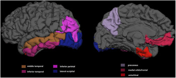
When separated for each ROI, seven of the 20 investigated ROIs showed a significant group effect, and remained significant after FDR correction (Fig. 1): entorhinal (F4,84 = 3.2, p = 0.017, η 2 = 0.133), inferior parietal (F4,84 = 4.4, p = 0.003, η 2 = 0.173), inferior temporal (F4,84 = 3.3, p = 0.015, η 2 = 0.136), lateral occipital (F4,84 = 3.6, p = 0.009, η 2 = 0.148), medial orbitofrontal (F4,84 = 3.8, p = 0.007, η 2 = 0.152), middle temporal (F4,84 = 3.3, p = 0.014, η 2 = 0.137), and precuneus (F4,84 = 3.5, p = 0.01, η 2 = 0.145) (for means and standard error of brain volumes, see online Supplementary Table S3).
Freesurfer-based visualization of the seven regions of interest with significant group effects in gray matter volume. The inferior parietal (p < 0.005), inferior temporal (p = 0.05), lateral occipital (p < 0.01), medial orbitofrontal (p < 0.01), middle temporal (p ⩽ 0.05), precuneus (p = 0.01) and entorhinal (p < 0.05) cortex, showed a significant group effect (FDR corrected).

Pairwise comparisons of GM volumes between diagnostic groups are depicted in Table 2 and Fig. 2. Six out of seven ROIs remained significant after Bonferroni correction. Three ROIs (inferior parietal, lateral occipital, and medial orbitofrontal) showed significantly lower GMV in UHR&BS and FEP, and another two ROIs (middle temporal, precuneus) showed significantly lower GMV in UHR&BS as compared to CC, whereas entorhinal cortex showed significantly higher GMV in UHR as compared to CC. For the inferior temporal cortex, pairwise comparisons did not yield any significant group difference (p > 0.05).
Gray matter volumes in diagnostic subgroups, pairwise comparisons. Controls = clinical controls, FEP = first-episode psychosis, BS = basic symptoms, UHR = ultra-high risk. Gray matter cortical volume in μl. Five areas (inferior temporal, inferior parietal, lateral occipital, medial orbitofrontal, precuneus) showed the lowest GMV in FEP and/or UHR&BS, whereas the entorhinal cortex showed highest GMV in UHR..*significant 2-group differences at p < 0.05, ** significant at p < 0.01 (Bonferroni corrected).

Pairwise comparisons of gray matter volumes of the seven ROIs, significant in overall ANCOVA

CC = clinical controls, FEP = first-episode psychosis, BS = basic symptoms, UHR = ultra-high risk, p-values refer to ANCOVA with gray matter volume as a dependent variable, group (UHR, BS, UHR&BS, FEP, CC) as a fixed factor and intracranial volume as a covariate, Cohen's d was calculated to measure effect size, n.s. not significant, * significant at p < 0.05, **significant at p < 0.01, Bonferroni corrected.
GMV correlations with COGDIS, COPER, and APS/BIPS scores
In CHR patients (n = 48), significant negative correlations were detected between COGDIS scores and GMV in two ROIs: inferior parietal (R = −0.36, p ⩽ 0.05) and middle temporal (R = −0.34, p ⩽ 0.05).
No significant correlations were detected between COPER or APS/BIPS scores and GMV in any ROI (R < 0.30, p > 0.05).
Discussion
To the best of our knowledge, this is the first neuroimaging study to differentially evaluate GMV reductions in patients fulfilling different CHR criteria – namely, the UHR and BS criteria, and the combination of both – in comparison to FEP and CC.
GMV group differences in total
As expected, when all 20 investigated ROIs were considered together, the GMV reductions of patients presenting with UHR&BS criteria and, hence, the highest risk of psychosis were found to be the most similar to those of FEP: both groups showed significantly lesser GMV than did the CC group. When the ROIs were separated, five regions – inferior temporal, inferior parietal, lateral occipital, medial orbitofrontal, precuneus – were both: (1) following our primary hypothesis of significantly lower gray matter in FEP and UHR&BS as compared to CC and (2) remaining significant after FDR correction. Importantly, the findings were of medium-to-large effect sizes. In line with what has been reported for conversion prediction (Schultze-Lutter et al., Reference Schultze-Lutter, Klosterkotter and Ruhrmann2014) and for clinical relevance of CHR symptoms and criteria (Schultze-Lutter, Michel, Ruhrmann, & Schimmelmann, Reference Schultze-Lutter, Michel, Ruhrmann and Schimmelmann2018a), our results indicate an additive effect of the two major evaluative approaches to CHR criteria also in detecting early neurobiological aberrations potentially related to the future development of psychosis, such as GMV reductions.
In line with the conceptualization of BS as the most immediate symptomatic expression of the neurobiological processes leading to psychosis (Schultze-Lutter et al., Reference Schultze-Lutter, Debbane, Theodoridou, Wood, Raballo, Michel and Uhlhaas2016) and of cognitive deficits as a core dimension of psychoses (Fluckiger et al., Reference Fluckiger, Michel, Grant, Ruhrmann, Vogeley, Hubl and Schultze-Lutter2019; Heckers et al., Reference Heckers, Barch, Bustillo, Gaebel, Gur, Malaspina and Carpenter2013; Insel, Reference Insel2010; Mollon, David, Zammit, Lewis, & Reichenberg, Reference Mollon, David, Zammit, Lewis and Reichenberg2018), the severity of the cognitive BS, included in COGDIS, correlated with GMV reductions; foremost in parietal and temporal regions. Thus, as the UHR&BS patients exhibited the most severe BS, their GMV reductions might be moderated by BS: especially the cognitive symptoms included in COGDIS, which have been recommended for inclusion in the definition of CHR status by the European Psychiatric Association (EPA) (Schultze-Lutter et al., Reference Schultze-Lutter, Michel, Schmidt, Schimmelmann, Maric, Salokangas and Klosterkotter2015a) in addition to the APS and BIPS UHR criteria.
BSs are a heterogeneous set of subtle self-experienced disturbances in cognition, body perception, motor action, affect, drive, and stress tolerance that are distinct from the attenuated psychotic symptoms included in the symptomatic UHR criteria (Fluckiger et al., Reference Fluckiger, Ruhrmann, Debbane, Michel, Hubl, Schimmelmann and Schultze-Lutter2016; Schultze-Lutter et al., Reference Schultze-Lutter, Ruhrmann, Fusar-Poli, Bechdolf, Schimmelmann and Klosterkotter2012, Reference Schultze-Lutter, Debbane, Theodoridou, Wood, Raballo, Michel and Uhlhaas2016). Among the BSs, 14 psychosis-specific cognitive and perceptual symptoms (Table S1) were included in COPER and COGDIS (Schultze-Lutter, Reference Schultze-Lutter2009, Reference Schultze-Lutter, Ruhrmann, Fusar-Poli, Bechdolf, Schimmelmann and Klosterkotter2012); under physiological conditions, the ROIs in which significant differences in GMVs were found are responsible for mental functions that, when disturbed, correspond to the disturbances in mental processes captured by these more specific BS (Schultze-Lutter, Reference Schultze-Lutter2009; Süllwold & Huber, Reference Süllwold and Huber1986). In the following discussion, we will focus on potential associations among ROIs, functions, and BSs, with special emphasis on regions that showed a significant group effect (FDR < 0.05) and followed our assumptions of lowest GMV in FEP and UHR&BS groups compared to CC.
Regional specifics of GMV
The parietal cortex shows the earliest and most extensive GMV changes in childhood-onset and chronic schizophrenia (Thompson et al., Reference Thompson, Vidal, Giedd, Gochman, Blumenthal, Nicolson and Rapoport2001; Weinberg et al., Reference Weinberg, Lenroot, Jacomb, Allen, Bruggemann, Wells and Weickert2016), respectively. In the inferior parietal cortex, volume (Borgwardt et al., Reference Borgwardt, McGuire, Aston, Gschwandtner, Pfluger, Stieglitz and Riecher-Rossler2008) and thickness (Cannon et al., Reference Cannon, Chung, He, Sun, Jacobson and van Erp2015) reductions have previously been reported in UHR samples, and a steeper rate of GMV loss in the parietal cortex has been associated with conversion to psychosis (Chung et al., Reference Chung, Haut, He, van Erp, McEwen and Addington2017). The inferior parietal cortex is a heteromodal association area that is essential to the assimilation and communication of visual and auditive information to higher sensory and motor areas; hence, this area is integral to sensory integration, perception, self-awareness, executive functioning, semantic processing, social cognition and emotion regulation: functions that are impaired in psychosis (Keefe & Harvey, Reference Keefe and Harvey2012; Penn, Sanna, & Roberts, Reference Penn, Sanna and Roberts2008). On a symptom level, BSs included in COGDIS (Table S1), such as the inability to divide attention, captivation of attention, thought blockages, interference or pressure, disturbance of receptive and expressive speech, and disruption of abstract thinking or unstable ideas of reference, might be closely related to parietal cortical functioning: this region not only yielded the highest effect size but also showed a negative correlation with COGDIS scores.
Further findings include GMV reduction in the medial orbitofrontal cortex, wherein volume reductions and reduced neuronal activity have previously been associated with UHR, FEP, and chronic schizophrenia (Borgwardt et al., Reference Borgwardt, McGuire, Aston, Gschwandtner, Pfluger, Stieglitz and Riecher-Rossler2008; Chakirova et al., Reference Chakirova, Welch, Moorhead, Stanfield, Hall, Skehel and McIntosh2010; Jung, Borgwardt, Fusar-Poli, & Kwon, Reference Jung, Borgwardt, Fusar-Poli and Kwon2012; Kindler et al., Reference Kindler, Schultze-Lutter, Hauf, Dierks, Federspiel, Walther and Hubl2018). This region is reportedly responsible for building associations among context, locations, events, and emotional responses; hence, it is implicated in decision-making and memory (Euston, Gruber, & McNaughton, Reference Euston, Gruber and McNaughton2012). Moreover, the orbitofrontal cortex is important for reward processing (Rolls, Reference Rolls2000). Specific attenuated psychotic symptoms such as ideas of reference, magical thinking, and suspiciousness, have been linked to orbitofrontal volume reductions in patients with psychosis (Borgwardt et al., Reference Borgwardt, McGuire, Aston, Gschwandtner, Pfluger, Stieglitz and Riecher-Rossler2008). Of these symptoms, only ideas of references is closely related to the BS of ‘unstable ideas of reference’: the patient immediately recognizes that inadequate salience is attached to random stimuli without – as in ideas of reference – considering a relation to him or herself for even a moment. More research on non-specific BS, such as memory disturbances, disturbance of the comprehension of visual or acoustic stimuli, reduced emotional responsiveness, increased indecisiveness, and reduced spontaneity, and their potential association with GMV in the orbitofrontal cortex is needed to better understand the relationship between neurobiology and human experiences (Schultze-Lutter, Schmidt, & Theodoridou, Reference Schultze-Lutter, Schmidt and Theodoridou2018b).
In the lateral occipital cortex (Reavis et al., Reference Reavis, Lee, Wynn, Engel, Jimenez and Green2017), cortical thickness is reduced in schizophrenia patients who exhibit abnormalities in visual perception. Disturbances of visual perception, as included in COPER (e.g. porropsia, micropsia, distorted shape-perception or colour vision), might be specifically associated with this area but were likely too infrequent to contribute to a significant correlation between the overall severity of all 10 COPER-BS and the GMV of the lateral occipital cortex. Thus, the GMV reduction may be principally moderated by, in terms of UHR, the more frequent attenuated hallucinations.
The precuneus cortex is involved in visuo-spatial imagery, memory, and experience of agency (Cavanna & Trimble, Reference Cavanna and Trimble2006), and plays an important role in the regulation of the default mode network (Kindler et al., Reference Kindler, Jann, Homan, Hauf, Walther, Strik and Hubl2015). Reductions in its GMV were associated with UHR and reported in youths at risk of developing psychosis (Borgwardt et al., Reference Borgwardt, Riecher-Rossler, Dazzan, Chitnis, Aston, Drewe and McGuire2007; Satterthwaite et al., Reference Satterthwaite, Wolf, Calkins, Vandekar, Erus, Ruparel and Gur2016); the present study found that these reductions were mainly observed in the UHR groups. BS associated with the integration of information (Gestalt) relating to perception of the environment and with recollection and memory, as well as with self-perception, such as derealization and the inability to discriminate ideas and perceptions, as well as non-specific auto- and somatopsychic depersonalization might be specifically related to precuneus cortical activity: a possible relation not evinced by the results of the total severity of COPER and COGDIS-BS.
Further, a significant group effect was detected in the middle temporal cortex, showing significantly lower GMV in the BS&UHR group than in the CC; this agrees with previous studies (Brent et al., Reference Brent, Rosso, Thermenos, Holt, Faraone, Makris and Seidman2016; Cullen et al., Reference Cullen, De Brito, Gregory, Murray, Williams, Hodgins and Laurens2013; Onitsuka et al., Reference Onitsuka, Shenton, Salisbury, Dickey, Kasai, Toner and McCarley2004; Ziermans et al., Reference Ziermans, Schothorst, Schnack, Koolschijn, Kahn, van Engeland and Durston2012). The middle temporal cortex is involved in functions such as semantic and memory processing as well as visual processing, e.g. recognition of familiar faces. Nevertheless, no difference was detected between FEP and CC; hence, our assumptions were not fully confirmed.
With regard to the entorhinal cortex, the highest GMV was detected in UHR, and the GMV of CC did not differ from those of FEP or BS&UHR. Thus, this result was not following the primary hypothesis of the current study.
Strengths and limitations
The performance of in-depth psychopathological assessments and the inclusion of FEP patients as a reference for neurobiological analyses constitute the principal strengths of our study. While the present study is subject to the limitation of a modest sample size, the sensitivity of the 3-T MRI was reported to be high enough to detect GMV differences of medium-to-large effect sizes in samples of n ~ 15–20 per group (Weinberg et al., Reference Weinberg, Lenroot, Jacomb, Allen, Bruggemann, Wells and Weickert2016); and several previous reports have used similar sample sizes (Borgwardt et al., Reference Borgwardt, McGuire, Aston, Gschwandtner, Pfluger, Stieglitz and Riecher-Rossler2008; Fusar-Poli et al., Reference Fusar-Poli, Broome, Woolley, Johns, Tabraham, Bramon and McGuire2011b; Smieskova et al., Reference Smieskova, Allen, Simon, Aston, Bendfeldt, Drewe and Borgwardt2012a,Reference Smieskova, Fusar-Poli, Aston, Simon, Bendfeldt, Lenz and Borgwardtb). The potential adequacy of our study population notwithstanding, investigations with larger sample sizes are warranted to reveal additional findings of smaller effect size that might have been missed in the current study. Another shortcoming of the present study is its cross-sectional nature, which prevents the drawing of causal conclusions.
In contrast with earlier studies, no healthy control group was included in this study. However, the CC included in the present study, who had initially been referred to our service on account of the clinical suspicion of psychosis, likely served as a much more effective comparison group: they were matched to the CHR patients for sex, age, and chlorpromazine equivalents, and did not differ from them in terms of SOFAS scores. Thus, the identified GMV differences can be considered to reflect both general findings related to ill mental health and disorder-specific aberrations.
Implications
Overall, our results support the hypothesis that combining UHR and BS criteria – in particular COGDIS – not only improves clinical predictive value but also achieves a better correlation with GMV reductions in many ROIs relative to the use of UHR or BS alone. Furthermore, as recently suggested (Schultze-Lutter et al., Reference Schultze-Lutter, Michel, Ruhrmann and Schimmelmann2018b), our results demonstrate that the detailed differential examination of the link between neurobiology and psychopathology can improve insight into the processes indicative of a risk of developing psychosis, as well as those involved in the early stages of the disorder. Studies on UHR samples usually do not evaluate BS. However, in line with previous studies (Schultze-Lutter et al., Reference Schultze-Lutter, Michel, Schmidt, Schimmelmann, Maric, Salokangas and Klosterkotter2015a), we found that more than half (53%) of the patients who fulfilled UHR criteria also satisfied BS criteria. It is therefore possible that the extent of GMV reductions reported in previous studies that exclusively evaluated UHR criteria are strongly influenced by patients who also fulfilled BS criteria that have gone undetected.
Conclusions
In summary, our study indicates that the combined UHR&BS was more similar to FEP with respect to GMV reductions than was either UHR or BS alone; thus, the GMV reductions observed in the UHR&BS patients were the most different from those of CC patients. To the best of our knowledge, this study is the first to demonstrate that patients who fulfill the criteria of both UHR and BS – a population already associated with higher conversion rates – exhibit more severe GMV reductions than do patients that satisfy BS or UHR criteria alone. This result appears to have been mediated by patients with more severe BS in the UHR&BS group, as only the severity of BS was linked to GMV reductions, and thus may provide evidence for the suggested link between BS and neurobiological aberrations (Schultze-Lutter et al., Reference Schultze-Lutter, Debbane, Theodoridou, Wood, Raballo, Michel and Uhlhaas2016).
Supplementary material
The supplementary material for this article can be found at https://doi.org/10.1017/S0033291720001282.
Acknowledgements
The FETZ Bern is a cooperation of the University Hospitals of Psychiatry and Psychotherapy and Child and Adolescent Psychiatry and Psychotherapy, University of Bern and the Soteria Bern. This study was supported by internal funding from the University Hospitals of Psychiatry and Psychotherapy and of Child and Adolescent Psychiatry and Psychotherapy, University of Bern and the Soteria Bern.
Conflict of interest
The authors report no biomedical financial interests or potential conflicts of interest relevant to this project.
Ethical standards
The authors assert that all procedures contributing to this work comply with the ethical standards of the relevant national and institutional committees on human experimentation and with the Helsinki Declaration of 1975, as revised in 2008.