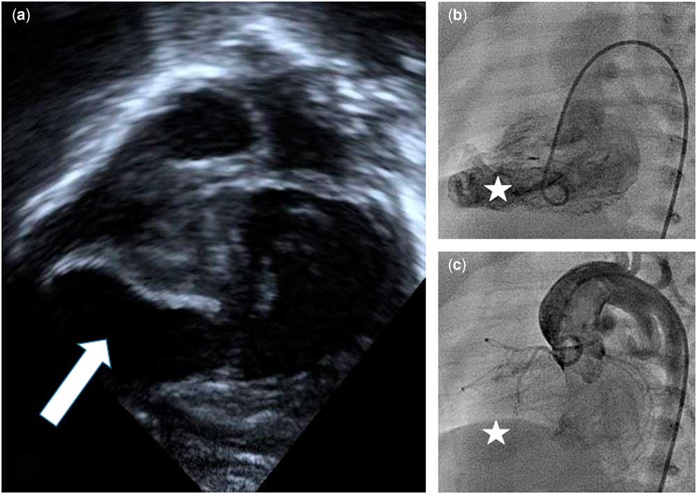
A male infant was born at 31 2/7 weeks of gestation after a prenatal diagnosis of pulmonary atresia with intact ventricular septum. His initial postnatal echocardiogram showed severe right ventricular hypoplasia with likely coronary sinusoids and bidirectional coronary flow. Owing to prematurity and size, he was maintained on prostaglandin infusion. On day of life 32, a routine echocardiogram showed a likely left ventricular aneurysm near the left ventricular apex and arising anteriorly and to the right (Fig 1). A review of previous transthoracic and fetal echocardiograms suggested that the aneurysm was probably present in utero, but not fully visualised owing to its very anterior and rightward position. On day of life 34, a cardiac catheterisation was performed to delineate the coronary artery anatomy, and to place a ductal stent. Angiography showed multiple right ventricle to coronary connections without clear evidence of right-ventricle-dependent coronary circulation. Coronary angiogram showed normal filling of the entire left coronary system. A left ventricular angiogram showed concern for a left ventricular aneurysm and described an area extending from the medial and inferior aspect of the left ventricle that lay underneath the right ventricular apex. It was smooth walled and hypokinetic, but with non-paradoxical movement seen in systole. The duct had significant tortuosity, and stent placement was not successful.
(a) Four chamber view showing large left ventricular aneurysm (white solid arrow). (b) Left ventricle angiogram showing aneurysm (white star). (c) Aortic root angiogram showing coronary distribution in relation to aneurysm (white star).

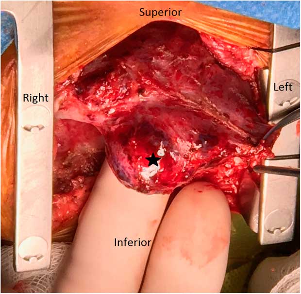
On day of life 39, he was taken to the operating room for placement of a central aorta to the pulmonary artery shunt. There was a large, thin-walled ischaemic aneurysm on the right side of the left ventricle (Fig 2), in an area thought to be in the distribution of the right coronary artery. The patient was subsequently considered for cardiac transplantation versus continuing with staged single-ventricle palliation. Although there was no clear right-ventricle-dependent coronary circulation seen on cardiac catheterisation, it was concluded that this aneurysm was the consequence of an in utero infarction, which could place this patient at risk for a future arrhythmia, cardiac dysfunction, or thrombus formation. The consensus was to list for heart transplantation. He remained clinically stable without arrhythmias or further ischaemic insult, and he underwent successful ABO-incompatible transplant at 4 months of age. He was discharged home after recovery, and remains well. The explanted heart had a thin-walled 2.5- by 1.3-cm aneurysm arising from the left ventricular apex. The aneurysm was lined by a thickened, grey–white endocardium consistent with endocardial fibroelastosis.
Intraoperative photo illustrating the left ventricular aneurysm (black star).

Discussion
Pulmonary atresia with intact ventricular septum represents <1% of CHD and is associated with abnormal coronary connections in 31–61% of patients.Reference Cheung, Richmond, Turner, Bacha and Torres 1 Right-ventricle-dependent coronary circulation is present when myocardial perfusion is derived from these abnormal connections, owing to proximal coronary stenosis and impaired antegrade flow. A number of studies have shown that the presence of right-ventricle-dependent coronary circulation is a risk factor for sudden death.Reference Cheung, Richmond, Turner, Bacha and Torres 1 This risk seems to be higher in the first 6 months of life, and decreases after bidirectional cavopulmonary anastomosis. In the setting of acute ischaemia, it is now recommended that such patients be evaluated for transplantation.Reference Cheung, Richmond, Turner, Bacha and Torres 1
Congenital left ventricular aneurysms and left ventricular diverticula are also quite rare, with a reported prevalence between 0.02 and 0.04%.Reference Ohlow 2 Left ventricular aneurysms commonly are dyskinetic structures with a wide opening into the left ventricle. Histologic examination of left ventricular aneurysms shows that a majority contain fibrous walls (89%), but in a small number of patients the wall of the left ventricular aneurysm may show no microscopic differences from normal left ventricle myocardium.Reference Ohlow, von Korn and Lauer 3 In contrast, left ventricular diverticula are sac-like outpouchings with normal contractility arising from the wall of the left ventricle.
While a majority of left ventricular aneurysms and diverticula are associated with other congenital anomalies, left ventricular aneurysms are more often isolated compared with diverticula. Left ventricular aneurysms have been reported in association with other CHD such as coarctation of the aorta, persistent left superior caval vein, hypoplasia of the pulmonary artery, and pulmonary valve atresia.Reference Ohlow, von Korn and Lauer 3 , Reference Kim, Kim and Min 4 The aetiology of congenital aneurysms is unknown, but some theorise that inadequate blood supply to the left ventricular apex in utero may lead to ischaemia and subsequent development of an aneurysm.Reference Ohlow 2 There are different strategies for the treatment of left ventricular aneurysms, including surgical options and non-surgical monitoring. Owing to the potential complications, some recommend surgical repair of the aneurysm even in the asymptomatic patient. It has also been shown in patients who have coexisting CHD; simultaneously repairing the heart defect along with repairing the left ventricular aneurysm may actually increase the risk for perioperative morbidity and mortality compared with an isolated aneurysm repair.Reference Ohlow 2
The association of left ventricular aneurysms and pulmonary atresia with intact ventricular septum has been documented in the literature, but all the previous case reports have involved children with acquired left ventricular aneurysms. One report described an acquired ventricular septal aneurysm in a 2-month-old with pulmonary atresia and intact ventricular septum, whereas another illustrates a left ventricular apex aneurysm discovered in a 12-month-old with pulmonary atresia with intact ventricular septum after cavopulmonary anastomosis.Reference Kim, Kim and Min 4 , Reference Patel, Farrel, Harris, Gaynor and Gillespie 5 To our knowledge, our patient is the first reported case of a congenital left ventricular aneurysm associated with pulmonary atresia with intact ventricular septum.
Acknowledgements
None.
Financial Support
This research received no specific grant from any funding agency or commercial or not-for-profit sectors.
Conflicts of Interest
None.