Introduction
Once spermatozoa have left the body, they gradually degenerate. However, they retain their motility for about 24 h at room temperature and can still fertilize oocytes (Christian et al., Reference Christian, Songsasen and Leibo1993), resulting in live offspring (Songsasen et al., Reference Songsasen, Tong and Leibo1998). Mouse spermatozoa from dead mice which were stored at 4–6°C have been found to retain fertility for up to 5 days (An et al., Reference An, Wada, Edashige, Sakurai and Kasai1999).
Generally, quiescence due to the lower temperature of the cauda epididymis (Djakiew & Cardullo, Reference Djakiew and Cardullo1986) allow the spermatozoa to save energy to traverse the female genital tract and to fertilize the oocytes. Kaneko et al. (Kaneko et al., Reference Kaneko, Fukumoto, Haruguchi, Kondo, Machida, Koga, Nakagawa, Tsuchiyama, Saiki, Noshiba and Nakagata2009) found that mouse spermatozoa could be short-term preserved via cold storage and the cold-preserved or transported spermatozoa in cauda epididymides at 4°C could still produce viable offspring. Thus the fertility of preserved or transported spermatozoa at 4°C was maintained, although motility gradually decreased with storage time.
In the present study, we preserved mouse cauda epididymis ligated at the both ends at 4–8°C for 1 day to 6 weeks, and cold-preserved spermatozoa were used as controls. From day 1 to day 10 of preservation, the motility and fertility of spermatozoa were daily examined to find the exact time of motility loss. From week 1 of cold storage, the immotile spermatozoa were used for ICSI at weekly intervals to test their fertility. The spermatozoa DNA was determined by comet assay in order to investigate the effect of cold preservation on DNA integrity.
Materials and methods
Humane care and use of animals
Test subjects were 6-week-old female and 10-week-old male BDF1 mice. All mice were bred at the Animal Center, Guangdong No. 2 Provincial People's Hospital, which also approved the animal procedures. This study was carried out in strict accordance with the Guide for Care and Use of Laboratory Animals of Guangdong No. 2 Provincial People's Hospital [No. 2014-KYLLM-065)].
Reagents
Pregnant mare serum gonadotropin (PMSG) and human chorionic gonadotropin (hCG) were purchased from Ningbo Sansheng Hormone Factory (Ningbo, China). All other chemicals were obtained from Sigma-Aldrich Chemical Co. (St. Louis, MO, USA), unless otherwise stated.
Collection and cold preservation of cauda epididymides
Male BDF1 mice were euthanized by cervical dislocation. A small incision was made in the scrotum, and the cauda epididymides were collected and ligated at both ends. These were then added to 0.2 ml tubes with 20 µl of M2 medium and placed into a refrigerator for preservation at 4–8°C.
In the control group, cauda epididymides of male mice were removed and squeezed with forceps to release a dense mass of spermatozoa into 100 μl of M2 medium, and finally these spermatozoa samples were transferred into 0.2 ml tubes and caped for preservation at 4–8°C immediately.
Capacitation of epididymal spermatozoa
From day 1 to day 10 of preservation, cold-preserved spermatozoa and the spermatozoa released from the cold-preserved epididymides were placed into 4-well dish containing 500 µl of human tubal fluid (HTF) medium (Millipore) pre-warmed to 37°C in 5% CO2 in air for 30 min, to facilitate capacitation.
Motility of cold-preserved spermatozoa
The motility of cold-preserved spermatozoa and the spermatozoa from cold-preserved epididymides were assessed immediately after capacitation. Motility was determined by microscopic analysis according to criteria described by Jequier & Crich (Reference Jequier and Crich1986), and the progressive motility scores were assessed using this method (three replicates per sample; 100 spermatozoa observed in five to six fields per replicate). The motility of progressive spermatozoa was scored on a scale of 0 to 4, where 0 indicates no motility; 1, weak or sluggish motility; 2, definite motility; 3, good motility; or 4, vigorous motility. Samples with scores of 2–4 were then used to calculate the percentage of actively motile spermatozoa.
Assessment of spermatozoa DNA integrity by comet assay
The integrity of spermatozoa DNA was assessed using an OxiSelect Comet Assay Kit (Cell Biolabs, San Diego, CA, USA) with minor modifications (Zou et al., Reference Zou, Yin, Chen, Yang and Huang2013). Spermatozoa were washed by centrifugation at 300 g for 10 min, and the pellet was washed with ice-cold PBS (without Mg2+ or Ca2+) at a final concentration of 10 × 105 cells/ml. A 10 μl sample was mixed with 100 μl OxiSelect Comet Agarose and 75 μl/well was immediately pipetted onto the slide. The slide was maintained horizontally at 4°C for 15 min, then immersed in the pre-chilled lysis buffer (25 μl/slide) for 30 min at 4°C, and finally in a pre-chilled alkaline solution (25 μl/slide) for 30 min at 4°C. The above procedures were all performed in the dark. After draining off the fluid, slides were placed in a mini-sub cell GT horizontal electrophoresis unit (BioRad Laboratories, Hercules, CA, USA), and were equilibrated in cold alkaline electrophoresis solution for 20 min before being electrophoresed at 15 V (1 volt/cm) and 300 mA for 30 min at room temperature. After electrophoresis, the slides were placed in 70% ethanol for 5 min, air dried and subsequently stained with 100 μl/well of propidium iodide solution (0.1 mg/ml) for 15 min at room temperature. Each slide was examined using an epifluorescence microscopy at ×400 magnification: band-pass filter, 510–550 nm; long-pass filter, 590 nm. Images were analyzed with Comet Assay Software (Comet Assay Internet Group, www.casp.of.pl). For each sample, the proportion of spermatozoa with damaged DNA in all cells was calculated to evaluate the level of DNA damage. At least 100 cells were observed to calculate the percentage of cell with damaged DNA.
In vitro fertilization (IVF)
Mature oocytes were obtained from superovulated BDF1 mice after intraperitoneal (IP) injection with 7.5 PMSG, followed 48 h later by 7.5 IU hCG. The oocytes were isolated in HTF medium and washed in the same medium three times. A few minutes before fertilization, the oocytes were placed in 500 µl pre-warmed HTF and covered with sterile mineral oil.
The capacitated BDF1 spermatozoa were used for IVF (Byers et al., Reference Byers, Payson and Taft2006; Sztein et al., Reference Sztein, Kastenmayer and Perdue2011). In brief, the spermatozoa were adjusted to about 1–2 × 10⁷ spermatozoa/ml, then 10 µl of spermatozoa were added to the HTF medium (Millipore Inc., Billerica, MA), a final concentration of about 2 × 10⁵ spermatozoa/ml. The mixture was incubated at 37°C for 5 h in a humidified atmosphere containing 5% CO2. Pronucleus formation was checked and the zygotes were incubated in KSOMaa medium (Millipore Inc.) at 37°C in an atmosphere of 5% CO2 in air for 72 h.
ICSI and embryo culture
ICSI was performed according to the method described by (Jiang et al., Reference Jiang, Zhu, Zhu, Sun and Chen2005). The surviving oocytes were incubated in KSOMaa medium at 37°C in an atmosphere of 5% CO2 in air. Pronucleus formation was checked 5 h after ICSI, and zygotes were incubated in KSOMaa medium at 37°C in an atmosphere of 5% CO2 in air.
Statistical analysis
Spermatozoa motility (%), progressive motility scores, integrity of spermatozoa DNA (%) and fertilization rates of two pronuclei (2PN) are presented as mean values ± SD. To identify significant differences, the results were compared by analyzed by one-way analysis of variance (ANOVA), and statistical significance was considered when the P-value was < 0.05.
Results
Motility of cold-preserved spermatozoa
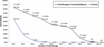
To evaluate the effects of storage time, we daily assessed the motility of cold-preserved spermatozoa and the spermatozoa from cold-preserved cauda epididymis for 1 to 10 days. The results showed that sperm motility of two groups both gradually decreased with storage time; the spermatozoa from cold-preserved cauda epididymis were all immotile at day 10 post-preservation, while cold-preserved spermatozoa became completely immotile until day 5; there were significant differences in spermatozoa motility between cold-preserved cauda epididymis and the control from day 1 to day 8 (P < 0.0001; Fig. 1).
Motility of cold-preserved spermatozoa and the spermatozoa from cold-preserved cauda epididymis for 1–10 days. Spermatozoa motility both gradually decreases with storage time between two groups, and all were immotile by day 10 post-preservation in cold-preserved cauda epididymis while cold-preserved spermatozoa became completely immotile until day 5 post-preservation. Error bars represent ± standard error of the mean (SEM).

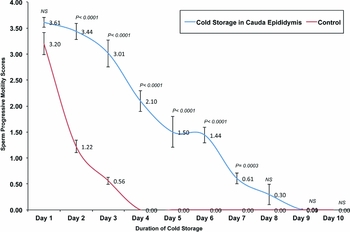
Spermatozoa progressive motility scores of both groups at day 1 to 10 also gradually decreased and there were significant differences between two groups on the same day post-preservation from day 2 to day 7 (P < 0.0005, Fig. 2).
Progressive motility scores of cold-preserved spermatozoa and the spermatozoa from cold-preserved cauda epididymis for 1–10 days. Progressive motility scores of cold-preserved spermatozoa and the spermatozoa from cold-preserved cauda epididymis at 4–8°C both gradually decreased from day 1 to day 10, and the decrease of progressive motility scores of cold-preserved spermatozoa were much faster than the spermatozoa from cold-preserved cauda epididymis. Error bars represent ± standard error of the mean (SEM).

Effect of cold storage duration on spermatozoa DNA integrity
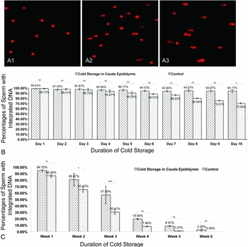
Spermatozoa with undamaged DNA, identified via comet assay, were considered viable (Fig. 3 A1). Spermatozoa with a low (Fig. 3 A2) and high (Fig. 3 A3) degree of DNA damage due to time in storage showed increasing migration from the nucleus to the anode. Through contrast analysis, we found that spermatozoa DNA integrity in cold-preserved cauda epididymis group was maintained in almost all spermatozoa from day 1 to day 10 (Fig. 3 B), but there was a significant increasing trend of spermatozoa with damaged DNA from week 1 to week 5 (Fig. 3 C). However spermatozoa DNA integrity of control group appeared a mild decreasing trend from day 1 to day 10 (Fig. 3 B); especially from week 1 to week 5, there was a more significant decreasing trend of spermatozoa DNA integrity comparing with cold-preserved cauda epididymis (Fig. 3 C).
DNA integrity of preserved spermatozoa at different storage times. (A) A1, A2 and A3 show the different percentages of spermatozoa with undamaged DNA: >80%, about 50% and <10%, respectively. (B) Almost all spermatozoa from cold-preserved cauda epididymis maintained DNA integrity from day 1 to day 10 after preservation while the DNA in 75.0% of spermatozoa was undamaged before day 9 post-preservation. Moreover, DNA integrity of cold-preserved spermatozoa appeared a more slowly decrease trend from day 1 to day 10 compared with the cold-preserved cauda epididymis. (C) From week 1 to week 5, the percentages of spermatozoa with damaged DNA showed a significant increase in both cold-preserved spermatozoa and cauda epididymis groups. Error bars represent ± standard error of the mean (SEM). Bars with different superscripts are significantly different P < 0.05. Asterisks on the bars represent a significant difference. NS means no significance.

Effect of cold storage on fertility of spermatozoa
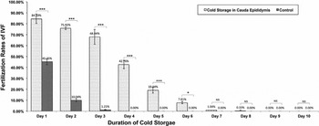
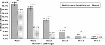
The pronuclei of oocytes were assessed to evaluate spermatozoa fertility at 5 h after IVF or ICSI. In cold-preserved cauda epididymis group, spermatozoa for IVF retained their fertility after cold storage for 6 days (Fig. 4); but fertility gradually decreased after day 3; there was a significant decrease in ICSI fertilization rates from over 94.6 % in week 1 to 7.48% in week 5 (Fig. 5). IVF fertilization rates dramatically decreased in control group and only 45.25% of spermatozoa could keep their ability of fertilizing oocytes at the first day of cold preservation (Fig. 4); ICSI fertilization rates also decreased from over 75.38 % in week 1 to 12.29% in week 3 (Fig. 5). There were significant differences in IVF fertilization rates between control group and cold-preserved cauda epididymis group from day 1 to day 6 post-preservation (P < 0.05); fertilization rates of ICSI were markedly different between two groups on week 1 to week 3 post-preservation (P < 0.0001).
Fertilization rates of IVF using cold-preserved spermatozoa and the spermatozoa from cold-preserved cauda epididymis from 1 to 10 days. In cold-preserved cauda epididymis group, spermatozoa for IVF retained their fertility after cold storage for 6 days, but the fertility gradually decreased after day 3; IVF fertilization rates dramatically decreased in control group and only 45.25% of spermatozoa could keep their fertility at day 1 of cold preservation. Values with different superscripts on the bars are significantly different at P < 0.05. Asterisks on the bars represent a significant difference. NS means no significance.

Fertilization rates of ICSI using cold-preserved spermatozoa and the spermatozoa from cold-preserved cauda epididymis from 1 week to 6 weeks. ICSI fertilization rates of spermatozoa from cold-preserved cauda epididymis gradually decreased from over 94.6% in week 1 to 7.48% in week 5 while these of cold-preserved spermatozoa dramatically from over 75.38% in week 1 to 12.29% in week 3. Values with different superscripts on the bars are significantly different at P < 0.05. Asterisks on the bars represent a significant difference. NS means no significance.

Discussion
Previous studies have found that spermatozoa can be cold stored for the short-term (Nichi et al., Reference Nichi, Goovaerts, Cortada, Barnabe, De Clercq and Bols2007; Takehito et al., Reference Takehito, Kiyoko, Yukie, Tomoko, Hiromi, Mika, Yoshiko, Shuuji, Kiyora and Shiho2009; Kaneko et al., Reference Kaneko, Fukumoto, Haruguchi, Kondo, Machida, Koga, Nakagawa, Tsuchiyama, Saiki, Noshiba and Nakagata2009). Nichi et al. (Reference Nichi, Goovaerts, Cortada, Barnabe, De Clercq and Bols2007) found that short-term storage of bull epididymides at 4°C provided spermatozoa of higher quality and IVF capacity than storage at 34°C. Fan et al. (Reference Fan, Li, Liu, Meng, Wang, Hou, Zhou and Zhu2008) stored mouse epididymides at 4°C in mineral oil or in the body for 4 days after death, and the recovered spermatozoa could be used to fertilize fresh oocytes. Moreover, several studies suggest that storing epididymides of various animals at 4–5˚C for several days can keep some spermatozoa viable (Kikuchi et al., Reference Kikuchi, Nagai, Kashiwazaki, Ikeda, Noguchi, Shimada, Soloy and Kaneko1998; Kaabi et al., Reference Kaabi, Paz, Alvarez, Anel, Boixo, Rouissi, Herraez and Anel2003; Soler et al., Reference Soler, Perez-Guzman and Garde2003; Ganan et al., Reference Ganan, Gomendio and Roldan2009). Even in humans, spermatozoa retrieved posthumously could be used in IVF (Rothman, Reference Rothman1980; Batzer et al., Reference Batzer, Hurwitz and Caplan2003), and indeed several pregnancies and births attributed to this type of spermatozoa have been reported (Check et al., Reference Check, Summers-Chase, Check, Choe and Nazari1999; Belker et al., Reference Belker, Swanson, Cook, Carrillo and Yoffe2001; Batzer et al., Reference Batzer, Hurwitz and Caplan2003). Kishikawa et al (Reference Kishikawa, Tateno and Yanagimachi1999) maintained mouse cadavers at 4°C for up to 20 days, and found that about 30% of the spermatozoa were viable 10 days after death; when the spermatozoa were injected into oocytes, the fertilization rate was more than 80%.
In the present study, in order to exclude the effect of medium on spermatozoa during storage, and prevent the leakage of epididymis fluid and spermatozoa out of cauda epididymis, the cauda epididymides were firstly ligated the both ends to completely prevent the samples from drying due to the moisture loss during storage, and then stored at 4–8°C from 1 day to 6 weeks. Our results showed that motility, progressive score and IVF rate of spermatozoa decreased progressively with storage time (Figs 1, 2 and 4). Examining these parameters from day 1 to day 10 showed that motility, progressive score and IVF rate of the spermatozoa decrease abruptly after day 4 (Figs 1, 2 and 4). Moreover, all the spermatozoa parameters from cold-preserved cauda epididymides were better than that of directly cold-preserved spermatozoa (Figs 1, 2 and 4). It suggests that if mouse epididymal spermatozoa cannot be collected immediately, motility and progressive score can be maintained up to 96 h after collection of the epididymis for later use in IVF, and cold preservation of cauda epididymides could be beneficial to short-term storage of spermatozoa. Therefore, this could be a useful method for preserving some valuable genetic resources or endangered species.
Our results in mice are similar to the study of Kikuchi et al (Reference Kikuchi, Nagai, Kashiwazaki, Ikeda, Noguchi, Shimada, Soloy and Kaneko1998) on boar spermatozoa. They suggests that cryopreservation of spermatozoa from boar epididymides stored at 4°C for 1–2 days can be used for conserving male germ cells when epididymal spermatozoa cannot be collected immediately and cryopreserved.
Although there was a decrease in motility and progressive score from day 1 to day 10, our results showed that DNA was undamaged in over 75.0% of spermatozoa before day 9 of cold preservation, irrespective of cold-preserved cauda epididymides or directly preserved spermatozoa (Fig. 3 B). This could explain why over 82.6% of oocytes could still be fertilized by spermatozoa that had been cold stored for up to 14 days in cauda epididymides (Fig. 5). Although most spermatozoa are classified as dead 10 days after death of the animal, the DNA is still intact and can be used to produce normal, fertile offspring by ICSI (Kishikawa et al., Reference Kishikawa, Tateno and Yanagimachi1999). Even though the spermatozoa plasma membrane is disrupted, the spermatozoa nuclei retains its genetic integrity and is able to fertilize an oocyte.
The present study found that 82.67% of spermatozoa had undamaged DNA, up to 2 weeks after preservation, when the cauda epididymis was cold stored at 4–8°C. It is well known that there are differences between spermatozoa and somatic cell chromatin (Ward & Coffey, Reference Ward and Coffey1991). Mammalian spermatozoa DNA is the most tightly compacted eukaryotic DNA, being at least six times more highly condensed than the DNA in mitotic chromosomes. To achieve this degree of condensation, spermatozoa DNA interacts with protamines to form linear side-by-side arrays of chromatin. This compacted DNA can resist various environmental and chemical factors (Ward & Coffey, Reference Ward and Coffey1991). Our present results indicate that this special structure of spermatozoa can resist DNA denaturation at 4–8°C. We therefore suggest that this ability of spermatozoa DNA to resist denaturation may help to improve routine methods of spermatozoa maintenance and to find new methods for long-term storage of spermatozoa such as cryopreservation.
In summary, short-term cold preservation of mouse spermatozoa in the epididymis at 4–8°C gradually lowers motility, fertility and the proportion of spermatozoa with undamaged DNA; however some spermatozoa are still able to fertilize an oocyte. Understanding the ability of spermatozoa DNA to resist denaturation may help to improve routine methods of spermatozoa maintenance and possibly to find new methods of storage.
Acknowledgements
This study was supported by Science and Technology Planning Project of Guangdong Province, China (No. 2014A020213025). We thank Drs Shen Li, Lei-Ning Chen and Ms Shao-Qing Chen for their technical expertises and valuable advice during study and manuscript writing.