Introduction
Deficits in the recognition of emotional facial expressions might lead to behavioral disturbances that often accompany Alzheimer disease (AD), and behavioral features are more distressing than cognitive deficits for caregivers of patients with AD (Donaldson et al., Reference Donaldson, Tarrier and Burns1998). Facial expressions are universally identified into six basic expressions: happiness, sadness, surprise, anger, disgust, and fear (Ekman et al., Reference Ekman and Friesen1971). The human face conveys non-verbal information about emotional states, the recognition of which is critical for appropriate social behavior.
In aged individuals, positivity recognition bias has been reported for facial expression (Mather and Carstensen, Reference Mather and Carstensen2003; Reference Mather and Carstensen2005). The positivity recognition bias was well-studied with memory; aged individuals remember a larger quantity of positive events than negative ones, and show more emotionally positive memory distortion for autobiographical information than younger adults do (Mather and Carstensen, Reference Mather and Carstensen2005). Such positivity bias in aged individuals has been consistently reproduced in experimental settings of various recognition modalities such as emotional facial recognition and visual stimuli as well as memory (Mather and Carstensen, Reference Mather and Carstensen2003; Reference Mather and Carstensen2005; Kapucu et al., Reference Kapucu, Rotello, Ready and Seidl2008; Spaniol et al., Reference Spaniol, Voss and Grady2008). However, studies on emotional facial recognition in AD patients have produced various results. First, it is controversial whether facial recognition itself is declined or not; some studies reported preserved ability of emotional facial recognition (Bucks et al., Reference Bucks and Radford2004; Luzzi et al., Reference Luzzi, Piccirilli and Provinciali2007; Guaita et al., Reference Guaita2009; Yamaguchi et al., Reference Yamaguchi, Maki and Yamaguchi2012), whereas others reported impairments (Spoletini et al., Reference Spoletini2008; Bediou et al., Reference Bediou2009; Drapeau et al., Reference Drapeau, Gosselin, Gagnon, Peretz and Lorrain2009). It is also controversial whether there were differences in the recognition of various emotions. Some studies reported no difference (Bucks et al., Reference Bucks and Radford2004; Luzzi et al., Reference Luzzi, Piccirilli and Provinciali2007), whereas others reported differences, e.g. selective impairment was reported in labeling the facial expression of sadness (Hargrave et al, Reference Hargrave, Maddock and Stone2002), and recognition of happy facial expressions was reported to be relatively preserved in comparison with angry facial expressions (Yamaguchi et al., Reference Yamaguchi, Maki and Yamaguchi2012). It was also reported that the most identified emotion was happiness among seven facial expressions (six basic expressions and boredom) in the moderate and severe stage of dementia (Guaita et al., Reference Guaita2009).
The controversy may be partly due to confounding factors. Some studies have suggested involvement of confounding factors such as deficits in spatial processing of non-emotional facial features and in verbal processing to express emotions (Cadieux et al., Reference Cadieux and Greve1997; Burnham et al., Reference Burnham and Hogervorst2004). The deficits shown in the experiments could be due to the decline of the spatial recognition and/or verbal processing, which were prominent in AD. Thus, in the present study, we demonstrated characteristics of emotional face recognition in AD patients, by adapting a new method that eliminated the influences of these confounding factors to reveal whether the recognition of positive expressions is relatively preserved in AD.
Methods
Participants
The participants were 12 outpatients with mild AD in Clinical Dementia Rating scale (CDR) 1, 17 aged normal control (ANC), and 25 young normal control (YNC). Participants were limited to mild AD patients to eliminate the influence of difficulties of understandings of the rules. The exclusion criteria were: prosopagnosia, psychiatric diseases, delirium, and verbal incomprehension including aphasia. Those who had weak in eyesight were also excluded; all the participants could distinguish a 2-pixel gap (0.58 mm) on a 15″ monitor screen of Landolt ring from 70 cm away. Subjects were diagnosed based on the criteria for AD by NINCDS-ADRDA (Dubois et al., Reference Dubois2007). Scores over 7 on the Japanese version of the Short Form of the Geriatric Depression Scale (Yesavage et al., Reference Yesavage, Brink and Rose1982) were also excluded because depressive tendencies could affect facial recognition. The Ethics Board of the Gunma University School of Health Sciences approved all procedures (No. 21-26), and written informed consent was obtained from all the participants.
Stimuli
Six hundred colored face images of six basic emotional expressions (happiness, surprise, anger, sadness, fear, and disgust) were used. To eliminate confounding factors related to individual difference in non-emotional facial features and ways to express emotions, we used standardized photos of four Japanese women (one neutral and six basic expression photos for each person) in database DB99 (Advanced Telecommunications Research Institute International, Inc. Nara, Japan); facial features and expressions of non-Japanese individuals could be confounding factors for Japanese. Then we made “averaged faces”, which canceled individual differences. We prepared one neutral and six emotional expression (100% expression faces) averaged faces by morphing photos of four women. For grading the ability, we prepared photos of 1%–99% intermediate expression levels of each emotion by morphing neutral and 100% expression faces with weight. In this way, the images of 600 emotional averaged faces were prepared; e.g. 38% happy image was made by morphing the 100% happy image and the neutral image with a ratio of 38–62. Each image was framed by an oval to avoid the influence of hairstyle and clothing.
Experimental setting
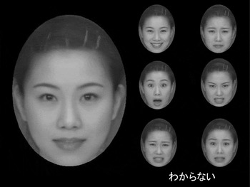
The experimental setting is shown in Figure 1A (stimuli were in color in the experimental setting). One of the images of intermediate expression levels was displayed on the monitor of touch panel screen in the left, and six small faces of 100% expression were displayed on the right. To eliminate the confounding factor of verbal processing, the participants were required to answer by touching the 100% face that corresponded to the expression of intermediate face. Using the choice of faces instead of verbal labels, even those who had difficulties in verbal processing could answer the question.
A stimulus shown on the monitor. On the left of the screen, 27% happy face was shown; recognition of 27% happy face corresponded to the sensitivity of 73%, which was the average sensitivity in patients with Alzheimer disease (AD). On the right, six kinds of 100% expressions were shown. The participants were required to choose and tough one of the 100% faces corresponding to the face on the left. The Japanese letters on the right bottom means to have no idea, and they could choose the option.

The sensitivity of expression was measured using staircase method. The orders of six expressions were randomized using a computer program, and the first stimulus was 100% expression faces in each expression. In each expression respectively, if the response was correct, the level of stimuli increased in the next trial (ex. 38%–35% expression face). Alternatively if the participant made an error, the level of stimuli decreased in the subsequent trial. When the sequence was switched from ascending to descending or vice versa, the level was recorded as a reversal point score. The levels were changed by 15% until the first reversal point, after that, by 3%. The experiment was continued until the four reversal points were obtained. The average of the third and fourth reversal point scores was used as the sensitivity of the expression. Sensitivity was the difference calculated by subtracting expression level from 100(%); the sensitivity corresponding to 38% expression face was 62. We used the screen of a 15″ touch panel connected to a PC running C++ software based on Windows XP. Before the experimental session, a practice session was conducted. In the practice session, 100% expression images were displayed as stimuli and the participants were confirmed to be capable to match the same expression on the right, where six small faces of 100% expression were displayed as choices. The participants were also required to explain the emotion verbally to confirm that they recognized each emotion.
Statistical analysis
AD patients, ANC, and YNC were compared by using repeated-measured analysis of variance (ANOVA; 3 groups × 6 basic expressions) followed by post hoc testing with Bonferroni correction. According to post hoc analysis, significantly higher sensitivity in YNC compared with ANC was defined as age effects, and significantly higher sensitivity in ANC compared with AD patients was defined as AD effects. The data were analyzed using the Japanese version of SPSS for Windows version 19.0 (IBM Corporation, New York). Significant differences are set for two-tailed p = 0.05 for all analyses.
Results
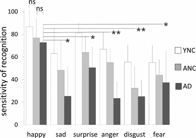
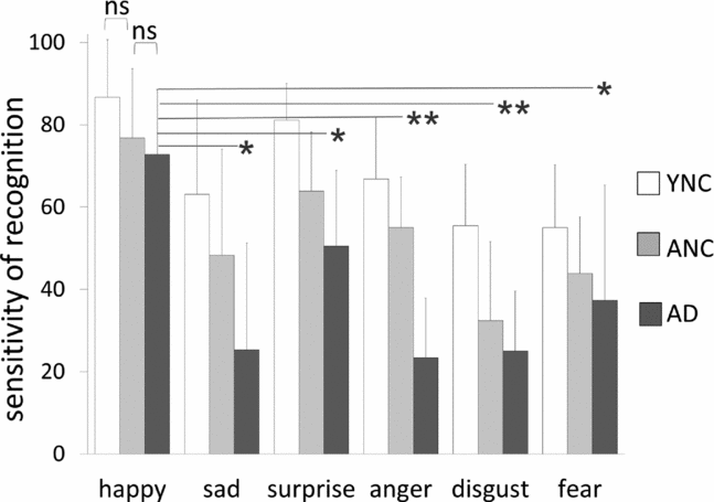
The ages of the participants were 81.1±9.2 years in mild AD, 76.8±3.5 years in ANC, and 18.9±1.1 years in YNC, and there was no significant difference between age of AD patients and that of ANC by two sample t-test. Sensitivities of the three groups and comparisons are shown in Figure 2 and Table 1. There was a significant difference among three groups in perception of facial expressions. According to the post hoc analysis, both age and AD effects were observed for anger and surprise (anger: age effects p = 0.031, AD effects p < 0.001; surprise: p < 0.001, p = 0.029), whereas for happiness and fear, neither age effects nor AD effects were observed (happiness: p = 0.138, p = 1.000; fear: p = 0.178, p = 1.000). For sadness, AD effects were observed (p = 0.048), whereas age effects were not (p = 0.183). However, for disgust, age effects were observed (p < 0.001), whereas AD effects were not (p = 0.718). Within AD patients, sensitivity of happiness was significantly higher than those of the other five expressions, and that of surprise was significantly higher than those of anger and disgust.
Age effects and Alzheimer disease effects

†YNC: young normal controls; ††ANC: aged normal controls; §age effects: significantly higher sensitivity of YNC in comparison with ANC; ¶AD effects: significantly higher sensitivity of ANC in comparison with AD. Both of the age and AD effects were shown by p values of intrasubject post hoc analysis with Bonferroni correction of 3 × 6 repeated measured ANOVA (three groups of YNC, ANC, and AD, and six expressions). *p < 0.05, **p < 0.001.
Results of sensitivities of the young normal controls (YNC), the aged normal controls (ANC), and the AD patients. Error bars indicate standard deviation. Regarding recognition of happy and fear faces, there was no significant difference between YNC and ANC, and ANC and AD patients. Regarding recognition of surprise and anger faces, there was significant difference between YNC and ANC, and ANC and AD patients. There was significant difference between ANC and AD in sad face recognition, and between YNC and ANC in disgust recognition. Within AD patients, sensitivity of happy face was significantly higher than that of other expressions. *p < 0.05, **p < 0.001.
Discussion
This study showed that recognition of happy facial expressions was relatively preserved in AD patients. Recognition of happiness was significantly easier than recognition of five other expressions and there were no age effects or AD effects. Regarding negative expressions, age effects were observed in recognition of anger and disgust, and AD effects were observed in recognition of sadness and anger. Surprise had a neutral emotional valence and both effects were observed in surprise recognition.
The results from this study should be reliable because the task used involved a sophisticated matching task that improved on problems in previous studies to cancel confounding factors. In previous experimental settings, participants were required to match the expression of photos of different people. Thus, impairment in the matching could be a result of visuospatial dysfunctions rather than deficits in processing emotions (Ekman et al., Reference Ekman and Friesen1971). Upon misunderstanding of individual differences in facial features, the participants might fail to extract the emotional implications. The stimuli used in the present study were averaged faces with different emotional valence, where non-emotional features were shared. Thus, differences in features are directly related to emotional differences. Another merit of this matching task was to eliminate the cognitive process to convert perception to abstract verbal expression; abstract thinking and verbal recognition also decline in AD patients. The use of images of Japanese individuals for Japanese participants also eliminated irrelevant cognitive load. Social recognition, including emotional facial expression, has sociocultural implications, and expression of facial emotions could be influenced by cultural backgrounds (Ekman et al., Reference Ekman1987; Shioiri et al., Reference Shioiri, Someya, Helmeste and Tang1999).
Adding to canceling confounding factors, another advantage of this method is the precise measurement of the sensitivity by using the intermediate level of expressions. In the often used experimental settings, the participants were required to classify the photos of typical emotional faces (100% in the present study) by emotional expression. According to a meta-analysis of 17 studies on emotion recognition and aging, the average of the stimuli of one emotion was around 7. Concerning happiness recognition, the magnitude of the difference between young and aged subjects is potentially masked by a ceiling effect, with young subjects scoring 98% or better in 15 out of 17 studies (Ruffman et al., Reference Ruffman, Henry, Livingstone and Phillips2008). Such ceiling effects could exist in the experiments comparing aged subjects and AD patients, thus more sensitive tests with subtle stimuli are desirable. In the present study, we applied 1%–99% intermediate levels of expression, which enabled precise measures of sensitivity.
After eliminating the confounding factors of deficits in spatial processing of non-emotional facial features and in verbal processing to express emotions, positivity bias in ANC was shown, in that recognition of happiness was spared in comparison with YNC. In AD patients, recognition of happiness was spared in comparison with ANC. Hargrave et al. (Reference Hargrave, Maddock and Stone2002) reported that AD patients showed selective impairment in labeling facial expressions of sadness compared with ANC. The results were not identical, as there were differences in the methods used to eliminate the confounding factors of facial features of different people. Hargrave et al. (Reference Hargrave, Maddock and Stone2002) tried to remove the factors by analysis. The experimental setting involved matching the emotion displayed on the reference face with one of six simultaneously presented alternatives, and all seven photographs were faces of different people. A multivariate analysis of covariance (MANCOVA) model was adapted using each subject's score on the facial identity matching task as a covariate. The advantage of the present study is eliminating the confounding factors at the experimental phase.
The mechanism of positivity recognition bias in aged individuals and AD patients remains unproven. Positivity bias in aged individuals was explained by lifetime perspective motivational changes; as the time perspective is reduced, current emotional goals associated with well-being become more important (Carstensen et al., Reference Carstensen, Isaacowitz and Charles1999). Consequently, aged individuals would tend to allocate more cognitive resources to improve emotion regulation, and their information processing was characterized by a positivity bias (Mather and Carstensen, Reference Mather and Carstensen2005; Mather and Knight, Reference Mather and Knight2005; Brassen et al., Reference Brassen, Gamer and Buchel2011). Within this framework, positivity bias in facial emotional recognition could be explained by shifts in attention allocation for positive stimuli (Mather and Carstensen, Reference Mather and Carstensen2005; Goeleven et al., Reference Goeleven, De Raedt and Dierckx2010).
Concerning such allocation of cognitive resources to emotion regulation, capacities of cognitive resources should be considered. Mather and Knight reported that aged individuals with superior cognitive abilities were more likely to exhibit positivity bias (Mather and Knight, Reference Mather and Knight2005). In line with the report, the positivity bias should be reduced in AD patients with cognitive decline. However, the experiment was conducted on memory, and if the allocation occurred only in the remembering phase, and not the memorizing phase, the explanation could not be applied to facial recognition. Goeleven et al. (Reference Goeleven, De Raedt and Dierckx2010) suggested that increased age is associated with reduced allocation of resources to negative stimuli, and the explanation could also be true in AD patients.
The present study showed decreases of negative emotion recognition and relatively preserved positive recognition. Our results are in line with the conclusions based on the meta-analysis of Murphy and Issacowitz, which revealed an age-related decrease of negativity preference as compared to an increased positivity preference (Murphy and Isaacowitz, Reference Murphy and Isaacowitz2008). The above explanations are still hypotheses, and specifying the interaction between cognitive decline and emotion processing would be a valuable topic for future research.
Regarding study limitations, it is possible that recognizing happy facial expressions was easier, as this was the only positive emotion in the study. The differentiation of the four negative expressions, sadness, anger, disgust, and fear, was more difficult. Thus, the results should be confirmed in an experimental setting using stimuli with three facial expressions: happiness, a negative emotion, and a neutral expression.
This study showed that recognition of happy facial expressions was relatively preserved in AD patients; the results could be generalized to other ethnicity because emotional facial recognition is basically universal. These experimental results may be useful if they are implemented in a way to improve the daily life of AD patients. Caregivers should take advantage of cues from happy facial expressions to provide beneficial care.
Conflict of interest
None.
Description of authors’ roles
Y. Maki designed the study, collected and analyzed the data, and wrote the paper. H. Yoshida designed the study and did the computer programming for the task. T. Yamaguchi collected and analyzed the data. H. Yamaguchi supervised the study and wrote the paper.
Acknowledgments
The authors thank all the study participants as well as their families, and Dr. Masamitsu Takatama for their contribution. No funding was provided for this work. However, H. Yamaguchi is supported by a grant-in-aid from the Ministry of Education, Science, Culture and Sports, Japan.