Case
A 31-year-old female was referred for fetal evaluation secondary to maternal Sjogren’s syndrome and anti-SSA (Anti-Sjögren’s-syndrome-related antigen A) antibodies. Her previous fetus developed atrioventricular valve regurgitation, echogenicity of the mitral valve annulus, and mild systolic ventricular dysfunction in utero. This was treated with two 7-day courses of dexamethasone. At birth, the previous fetus had normal cardiac function and anatomy and needed no further follow-up.
The current fetus was followed-up by echocardiogram prenatally, beginning at 18 weeks of gestation. At 22 weeks of gestation, mild mitral regurgitation and mild echogenicity of the left atrium developed. The ductus arteriosus was patent and normal. Within 1 week, the mitral valve became markedly echogenic causing concern for endocardial fibroelastosis or inflammation from the anti-SSA antibodies. The ductus arteriosus was normal.
At 24 weeks of gestation, the right atrium and ventricle were mildly dilated, with flow reversal in the aortic arch. There was also mild tricuspid regurgitation and continued echogenicity of the mitral valve and left atrium. There was no change in the ductus arteriosus. By 27 weeks of gestation, given these continued findings and a previous history of a fetus with cardiac neonatal lupus erythematosus, the mother was started on dexamethasone (4 mg daily). At this point, before starting the steroids, the ductus arteriosus pulsatility index was 2.1, with a peak systolic velocity of 0.82 m/second and diastolic velocity of 0.07 m/second.
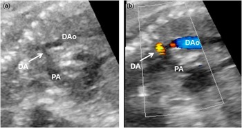
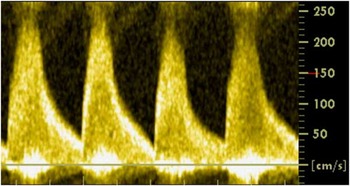
A week after steroids were initiated, the ductus arteriosus showed signs of significant constriction measuring 2 mm in diameter (Fig 1). The pulsatility index was 1.4, with peak systolic velocity of 2.3 m/second and diastolic velocity of 0.4 m/second (Fig 2). Steroids were discontinued, but again re-initiated after review of the literature showed no other reports of ductal constriction. They were initiated at a higher dose of 8 mg with rationale that the ductal constriction may have been related to inflammation. It is unclear how many doses of the higher dose were administered between the two echocardiograms.
Ductus arteriosus at 28 weeks of gestation. ( a ) Note the serpiginous ductus arteriosus with significant constriction, measuring 2 mm in diameter. ( b ) Colour flow through the serpiginous ductus arteriosus with turbulence noted due to ductal constriction. DA=ductus arteriosus; DAo=descending aorta; PA=pulmonary artery.

Ductus arteriosus pulse Doppler at 28 weeks of gestation. Note the peak systolic velocity of 2.3 m/second and diastolic velocity of 0.4 m/second.

On the next echocardiogram, 4 days after the ductal constriction was first noted, the fetus had periods of supraventricular tachycardia, with maximum heart rate of 215 beats/minute. Echocardiography at this time – after discontinuation of steroids followed by re-initiation of steroids at 8 mg – showed normal pulsatility index of 2.3 with a serpiginous ductus arteriosus. Peak systolic velocity was 0.7 m/second, and diastolic velocity was 0.05 m/second. Before this, all P-R intervals had been measured and were within normal limits for gestational age. The mother was started on flecainide because of concern that arrhythmia represented worsening inflammation. She completed a 10-day course of high-dose steroids followed by a taper to 2 mg over 2 weeks. This dose was continued for the remainder of pregnancy. By the end of the taper, at 30 weeks of gestation, the ductus arteriosus remained serpginous with normal flow patterns. The pulsatility index was 2.5, with a peak systolic velocity of 1.1 m/second and diastolic velocity of 0.1 m/second.
At 32 weeks of gestation, the ductus arteriosus had a pulsatility index of 2.1, with a peak systolic velocity of 2.5 m/second and diastolic velocity of 0.4 m/second. By 34 weeks of gestation, the ductus arteriosus had a pulsatility index of 2.3, with a peak systolic velocity of 1.9 m/second and diastolic velocity of 0.1 m/second. Findings were stable with no resolution of right heart changes until 35 weeks of gestation. At 36 weeks of gestation, the ductus arteriosus had a pulsatility index of 2.4, with a peak systolic velocity of 1.2 m/second and diastolic velocity of 0.1 m/second.
The infant was born at 38 weeks of gestation due to spontaneous labour and was intubated briefly for respiratory distress, but weaned to room air within 24 hours. Postnatal echo confirmed the fetal findings of right heart dilation and right ventricular dysfunction without increased endomyocardial echogenicity. The infant was observed without medications and no arrhythmias occurred.
Given the in utero concern for myocardial inflammation – arrhythmia, valve regurgitation, right heart abnormality, and echogenicity of the mitral valve annulus – a cardiac MRI was ordered on day of life two. The rationale was that it could aid in determining prognosis and making therapeutic decisions. Transthoracic echocardiography is not sensitive for myocardial inflammation in cardiomyopathy or infectious myocarditis, although the sensitivity in SSA-mediated disease is unknown. There was concern that evidence of inflammation or fibrosis may not necessarily be seen on transthoracic echocardiogram but detectable by MRI. The MRI was performed on a Siemens Aera 1.5T scanner (Siemens Healthcare, Erlangen, Germany) with a feed-and-bundle technique using infant immobiliser; therefore, sedation was not required. Volumetric analyses and early and late gadolinium enhancement imaging were acquired, using free-breathing techniques that were optimised for infant imaging.Reference Tham, Hung and Myers 1
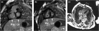
The MRI showed no areas of late gadolinium enhancement to suggest fibrosis, infarct, or inflammation. Biventricular function was normal, with mild enlargement of the right ventricle. Echocardiogram before discharge showed mildly decreased right ventricular function and moderate tricuspid regurgitation. With no arrhythmias and improving cardiac function, the infant was discharged home. In 2 weeks, an echocardiogram revealed normal structure and function.
Discussion
This case highlights cardiac findings in an anti-SSA-antibody-exposed fetus and explores a role of postnatal cardiac MRI in fetuses with concern for SSA-mediated myocardial inflammation and fibrosis. In utero echogenicity of the mitral valve annulus and left atrium, mitral regurgitation, and atrial tachycardia may have represented transient inflammation even without definite endocardial fibroelastosis or cardiomyopathy. Reversal of flow in the aortic arch may reflect lower left cardiac output and early signs of cardiomyopathy. It also may reflect inflammation of the left heart causing diastolic dysfunction and under filling. Although ductus arteriosus constriction can occur spontaneously, in this case, dexamethasone therapy may have been associated with ductal constriction and the marked changes in the right heart. No other known causes such as use of non-steroidal anti-inflammatory drugs or high intake of polyphenol-rich foods were present.
Maternal anti-SSA antibodies can cause fetal heart block, cardiomyopathy, arrhythmias, valvar regurgitation, and endocardial fibroelastosis.Reference Krishnan, Pike and Donofrio 2 , Reference Krishnan, Arya, Moak and Donofrio 3 No consensus exists for surveillance and follow-up of exposed fetuses, partially because the risk of disease is low (~2%). Nevertheless, expert opinion calls for prenatal echocardiograms every 1–2 weeks between 16 and 28 weeks of gestation.Reference Donofrio, Moon-Grady and Hornberger 4 Siblings of fetuses with previous congenital heart block are at higher risk for cardiac neonatal lupus erythematosus (25% risk); however, the risk for siblings with more subtle disease is unknown.
The indications for maternal dexamethasone therapy in antibody-mediated cardiomyopathy are controversial. Maternal anti-SSA antibodies cross the placenta and can cause immune-mediated myocarditis, with histological changes seen within the myocardium, including diffuse deposition of immunoglobulin G and T-cell infiltration.Reference Nield, Silverman and Smallhorn 5 Therefore, prenatal echogenicity of the left atrial wall and mitral valve may represent endocardial fibroelastosis or inflammation. Reports have linked maternal fluorinated steroids with improved postnatal outcome for immune-mediated cardiomyopathy, and some have suggested that in utero abnormalities may be reversible.Reference Saleeb, Copel, Friedman and Buyon 6
In one study, the combination of betamethasone and indomethacin in human fetuses was associated with higher rates of ductal constriction than indomethacin alone.Reference Levy, Matitiau and Ben Arie 7 In rats, steroids alone have been shown to cause ductus arteriosus constriction.Reference Takami, Momma and Imamura 8 In our patient, constriction of the ductus arteriosus may have been related to maternal steroids. We acknowledge that it is also possible that it was an idiopathic or unrelated finding. This is the first published report of ductal constriction temporally associated with maternal dexamethasone therapy in an anti-SSA-antibody-exposed fetus.
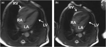
In addition, we utilised cardiac MRI to characterise the myocardium as it has become a mainstay for diagnosing paediatric conditions causing inflammation and fibrosis of the myocardium. Studies have cited the diagnostic and prognostic utility of late enhancement imaging in paediatric cardiac disease, predominantly in the setting of CHD and cardiomyopathies.Reference Latus, Gummel and Klingel 9 Typically, this technique requires suspending respiration to acquire images. A recent study, currently in press, showed that MR single-shot late enhancement imaging with motion-corrected averaging is feasible in children, at high heart rates and with variable R-R intervals.Reference Olivieri, Cross and O’Brien 10 It can also be performed without breath-holding with higher image quality ratings than standard breath-held techniques.Reference Olivieri, Cross and O’Brien 10 This technique, therefore, can be used in patients who cannot hold their breath. In this case, imaging was performed with a fast acquisition, free-breathing technique, in combination with a feed-and-swaddle technique to reduce motion artefact and avoid sedation. The MRI was timed for the newborn feeding schedule and took 30 minutes to complete. The newborn was fed and swaddled, and then placed in the MRI machine. The free-breathing MRI techniques are optimised to minimise repetitive movement artefacts, such as respiratory motion. Thus, there were no respiratory movement artefacts seen (Fig 3). This produced diagnostic quality imaging, indicating no major myocardial fibrosis or inflammation (Fig 4). This aided clinicians in making the decision to hold all medications and simply observe the baby on telemetry. Follow-up was arranged for 2 weeks after discharge. To our knowledge, this is the only case where a non-sedated MRI using these techniques was used to assist in treatment planning for this disease. Future studies are needed to validate this finding and explore the role of MRI in diagnosis and treatment of anti-SSA-mediated cardiac disease in fetuses.
Real-time MRI images acquired that were used for the volumetric analysis. ( a ) Still frame in diastole. ( b ) Still frame in systole. LA=left atrium; LV=left ventricle; RA=right atrium; RV=right ventricle.

MRI late gadolinium enhancement still frames, showing a normal healthy myocardium with no evidence of myocardial fibrosis or enhancement. ( a and b ) Short-axis views. ( c ) Vertical long-axis view. LA=left atrium; LV=left ventricle; RA=right atrium; RV=right ventricle.

Acknowledgements
None.
Financial Support
This research received no specific grant from any funding agency, commercial, or not-for-profit sectors.
Conflicts of Interest
None.