


Attitudinally appropriate nomenclature
The basic rules of human anatomy state that all structures should be described relative to the so-called anatomic position, this being the position taken by the subject whilst standing upright and facing the observer. In terms of description for clinical examination and diagnosis, this convention is followed for virtually every organ except the heart. Although the reasons for this discrepancy are not clear, it is conventional to describe the heart as though removed from the body, and positioned on its own apex. In the past, this convention was of relative insignificance, but the advent of new diagnostic techniques that display the heart in the context of the body continually emphasize its logical deficiencies. For modern-day description, it makes more sense to put the heart back within the body, and to describe the cardiac components as seen relative to the bodily coordinates, as shown in Figure 1. Structures adjacent to the diaphragm, therefore, should logically be described as being positioned inferiorly, whilst it is those closest to the spine which should be described as being posterior.3

Figure 1. The left hand panel shows the coordinates for description of all structures within the body. The subject is viewed upright and facing the observer. All structures are described relative to the three orthogonal planes, using the adjectives as illustrated in the figure. The right hand panel shows the short axis view of the heart relative to the sagittal plane. As is obvious from the relations to the thorax, it is structures closest to the diaphragm that are inferior, whilst those closest to the spine are posterior.
Anatomy of the normal morphologically tricuspid valve
The morphologically tricuspid valve, in the normally structured heart, guards the so-called right atrioventricular junction, which in the setting of the heart itself is positioned anterior and relatively inferiorly, as is well seen in short axis sections viewed from the cardiac apex (Fig. 2). The ventricles themselves are best described in terms of three component parts, the inlet, the apical trabecular component, and the outlet. The inlet extends from the atrioventricular junction, and is contained and limited by the tension apparatus of the tricuspid valve. It is because the atrioventricular valves must withstand the full force of ventricular contraction when in their closed position during ventricular systole that they are furnished with their tension apparatus, which stops them turning inside-out. The arrangement of this tension apparatus relative to the skirt of leaflet tissue suspended from the atrioventricular junction then gives the atrioventricular valves their characteristic appearances. The essence of normality for the tricuspid valve is that, apart from the small area related to the membranous septum, the entire circumference of the skirt of leaflet tissue is suspended from the muscular atrioventricular junction, this being the area where the fibro-fatty tissues of the atrioventricular groove insulate the atrial from the ventricular myocardial masses. Distinguishing the presence of boundaries within the overall skirt of leaflet tissue, however, can be difficult, and there is marked individual variation. We distinguish the location of individual leaflets within the skirt according to the general pattern of closure of the valve, distinguishing three leaflets. When considered relative to the landmarks of the body, these leaflets are located in septal, anterior, and inferior positions (Fig. 2). The papillary muscles supporting the zones of apposition between the three leaflets, or the commissures, show marked difference in structure. A small, but particularly characteristic, papillary muscle, the medial papillary muscle, or the papillary muscle of the conus, supports the zone of apposition between septal and anterior leaflets. A much larger papillary muscle, the anterior papillary muscle, supports the zone of apposition between the anterior and inferior leaflets in some hearts, but this muscle is not seen to supply tendinous cords to both leaflets in all hearts. Indeed, in perhaps half of all normal individuals, the large anterior papillary muscle supports only the leading edge of the anterior leaflet, with a smaller muscle providing cords to the zone of apposition with the inferior leaflet. It is then usual for a smaller inferior muscle, or multiple muscles, to support the zone of apposition between the inferior and septal leaflets. The most constant, and characteristic feature of the normal tricuspid valve, nonetheless, is the presence of multiple attachments of cords from the septal leaflet to the ventricular septum.

Figure 2. The short axis of the ventricular mass is shown as viewed from the apex and looking towards the base, replicating the left anterior oblique projection. The three leaflets of the tricuspid valve are positioned septally, antero-superiorly, and inferior or murally. Note the location of the membranous septum (double headed arrow). In this heart, the anterior papillary muscle (asterisk) is attached to the midpoint of the antero-superior leaflet.
Congenital malformations of the tricuspid valve
Although relatively rare in the overall context of congenital cardiac malformations, it is congenital deformities that now constitute the commonest reason for surgical correction involving the tricuspid valve, having overtaken post-inflammatory disease in this context.4 The most important lesion is Ebstein's malformation,5 and because of the constraints of space, we will concentrate our attention on this entity. An integral part of Ebstein's malformation, nonetheless, is valvar dyplasia,6 so it is important to distinguish isolated valvar dysplasia from the congenital malformations itself, since isolated dysplasia can also have a poor prognosis when developing during fetal life even in the setting of a normally structured valve.7
Ebstein's malformation is, in essence, a manifestation of abnormal development of both the myocardium and the valvar components. During development of the normal heart, the leaflet tissue of the valves evolves from the supporting ventricular myocardium in a process that has been termed “delamination”. Simply put, a layer of endomyocardium separates, or delaminates, from the underlying ventricular myocardium, and loses its muscular character. This separation begins within the cavity and proceeds back toward the atrioventricular junction, eventually forming what we recognize as the valvar complex. The development of the myocardium in Ebstein's malformation, and the process of delamination, are both abnormal. As a result, the attachment of the skirt of leaflet tissue guarding the entrance to the right ventricle is not suspended from the atrioventricular junction. Rather, the delamination is incomplete, and the hingepoints of the septal and inferior leaflets are displaced. The skirt of leaflet tissue attaches within the cavity of the ventricle itself, not at the atrioventricular junction.
In the past, it has been conventional to describe this finding as “downward displacement” of the valvar hinges. There are two problems with this approach. First, rather than representing “displacement” of the hinges of the valvar leaflets, the anomaly is the consequence of failure of delamination of the leaflets from the ventricular inlet during fetal development.8 Second, and perhaps more important, when considered in attitudinally appropriate fashion, the normal right atrioventricular junction is positioned vertically rather than horizontally. The abnormal location of the leaflets as seen in Ebstein's malformation, therefore, is better conceptualised as rightward and anterior rotational displacement,9 rather than simple “downward” displacement (Fig. 3). Analysis in this fashion then shows that the proximal valvar attachments are typically located at the junction of the right ventricular inlet with the ventricular apical component. These attachments can be recognized as the functional hinge points of the mobile leaflet tissue. As a result of this rotational displacement, the valve affected by Ebstein's malformation has a bifoliate pattern of closure, rather than forming the expected trifoliate valvar complex. There are gross malformations of the tension apparatus in more severely afflicted individuals. This is because it is primarily the septal and inferior leaflets that fail to delaminate from the walls of the inlet component. The anterior leaflet, in contrast, usually retains its hinge point from the atrioventricular junction. In less severely afflicted patients, this leaflet can retain relatively normal support of its leading edge from medial and anterior papillary muscles. In the more severely afflicted patients, the entire leading edge of this leaflet can be anomalously attached at a muscular shelf formed at the junction on the inlet and apical components, often with marked abnormal tethering of the ventricular surface of the leaflet to the parietal wall of the ventricle. There is a spectrum between these two extremes.10 In clinical practice, it is the duty of the echocardiographer to delineate the precise anatomy and functional state of both the myocardium and the malformed valvar complex in these patients, as both have important influences on management and outcome.11–15

Figure 3. The cartoon shows the location of the orifice of the abnormal valve, shown by the green ovals, as observed in the series of hearts examined by Schreiber et al.9 The valvar orifice is displaced rotationally, rather than in a downward fashion.
Echocardiographic evaluation of the tricuspid valve
Thorough echocardiographic interrogation of the tricuspid valve involves evaluation of both its anatomy and function. Although both reconstructed and real-time multi-dimensional approaches are now available, the standard cross-sectional Doppler echocardiogram is most widely applied in clinical settings. The parasternal long axis and apical four chamber views show well the anterior and septal leaflets16 (Fig. 4). Angulation of the plane of sound from the long axis toward the right ventricular inlet and lateral wall also allows visualization of the inferior leaflet (Fig. 4 – left panel). Parasternal short axis, modified subcostal views when available, transoesophageal transgastric views, and three-dimensional echocardiography (Fig. 5) may all be utilized to show the three leaflets. Transoesophageal echocardiography is then useful in providing still further views of the valve. The anterior and septal leaflets can be shown via the mid-oesophageal four chamber view (Fig. 6, left), and in the basal horizontal plane (Fig. 6, right); demonstrating inflow to and outflow from the right ventricle.17, 18

Figure 4. The parasternal long axis view through the right ventricular inflow (left hand panel) shows the anterior and inferior leaflets of the tricuspid valve, while the apical four chamber section (right hand panel) shows the anterior and septal leaflets. Abbreviations: RA: right atrium; RV: right ventricle; CS: coronary sinus.
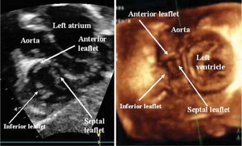
Figure 5. The short axis echocardiographic image (left hand panel), and the three-dimensional echocardiographic reconstruction (right hand panel) show well the locations of the three leaflets of the tricuspid valve.

Figure 6. These transoesophageal views at the level of the middle of the oesophagus show the septal and anterior leaflets as seen in four chamber projection (upper panel) and the view showing the inlet and outlet of the right ventricle (lower panel).
Doppler interrogation frequently reveals trivial tricuspid regurgitation. It is regarded as a normal variant, being seen in up to nineteen-twentieths of normal patients. Features suggesting a normal physiological variant are an otherwise normal valve, a small jet area, and a peak velocity of 1 to 2 metres per second.19 When tricuspid regurgitation is significant, the valve will be seen to be abnormal, the jet area will be larger, and although the peak velocity may still be 1 or 2 metres per second, it can be higher (Fig. 7).

Figure 7. The upper panel, in four chamber orientation, shows physiological mild regurgitation across the tricuspid valvar orifice. Note that the leaflets of the valve are normal. The bottom panel, in contrast, shows severe regurgitation across a valve with thickened and dysplastic leaflets.
The presence of tricuspid regurgitation of any degree when seen on a fetal echocardiogram is more likely to be abnormal.20 Most examples of tricuspid valvar dysplasia or Ebstein's malformations detected prenatally are associated with significant regurgitation.21 In cases of significant regurgitation, and/or fetal congestive heart failure, there are usually venous pulsations noted in the umbilical vein and venous duct, possibly associated with hydrops fetalis (Fig. 8).

Figure 8. These two panels, from fetuses imaged in four chamber projection, compare mild (left hand panel) as opposed to severe (right hand panel) regurgitation across the tricuspid valve.
Ebstein's malformation
Echocardiography is the procedure of choice for both the diagnosis and long term assessment of patients with Ebstein's malformation. The previously described anatomic abnormalities can all be demonstrated with cardiac ultrasound. In fact, due to the enlargement of the right heart, lung tissues are often displaced, thus creating windows for high quality transthoracic imaging even in adults.
The single most sensitive, and specific, diagnostic feature is excessive displacement of the hinge point of the septal leaflet of the valve into the ventricular cavity. This displacement is most easily appreciated when the insertion of the leaflet is imaged relative to the opposing insertion of the mitral leaflet in the apical four-chamber view.22–24 Normally (Fig. 9) this is slightly apical to the insertion of the mitral valve. In patients with Ebstein's malformation, this displacement is exaggerated (Fig. 9 – right panel). The increased off-setting can easily be measured, and indexed to body surface area, producing the so-called displacement index. A value greater for the index of greater than 8 millimetres per metre squared distinguishes reliably the malformation from other lesions associated with tricuspid regurgitation or enlargement of the right ventricle.23

Figure 9. These four-chamber sections compare the normal off-setting of the hingepoints of the tricuspid (dashed arrow) and mitral valves (left hand panel). The right hand panel shows the arrangement as typically seen in Ebstein's malformation. The solid arrows mark the excess displacement of the septal tricuspid hingepoint relative to the hingepoint of the mitral leaflet in this plane.
Occasionally, nonetheless, it may prove difficult to assess the internal crux. In these situations, Ebstein's malformation may be suggested by displacement of the hingepoint of the inferior leaflet into the cavity and away from the atrioventricular junction, elongation of the anterior leaflet, tethering of any of the leaflets to the underlying myocardium, shortening of the tendinous cords, attachment of the leading edge of the anterior leaflet to the right ventricular myocardium, apical displacement of the hinge of the anterior leaflet, which is very uncommon, absence of the septal or inferior leaflets, also uncommon, or fenestration of any of the leaflets. If present, these findings must be associated with some evidence of incomplete delamination of the leaflets in order to confirm the presence of Ebstein's malformation.
Ebstein's malformation is not the only cause of enlargement of the right heart and tricusupid regurgitation. Other notable disorders include congenital valvar dysplasia, and traumatic injury to the valvar complex. Although the tricuspid valve is abnormal in these other situations, calculation of the displacement index, along with the other characteristic morphologic features of the malformation, should permit clear separation of the diagnostic groups.
Echocardiographic features indicating a potentially good outcome from surgical repair focus on the mobility of the anterior leaflet, since repairs rely heavily on this leaflet. Often the “repaired” anterior leaflet acts as a solitary leaflet that coapts with the septum rather than with the other leaflets.25 The assessment of mobility is made predominantly in the apical four-chamber view. Identification of a free leading edge is especially important to the success of the repair (Fig. 10). An edge that is tethered to the myocardium (Fig. 11), either by direct attachment or by papillary muscles, will not coapt with either the septum or another leaflet, resulting in a significant residual regurgitant orifice.

Figure 10. These four chamber sections show a free leading edge to the anterior leaflet of the tricuspid valve in a patient with Ebstein's malformation – the left hand panel is a diastolic frame, while the right hand panel is taken during systole. This free leading edge is a good sign for a potentially successful surgical repair.

Figure 11. In this anatomic specimen, reproduced by permission of Dr W.D. Edwards, and a comparable four-chamber echocardiographic section, the anterior leaflets of the tricuspid valve are tethered to the ventricular wall (arrows on the anatomic image and asterisks on the echocardiographic image show points of tethering). The asterisk on the anatomic image marks the true right atrioventricular junction.
The preoperative examination should determine whether manoeuvres that advance the anterior leaflet toward the septum, and reduce the size of the right ventricle and the tricuspid valvar orifice, can allow the leading edge of the anterior leaflet to reach another surface for coaptation without creating stenosis. Other favourable features include the presence of only one, central regurgitant orifice, and the lack of direct muscular insertions into the anterior leaflet (Figs. 10 and 12).

Figure 12. This cross-sectional image shows an anterior leaflet with a free leading edge, and Doppler interrogation reveals a single central jet of regurgitation, also a good sign for potentially successful surgical repair.
Valvar abnormalities that, when present, make repair unlikely include inadequate delamination and mobility of the anterior leaflet, a tethered leading edge of the anterior leaflet, multiple separate jets of tricuspid regurgitation (Fig. 13), and direct muscular insertions into the leading edge of the anterior leaflet. If the functional orifice of the valve is positioned at the entrance to the infundibulum, or within the outflow tract rather than within the body of the right ventricle, this also usually militates against successful repair, since the volume of the functional ventricle is not adequate independently to support the pulmonary circulation.

Figure 13. In this patient, the anterior leaflet is tethered to the ventricular wall (arrow, left hand panel), while Doppler interrogation reveals multiple jets of regurgitation (arrows, right hand panel).
The echocardiographer should also establish the functional impact of the abnormalities of the right ventricle and tricuspid valve. Anatomical and functional severities are not necessarily similar. For example, a patient can have severe displacement of the leaflets with minimal delamination, but show only mild functional disease, with preserved ventricular performance and only mild dysfunction of the displaced valve. Both the anatomic and functional states play an important role in determining symptoms, prognosis, and reparability of the tricuspid valve.23, 26–28 The degree of enlargement of both right atrium and ventricle, the performance of the right ventricular myocardium, should be defined. Right ventricular function can be difficult to evaluate by echocardiography. It is preferable to include all of the right ventricular myocardium in this assessment, especially if the atrialized right ventricle is of normal thickness, and retains systolic thickening with appropriate systolic motion. We advocate an approach combining several qualitative and quantitative techniques. Relative wall motion should be assessed visually, and by measurement of the reduction in size of the systolic cavity. Although there are no satisfactory geometric formulas for assessment of right ventricular volume, the area of the cavity can be measured in both systole and diastole. From these values, it is possible to calculate the fractional change in area. At least two planes should be assessed, usually parasternal short axis and apical four-chamber views. The median normal area change value is 49 percent, with a range from 40 to 60 percent. Non-geometric parameters of performance are also useful in these patients, such as the myocardial performance index.29
The left ventricle has also been described as being abnormal in a subset of the patients with Ebstein's malformation, with abnormalities noted in shape, global function, and development, non-compaction also having been observed.11–15 Quantitative evaluation of left ventricular performance, therefore, should also be a routine component of the echocardiographic evaluation. This is especially important due to the association of left ventricular dysfunction with early post-operative mortality.30 Defects such as atrial or ventricular septal defects, and pulmonary stenosis, may also be found in association with Ebstein's malformation, and should be evaluated using standard techniques.23, 26, 31 Systolic venous reversal, nonetheless, is often absent in Ebstein's malformation, even when transvalvar regurgitation is severe. This is because the regurgitant jets are almost always under a low driving pressure, and the functional right atrial chamber is often very large and compliant. The key to appropriate analysis, therefore, is to possess a thorough understanding of the anatomy of the tricuspid valvar complex, and the disorders that may affect it. The recommended approach is also used to examine patients with other abnormalities of the tricuspid valve abnormalities. Attention to mobility, tethering of the leaflets, and ventricular function, by transthoracic echocardiography will usually provide enough information to guide clinical and surgical care.
Transoesophageal echocardiography has primarily been used as a tool for intraoperative monitoring tool.32 When images obtained from transthoracic windows are poor, the anatomic features described above can usually be clearly displayed by transoesophageal examination. Transgastric views are particularly good at displaying the anatomy of the valvar leaflets in their short axis (Fig. 14), and defining the mechanism of transvalvar regurgitation.

Figure 14. Transgastric interrogation shows the valvar leaflets in their short axis. The inferior leaflet is essentially absent, the dashed line on the right panel indicates where this leaflet “should” be found. The anterior and septal leaflets (arrows) were the substrate for repair of the valve in this patient. The asterisk indicates the anterior papillary muscle in this patient.
Acknowledgements
Robert H. Anderson is supported by grants from the British Heart Foundation together with the Joseph Levy Foundation. Research at the Institute of Child Health and Great Ormond Street Hospital for Children NHS Trust benefits from R&D funding received from the NHS Executive.



