Introduction
Among the different types of neoplasms, bone cancer is one of the most common form of the disease in the world, an event that is attributed to the occurrence of metastases that reach the skeleton [Reference Ouyang, He, Zhang, He, Jiang, Wang, Chen, Pan, Li and Guo1, Reference Buijs and Pluijm2]. Chemotherapy using specific drug delivery to the bone is still rather limited [Reference Hirabayashi and Fujisaki3]. The structural similarity between the bone and the synthetic hydroxyapatite (HAp) has led to research aimed at its use as a drug delivery system, since synthetic HAp is an ideal support for applications in bone tissue engineering as it can be used as a graft and can be loaded with either a therapeutic agent or a diagnostic agent [Reference Cawthray, Wasan and Wasan4].
In the case of bone cancer, a surgical intervention for removing the tumor tissue is usually required, causing a deformity at the site that needs filling [Reference Andronescu, Ficai, Voicu, Ficai, Maganu and Ficai5]. A major problem related to implant surgeries is infections associated with microbial colonization. Thus, using bone grafts with antimicrobial action is a better alternative to eliminate potential inflammation; furthermore, vectorization at a specific site of an antibiotic typically administered systemically will have reduced toxicity [Reference Marques, Ferreira, Adronescu, Ficai, Sonmez and Ficai6, Reference Kuehl, Brunetto, Woischnig, Varisco, Rajacic, Vosbeck, Terracciano, Fromm and Khanna7].
The need for early detection of tumors and the strong side effects caused by conventional therapy in cancer treatment are some of the concerns that drive research in the area. The development of nanoparticles capable of uniting in a single system the delivery of a drug to the specific site with controlled release and, also, the possibility of obtaining images from the affected region and, thus, detect potential occurrences related to it define the field of study of theranostic nanomedicine. It aims to bring together three different functions in the same system: targeting, diagnostic imaging, and therapy [Reference Costa and Silva8, Reference Liu, Miyoshi and Nakamura9].
In recent years, HAp has aroused interest in the field of drug vectorization because in addition to its properties as a biomaterial, it has a high molecular adsorption capacity, making it capable of serving as a support for drugs [Reference Lopes, Oliveira and Esteves10]. The incorporation of certain elements with magnetic and/or luminescent properties to the HAp matrix has attracted research due to its diagnostic capacity through molecular imaging techniques [Reference Lin and Chang11]. An example is presented in the study of Ashokan et al. [Reference Ashokan, Menon, Nair and Koyakutty12], which developed a multimodal contrast agent based on HAp nanocrystals doped with Eu3+ and Gd3+, indicating simultaneous contrast for three important molecular imaging techniques, such as magnetic resonance imaging (MRI), X-ray and fluorescence imaging. Chen et al. [Reference Chen, Huang, Zhu, Wu, Zhang and Cui13] observed a high adsorption capacity of a drug in HAp nanoparticles, and, additionally, the material doped with Eu3+ and Gd3+ ions displayed application potential as an MRI, photoluminescence, and computed tomography agent.
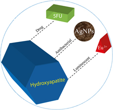
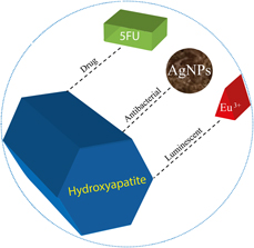
In this context, this paper proposes the synthesis of a material with multifunctional properties. HAp is used as a matrix that consists of a biomaterial fully compatible with the human body. The incorporation of europium ions aims to introduce luminescent characteristics to the material and the adsorption of silver nanoparticles (AgNPs) intends to provide antimicrobial properties. Given that the main purpose of the application of the proposed multifunctional system is drug vectorization for cancer treatment, a 5FU antineoplastic model was used to evaluate its loading in the HAp matrix as well as to study the rate of release. A schematic of the proposed multifunctional material is shown in Fig. 1.
Representative scheme of multifunctional biomaterial as support for antineoplastic drug.

Results and discussion
HAp, HAp/5FU, and HAp/Eu/5FU/AgNPs
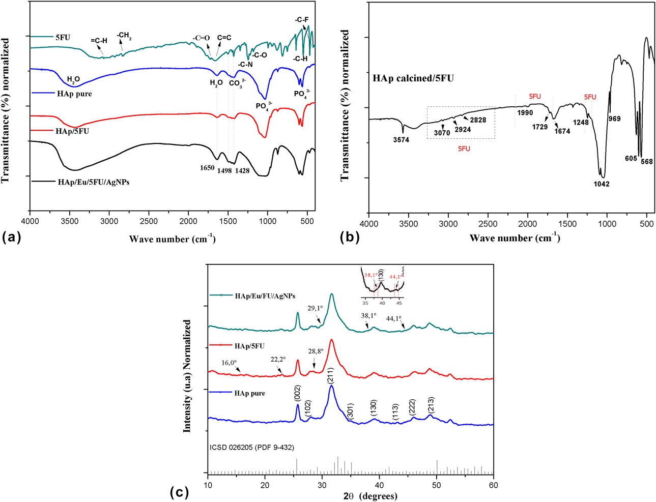
In the spectra of Fig. 2(a), the main absorption bands typical of HAp [Reference de Oliveira, Cavalcanti, Rabelo, Araújo and Fook14] and 5FU [Reference Tseng, Chen, Fang, Lin and Tang15] can be observed. In the same figure, there is a spectrum referring to the sample of HAp with the incorporation of 5FU, where it was not possible to observe the absorption bands attributed to the drug. However, it appears that absorption bands of 5FU appear in the same region as some groups of HAp, as highlighted in 1650, 1498 and 1428 cm−1. Bands featuring 5FU in 2830 and 3115 cm−1 are probably not visualized because of the excessive amount of water present in the material, as observed in the sample spectrum HAp/Eu/5FU/AgNPs. This conclusion can be confirmed with the HAp/5FU spectra with calcination heat treatment for water elimination [Fig. 2(b)]. In this case, many characteristic 5FU of the HAp can be visualized. Thus, it can be stated that the material consists of a carbonated HAp with the drug incorporated in its structure.
(a) Fourier Transform InfraRed (FT-IR) spectra for HAp pure, 5FU, HAp/5FU, and HAp/Eu/5FU/AgNPs; (b) HAp/5FU calcined; and (c) X-ray diffractograms of HAp, HAp/5FU, HAp/Eu/5FU/AgNPs, and HAp matrix standard.
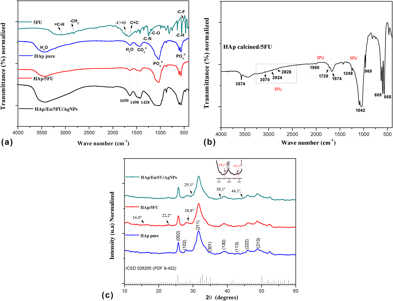
Fig. 2(c) shows X-ray diffractograms of HAp, HAp/5FU, and HAp/Eu/FU/AgNPs samples. Both diffractograms are similar and display the main crystalline peaks attributed to the majority phase of HAp, according to the ICSD 026205(PDF 9-432) standard for the hexagonal HAp. No changes in intensity or displacement of peaks were observed in the diffractograms in the presence of the drug. Characteristic diffraction peaks for 5FU are located in 2θ = 16°, 22°, and 29° [Reference Singh, Tyagi, Mehrotra and Bakhshi16] in the diffractograms in Fig. 2(c). Although these peaks are highlighted, their identification is difficult due to low sample crystallinity. Despite the poor definition of the crystalline peaks of the HAp/Eu/FU/AgNPs sample, it is possible to observe low-intensity peaks typical of Ag identified at 2θ = 38.1°, and 44.1° according to the ICSD 010870720 (PDF 4-783) standard for metallic silver, best viewed in the area highlighted in the graph. The presence of 5FU is also detected from the peak at 2θ = 29.1°, typical of 5FU [Reference Singh, Tyagi, Mehrotra and Bakhshi16].
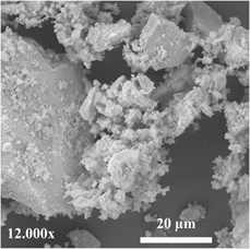
The morphology of HAp/Eu/5Fu/AgNPs powder is shown in Fig. 3. In the micrograph, it is possible to observe clusters of round particles of nanometric nature, uniformly distributed in the material.
Micrographs obtained by scanning electron microscopy for HAp/Eu/5Fu/AgNPs.

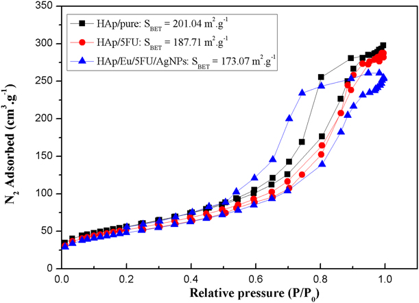
Figure 4 shows the adsorption isotherms for the HAp, HAp/5FU, and HAp/Eu/5Fu/AgNPs samples obtained by the Brunauer–Emmett–Teller (BET) technique. According to IUPAC, these curves can be identified as type-IV isotherms, characterized as mesoporous material [Reference Sing, Everett, Haul, Moscou, Pirotti and Rouquerol17]. Comparing the surface area values (S BET) of the samples, it can be verified its reduction as a function of the addition of 5FU. This observation may indicate the presence of the drugs in the pores of HAp, since it has an 8.6-nm-diameter pore, whereas 5FU has a 0.81-nm-diameter pore and can be totally retained in the bioceramic material [Reference Jin, Wang, Wu, Yin, Liu, Chen, Liu and Li18]. This explains the decrease in the surface area of the samples and confirms the incorporation of 5FU into the HAp matrix.
Nitrogen adsorption isotherms and BET surface area of HAp, HAp/5FU, and HAp/Eu/5Fu/AgNPs.
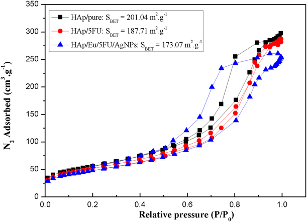
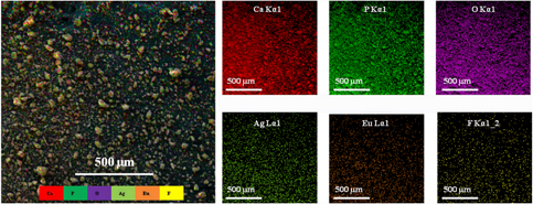
A reduction in S BET of HAp/Eu/5FU/AgNPs can also be observed when compared with pure HAp, indicating that dopant was introduced into the matrix from the pores and interstices of HAp. The observation of the mapping of the simultaneous distribution of the chemical elements from the scanning of much of the materials presented in Fig. 5 corroborates the BET data since this analysis presents the highly uniform distribution of all the characteristic elements of the dopant inserted into the HAp matrix, ensuring the success of the material synthesis.
Mapping of the simultaneous distribution of elements based on the scan of the HAp/Eu/Fu/AgNPs sample obtained from the energy-dispersive spectroscopy analysis.
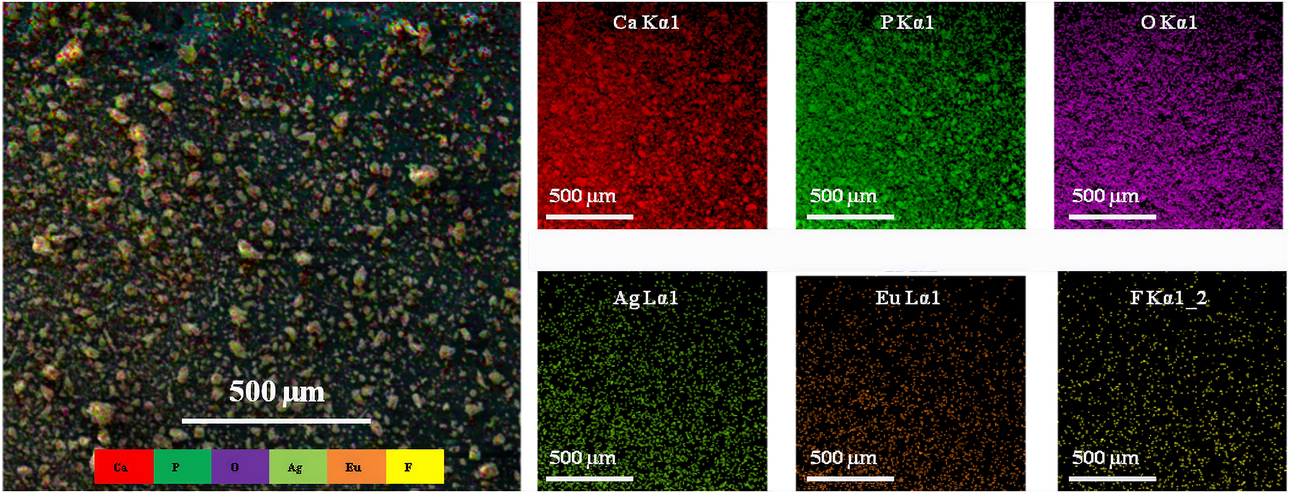
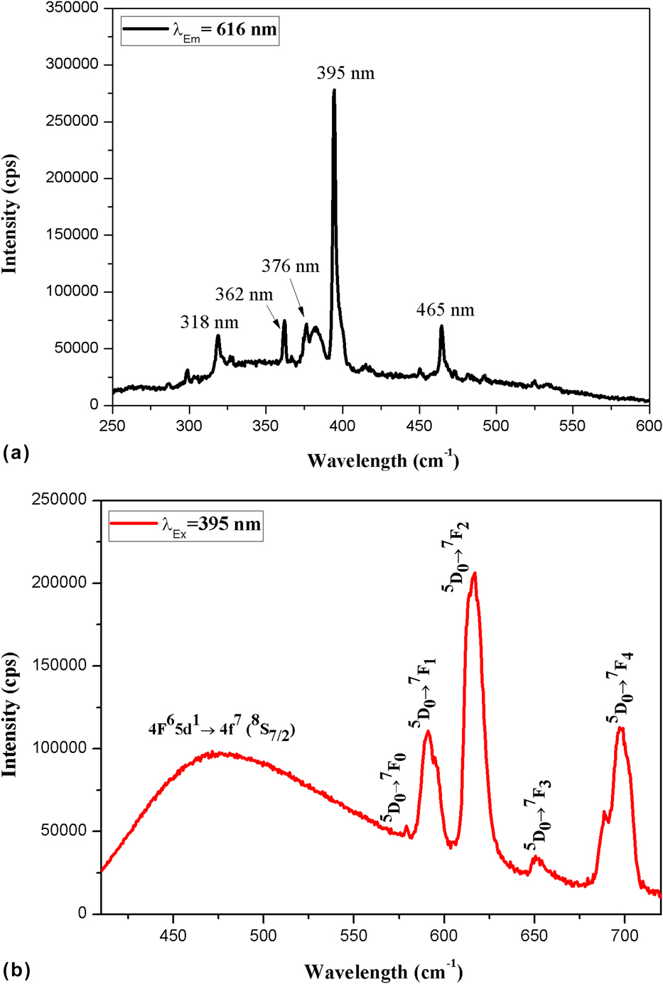
Figure 6 shows the excitation and emission spectra of the HAp/Eu/5FU/AgNPs sample. The material was excited using the maximum excitation wavelength at 395 nm [Fig. 6(a)]. In the emission spectrum, Fig. 6(b), a wide band with emission in the blue spectral region (440–485 nm) with transition 4F65d 1 → 4f 7 (8S7/2) typical of Eu2+ ions is observed, besides the f–f transitions attributed to the Eu3+ ion: 5D0 → F0 (579 nm), 5D0 → F1 (590 nm), 5D0 → F2 (616 nm), 5D0 → F3 (650 nm) and 5D0 → F4 (696 nm).
(a) Excitation spectrum and (b) emission spectrum for the HAp/Eu/5FU/AgNPs sample.
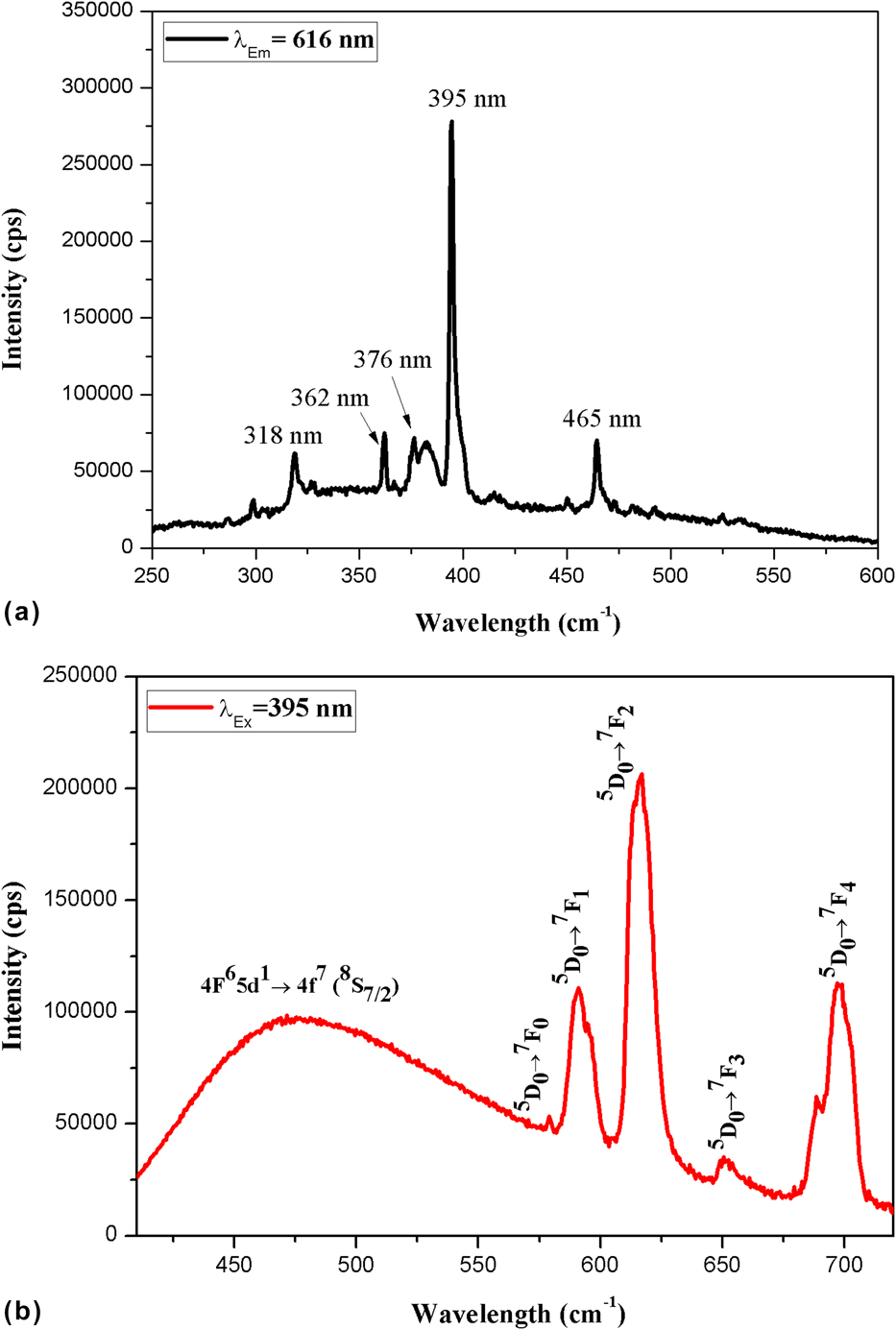
The Eu3+ ions can be present in two different environments in the host matrix due to the existence of two different sites for the Ca2+ ions in the HAp crystalline structure named CaI and CaII. The CaI site is of high symmetry (C 3), surrounded by nine oxygen atoms, and the CaII site is of low symmetry (C s), surrounded by six oxygen atoms and one OH−. In order for the Eu3+ ions to be replaced with Ca2+ ions, a charge compensation mechanism is proposed in Eq. (1) [Reference Martin, Carlot, Chevarier, Den-Auwer and Panczer19]:
 $${\rm{E}}{{\rm{u}}^{3 + }} \!+ {{\rm{O}}^{2 - }} \leftrightarrow {\rm{C}}{{\rm{a}}^{2 + }} \!+ {\rm{O}}{{\rm{H}}^ - } \leftrightarrow {\rm{C}}{{\rm{a}}_{10 - x}}{\rm{E}}{{\rm{u}}_x}{\left( {{\rm{P}}{{\rm{O}}_4}} \right)_6}{\left( {{\rm{OH}}} \right)_{2 - x}}{{\rm{O}}_x}\quad .$$
Based on Fig. 6(b), it is possible to observe that many emissions of Eu3+ ions are attributed to its location in the CaI site of the host network: 579, 590, 650, and 696 nm and for CaII: 616 nm. The emission in the blue spectral region attributed to the occurrence of transitions of Eu2+ ions can be explained by the influence on the valence state of europium ions generated by the calcium deficiency in the matrix structure. This occurs due to the destabilization of the crystal resulting from the replacement of Ca2+ ions with Eu3+ ions; thus, a huge amount of energy is required for the occurrence of this charge compensation, and in the absence of this energy—which is the case of the non-calcined material (HAp/Eu/5FU/AgNPs), a mix of Eu2+ and Eu3+ ions is observed. On the other hand, the heat treatment of the material supplies energy to the system and eliminates emissions related to the Eu2+ ion, indicating oxidation for Eu3+ [Reference Zavala-Sanchez, Hirata, Novitskaya, Karandikar, Herrera and Graeve20, Reference Graeve, Kanakala, Madadi, Williams and Glass21].
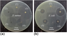
Figure 7 shows the photographic images obtained after the incubation period of the different samples in two different bacterial media. The formation of inhibition halos around the pellets is an indication of the antiproliferative action of bacteria [22]. In this case, the pure HAp serves as a negative control, since no effect is expected in relation to the bacteria, as verified in Figs. 7(a) and 7(b), where it is observed that there was no formation of inhibition halos for pure HAp. On the other hand, zones of inhibition were observed for all the samples studied. The effect of silver as an antibacterial can be confirmed for both bacteria; however, the different amounts of silver added did not indicate perceptible differences in the size of inhibition halos.
Assay in a bacterial media. (a) Staphylococcus aureus and (b) Escherichia coli: (1) Hap pure; (2), (3), and (4) HAp/AgNPs for 0.1, 0.5, and 1.0% of Ag, respectively; and (5) HAp/Eu/5FU/AgNPs.
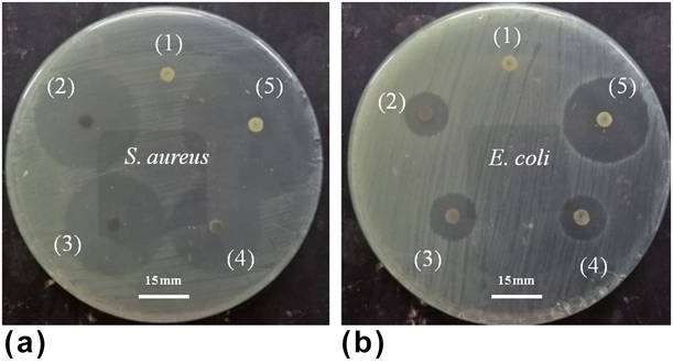
In Figs. 7(a) and 7(b), it can also be verified that the largest inhibition halo is assigned to the sample (5) HAp/Eu/5FU/AgNPs. Thus, we conclude that large halos around pellets must be attributed to the action of another doping component in the material, and thus, knowing the mechanism of action of 5FU, which interferes with the synthesis of deoxyribonucleic acid (DNA) [Reference Nair, Jagadeeshan, Nair and Kumar23], the formation of halos can be attributed to the drug.
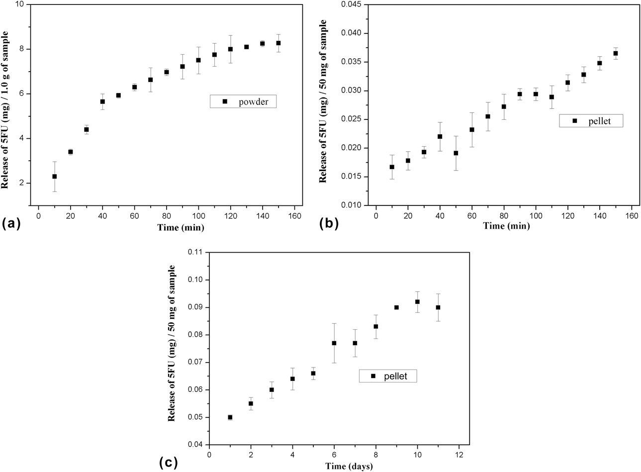
In the study of the release of the drug supported in nanoparticles, we observed the behaviors of the material in powder form and also in pellet form, since the material can be employed in two different ways. In Fig. 8(a) it is possible to verify the amount (in mg) of 5FU released every 10 min by HAp/Eu/5FU/AgNPs powder. The release of 5FU is increasing and gradual; thus, the value starts at 2.3 mg and ends at approximately 8.2 mg after 150 min (2.5 h). According to the total release, we calculated that 82% (±2.8) of 5FU was incorporated into the HAp/Eu/5FU/AgNPs matrix. For the 5FU release from the sample in pellet form [Fig. 8(b)], it was verified a considerable lower release in relation to the powder form, since the surface area exposed to the medium is consequently smaller; thus, for 1.0 g of the material, only 0.33 mg was released in the first 10 min, reaching 0.73 mg at the end of 150 min and confirming that the release of 5FU in the pellet form is rather slow, presenting extremely low although increasing values. The release of the drug in the pellet form was also measured every 24 h, as displayed in Fig. 8(c). In this case, after the first 24 h, 1.0 g of the material released 1.0 mg of 5FU, and after 10 days, the value was 1.84 mg.
5FU release study in a buffer solution: (a) HAp/Eu/5FU/AgNPs in powder form, (b) HAp/Eu/5FU/AgNPs in pellet form, measured in minutes, and (c) HAp/Eu/5FU/AgNPs in pellet form, measured in days.

From the obtained data, it is observed that 5FU was incorporated in an intercalated way between the structural layers of HAp or in its interstices, corroborating the BET data displayed in Fig. 4, where a reduction in the surface area as a function of the addition of the drug is observed, corroborating the data found in the literature [Reference Singh, Tyagi, Mehrotra and Bakhshi16, Reference Luo, Ji, Li, Zhu, Xiong and Wan24]. Considering that biological half-life of 5FU is 10–20 min, it can be stated that the HAp/Eu/5FU/AgNPs developed in this study has a high potential as a support for the drug, since this bioceramic material can extend the permanence of 5FU in the organism from minutes to hours—or even days, as verified in Fig. 8(c) [Reference Hardman, Limbird and Gilman25].
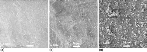
The ability of the material to bind to bone tissue was assessed from the formation of an apatite layer on its surface. The assay was performed employing a solution that mimics blood plasma and body conditions. In Fig. 9, micrographs are presented, where it is possible to observe the formation of a layer of globular apatite crystals on the surface of the sample, indicating its potential for osseointegration.
Surface micrographs of HAp/Eu/5FU/AgNPs after immersion in solution of simulated body fluids at different periods: (a) 0 days, (b) 3 days, and (c) 21 days.

Conclusion
Based on the similarities between HAp and bone tissue already widely described in literature and its confirmed bioactivity in this work, we suggest that the system developed here may be successful when applied as a support or graft in the treatment of bone cancer. This is because from the characterizations carried out, it was verified that the material has potential to sustain and progressive release of an antineoplastic drug, and the HAp/Eu/5FU/AgNPs system has also shown efficiency against bacteria and consequently possible infections that arise from the use of implants. In addition, the behavior of the material can be monitored through images generated by luminescent europium emissions.
Materials and methods
HAp and HAp/Eu/5FU/AgNPs syntheses
HAp synthesis
The experimental procedure adopted for the HAp synthesis was the precipitation method. For the production of the HAp bioceramic powder, we used gypsum (calcium sulfate hemihydrate, CaSO4·1/2H2O—purity between 80 and 95%) as a starting material, supplied by the Gesso Mineral Inc. industry, Araripina, Brazil; diammonium phosphate [(NH4)2HPO4], supplied by Vetec, Rio de Janeiro, Brazil; and ammonium hydroxide (NH4OH), supplied by Êxodo científico, São Paulo, Brazil, all three of analytical grade. The synthesis reaction was performed using a mixture of the sources of calcium and phosphorus, maintaining a molar ratio of 1.66 Ca/P. Thus, to 0.1 mol/L CaSO4·1/2H2O solution, we added 3.0 mol/L NH4OH to set the pH of the reaction medium to 10. Then, 0.06 mol/L (NH4)2HPO4 solution was added at a rate of incorporation of 20 mL/min until the desired final volume was attained. Throughout the entire process, the pH was controlled by adding NH4OH. At the end of the addition of the reactants, the system remained under stirring for one hour. Then, it was put to rest for 48 h. Subsequently, the material produced underwent vacuum filtration and washed with deionized water to reach a neutral pH; then, it was oven dried at 70 °C for 24 h; once dried, it was crushed in a mortar to obtain a fine powder. The chemical equation [Eq. (2)] represents HAp formation reaction:
 $$\eqalignb{ {& 10\;{\rm{CaS}}{{\rm{O}}_4}{1 / 2}{{\rm{H}}_2}{\rm{O}} + 6{\left( {{\rm{N}}{{\rm{H}}_4}} \right)_2}{\rm{HP}}{{\rm{O}}_4} + 8{\rm{N}}{{\rm{H}}_4}{\rm{OH}} \to \mathop }\cr{{\bf{C}}{{\bf{a}}_{{\bf{10}}}}{{\left( {{\bf{P}}{{\bf{O}}_{\bf{4}}}} \right)}_{\bf{6}}}{{\left( {{\bf{OH}}} \right)}_{\bf{2}}}}\limits_{{\rm{Hydroxyapatite}}} & \cr+ 10{\left( {{\rm{N}}{{\rm{H}}_4}} \right)_2}{\rm{S}}{{\rm{O}}_4} + 11\;{{\rm{H}}_2}{\rm{O}}\quad {\rm{.}} \cr}$$
HAp/5FU synthesis
For comparative purposes, the synthesis of HAp with 5FU was performed. The sequence of the HAp synthesis reaction with the incorporation of the drug followed the same procedure described in “HAp synthesis” section; however, the 5FU powder (99%) supplied by Sigma-Aldrich Inc. (St. Louis, Missouri) was solubilized in deionized water (solubility of 12 g/L) and subsequently added to the calcium sulfate solution during the stirring step. The amount of 5 FU added was 1.0% relative to the total mass of HAp (amount chosen only for evaluation of the release profile of the drug when on the carrier).
HAp/Eu/5FU/AgNPs synthesis
From the reaction of Eu2O3 (99.99%; Aldrich) with excess HCl (Vetec PA), under heat treatment (100–150 °C) and stirring, the EuCl3 was obtained after washing with deionized water until pH = 6.0 and subsequent evaporation of the liquid, as shown by Eq. (3):
 $${\rm{E}}{{\rm{u}}_{\rm{2}}}{{\rm{O}}_3}\left( {\rm{s}} \right) + 6{\rm{HCl}}\left( {{\rm{aq}}} \right) + 9{{\rm{H}}_2}{\rm{O}} \to 2{\rm{EuC}}{{\rm{l}}_3} \cdot 6{{\rm{H}}_2}{\rm{O}}\left( {\rm{s}} \right)\quad .$$
Soon after, AgNPs were synthesized based on the methodology previously described in the literature [Reference Solomon, Bahadory, Jeyarajasigam, Rutkowsky and Boritz26]. For the reaction, sodium borohydride (NaBH4) (Sigma-Aldrich) and silver nitrate (AgNO3) (PLAT-LAB) solutions were used, both of analytical grade, according to Eq. (4):
 $$2{\rm{AgN}}{{\rm{O}}_3} + 2{\rm{NaB}}{{\rm{H}}_4} \to 2{\rm{Ag}} + 2{\rm{NaN}}{{\rm{O}}_3} + {{\rm{H}}_2} + {{\rm{B}}_2}{{\rm{H}}_6}\quad .$$
For synthesis of multifunctional system, pure HAP production method (see “HAp synthesis” section) was used; in this case, 1% by mass of europium(III) chloride (quantity chosen based on the intense luminescence peaks shown in the spectrum of the HAp/Eu sample) was added to the calcium sulfate solution during stirring. For incorporation of the drug, 5FU (99%) powder was solubilized in deionized water and subsequently added to the solution. After the rest and drying of HAp/Eu/5FU powder, it was added to an AgNPs solution with 1.0% concentration calculated in relation to the mass of HAp. In this step, the HAp/Eu/5FU powder remained under stirring for 30 min to the adsorption of AgNPs to its surface, and subsequently, the material was filtered and oven dried at 70 °C for 24 h, resulting in the HAp/Eu/5FU/AgNPs system.
In vitro 5FU release study
The release of the drug was studied based on the methodology previously described in the literature [Reference Tseng, Chen, Fang, Lin and Tang15]. In this case, a phosphate-buffered saline (PBS) solution, 0.13 mM, pH = 7.4 at 37 °C, was used, with 5 mL of this solution being added to a 15 mL Falcon tube, to which 20 mg of HAp/Eu/5FU/AgNPs powder was added. This system was centrifuged in different periods at a rotation rate of 200 rpm, with 3 mL of the supernatant collected every 10 min for the quantification of the released drug. The measurements were made with the ultraviolet–visible (UV-Vis) spectrophotometer (model IL-592) with an absorbance of 240 nm for 5FU. For every aliquot withdrawn, the same amount of a new solution was replaced. Each sample was prepared in triplicate and both the mean and standard deviation were calculated.
The release of the drug incorporated to the system was also evaluated for the material molded in the pellet form. To do so, 50 mg of the HAp/Eu/5FU/AgNPs powder sample was added to a steel mold (6 mm diameter), where approximately 9.8 Pa/m2 of force was applied for material compaction and disk formation. Subsequently, the pellets were placed in different beakers with 5 mL of PBS at 37 °C and pH = 7.4. Then, after a mild stirring, aliquots of 3 mL of the supernatant were collected every 10 min for the quantification of the released drug and, later, every 24 h, following the same procedure described for the analysis of the powder material.
Antimicrobial assay
The tests were performed using two types of bacteria: the Gram-positive (Staphylococcus aureus ATCC 25923) and the Gram-negative (Escherichia coli ATCC 25992) bacteria. To perform the assay, bacterial cultures (stock solution) were grown in nutrient-rich agar at 4 °C, with isolated colonies being selected subsequently for the formation of a 0.5 McFarland turbidity standard. Plating was performed using 10 μL of the resulting solution, where the bacteria were inoculated in 150 mm plates containing Mueller–Hinton Agar for microorganism growth. Subsequently, pellets (50 mg and 6 mm diameter) from the different samples (HAp/pure, HAp/AgNPs, and HAp/Eu/5FU/AgNPs) were deposited aseptically onto the plate surface. The resulting plates were incubated at 37 °C for 24 h, and after this time, the growth inhibition halos formed around each sample were observed and recorded in the photographic images.
In vitro bioactivity assay
The bioactivity assay was performed using the methodology proposed by Kokubo and Takadama [Reference Kokubo and Takadama27]. For the preparation of the simulated body fluids solution, the following reagents were used: sodium chloride (NaCl), sodium hydrogen carbonate (NaHCO3), potassium chloride (KCl), dipotassium hydrogen phosphate trihydrate (K2HPO4·3H2O), magnesium chloride hexahydrate (MgCl2·6H2O), calcium chloride (CaCl2), sodium sulfate (Na2SO4), tris(hydroxymethyl)aminomethane [(HOCH2)3CNH2], hydrochloric Acid (HCl, supplied by Vetec), all of analytical grade. The assay was performed for different periods of time (0–21 days), and the samples were kept under constant stirring and fixed temperature equal to 37 °C.
Characterizations
The morphology of the different samples was observed by scanning electron microscopy (High Voltage (HV) = 10 kV, Working Distance (WD) = 5.31 mm), and the mapping of the chemical elements was obtained through energy-dispersive X-ray analysis (Hitachi TM1000 and Vega 3 Tescan, Tescan, Brno, Republica Checa). The samples were covered in a thin gold layer (metallization process) to increase conductivity in its surface. The materials were characterized based on their functional groups by infrared spectra obtained by the Fourier Transform InfraRed (FT-IR) equipment (model Spectrum Two, PerkinElmer do Brasil Ltda., São Paulo, Brazil), at a wave number range of 4000–400 cm−1. The samples were prepared in the form of KBr pellets using mechanical pressing. The analysis of the crystalline phases was performed using X-ray powder diffraction with the Diffract ACT Series 1000 (Siemens, Nürnberg, Germany) equipment, using the K-alpha line of copper (λ = 1.54056 Å) at 40 kV and 40 mA. The 2θ range was 10°–60° at a 0.02° step size and a time step of 1 s. The phases were identified using the data from ICSD 026205 (PDF 9-432) files for the hexagonal HAp, using the X’pert HighScore Plys software (version 2.0a). The BET surface area measurements were performed by the surface area analyzer Micromeritics ASAP 2420, Micromeritics Instrument Corp., Norcross, Georgia. The UV-Vis region absorption measurements were performed in a UV spectrophotometer (IONLAB, Curitiba, Brazil), model IL-592, using quartz cuvettes and the sample in the liquid form. Measurements of excitation and emission of the doped samples were carried out in a Jobin Yvon Ramanor U-1000 double monochromator spectrofluorometer (Horiba, USA). For the excitation, a Jobin Yvon H-10 monochromator (Horiba, USA) was used with a Xe–Hg (150 W) lamp. The analyses were performed for the solid samples deposited on carbon ribbons.
Acknowledgment
This research was supported by Foundation for protection of research in the state of Bahia—FAPESB.