Introduction
Pneumoceles are rare, air-containing structures that arise within the aerated cavities of the cranium. They have a pathological tendency to expand gradually, destroying and thinning surrounding bone.Reference Martin, Bhargava, Ashforth and Russell 1 , Reference Lichtenberg and Russell 2 They arise most commonly in the frontal sinus, but have also been described in the other paranasal sinuses.Reference Urken, Som, Lawson, Edelstein, Weber and Biller 3 A pneumatocele occurs when air extends from an aerated cavity to form a second air-filled cavity within the adjacent soft tissues.Reference Martin, Bhargava, Ashforth and Russell 1 , Reference Lichtenberg and Russell 2
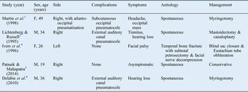
Pneumoceles of the mastoid bone are rare, with only five previously described cases (Table I).Reference Martin, Bhargava, Ashforth and Russell 1 , Reference Lichtenberg and Russell 2 , Reference Ivers, Roche and Fagan 4 – Reference Delabie, Gordts and Clement 6 Of these, one case was secondary to previous cranial trauma and subsequent surgical explorationReference Ivers, Roche and Fagan 4 and the other four were spontaneous in origin.Reference Martin, Bhargava, Ashforth and Russell 1 , Reference Lichtenberg and Russell 2 , Reference Patnaik and Mahapatra 5 , Reference Delabie, Gordts and Clement 6 Two of these were associated with pneumatoceles of the external auditory canal and one was associated with a subcutaneous tissue occipital pneumatocele (Table I).
Location, aetiology, associated complications and management of previously reported temporal bone pneumoceles

F = female; M = male
Given the close proximity of the mastoid cavity to several intracranial structures, the potential exists for mastoid pneumoceles to produce a range of intracranial complications. We describe a case of hypoglossal palsy secondary to trauma in a patient with a pre-existing diffuse temporal bone pneumocele, with further expansion over time and subsequent development of an intracranial middle fossa pneumatocele.
Case report
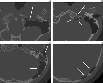
A previously well 46-year-old man presented with head and neck pain, dysphagia, dysarthria and impaired glossal mobility after heading a football. Examination showed left hypoglossal palsy and a bullous left tympanic membrane, but was otherwise unremarkable. Computed tomography (CT) scans of the head showed a diffuse left temporal bone pneumocele with extensive pneumatisation of the left temporal bone extending to the occipital condyle, and involving bone on both sides of the hypoglossal canal (Figure 1). In addition, a fracture of the hypoglossal canal was identified, presumed due to recent minor trauma, with associated adjacent blood within the pneumocele. This was confirmed on subsequent magnetic resonance imaging (MRI). The patient was treated conservatively.
Axial computed tomography head scans (a – d), demonstrating extensive left temporal bone pneumocele (arrows), extending from the occipital bone/atlas (a) to above the petrous apex level (d). The pneumocele is seen to extend, involving bone on either side of the left hypoglossal canal (b).

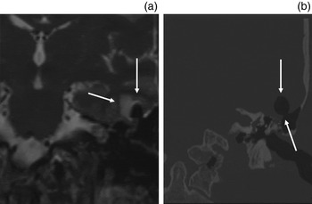
Initial MRI also showed an incidental 2 mm left intracanalicular vestibular schwannoma requiring clinical and imaging follow up. Follow-up MRI at two years demonstrated a new, small left middle fossa pneumatocele with minor adjacent temporal lobe oedema (Figure 2a). The CT scans demonstrated a defect in the left tegmen mastoideum adjacent to the diffuse mastoid bone pneumocele (Figure 2b). The patient was asymptomatic. Potential intracranial complications, including the risk of meningitis, and possible surgical options for repair of the tegmen defect were discussed; however, the patient declined surgery and opted for conservative management and routine ongoing clinical and imaging follow up.
Coronal, T2-weighted magnetic resonance imaging scan (a) and coronal computed tomography scan (b) of the head, showing a small left middle fossa pneumatocele (arrows), with minor adjacent left temporal lobe oedema secondary to a defect in the tegmen mastoideum.

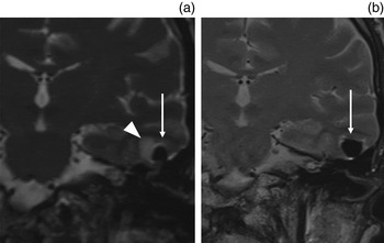
Subsequent routine follow-up MRI over five years showed a gradual increase in size of the left middle fossa pneumatocele for the first three years, albeit with resolution of oedema in the adjacent brain parenchyma (Figure 3), then stabilisation for the last two years.
Baseline (a) and follow-up (b) coronal, T2-weighted magnetic resonance imaging scans, showing interval enlargement of the left middle fossa pneumatocele (arrows). Temporal lobe oedema adjacent to the pneumatocele, as observed on baseline imaging (a, arrowhead), had resolved on subsequent imaging.

The patient remains clinically well, with no features of cerebrospinal fluid (CSF) leak within the mastoid or middle ear, either clinically or radiologically. He continues to decline surgical repair of the left middle fossa floor, despite the ongoing risk of intracranial infection. He has been advised to avoid vigorous nose-blowing following episodes of headache associated with this.
Discussion
Aetiology
Current thinking identifies three distinct entities that involve air within the cranium and which have the potential for spread to adjacent structures as a result of deossification and bony erosion. Pneumoceles are expansions of the bony cavities of the skull, usually associated with the sinuses, where the bony walls are thinned or eroded.Reference Martin, Bhargava, Ashforth and Russell 1 – Reference Delabie, Gordts and Clement 6 Pneumatoceles are extraosseous gas collections adjacent to bony cavities where the air is contained by the surrounding soft tissues.Reference Martin, Bhargava, Ashforth and Russell 1 , Reference Lichtenberg and Russell 2 They may be extracranial or intracranial, but when intracranial they may sometimes be called pneumocephalus.Reference Añorbe, Aisa and Saenz de Ormijana 7 Pneumocephalus is considered to be air anywhere within the cranial cavity, either involving the meninges, the ventricles or the cerebrum itself.Reference Markham 8 However, Markham notes that, historically, terminology referring to pathologies involving air in the cranium made no distinction between these three entities, which may cause confusion in some of the literature.Reference Markham 8 The pathogenesis of these entities has been postulated to occur in a number of similar ways, notably as a result of cranial trauma, neoplasm, infection, surgery or spontaneously; pneumoceles have also been described as a direct cause of pneumatoceles and pneumocephalus.Reference Martin, Bhargava, Ashforth and Russell 1 , Reference Lichtenberg and Russell 2 , Reference Delabie, Gordts and Clement 6 , Reference Park and Kim 9
The theory currently favoured regarding the development of spontaneous pneumoceles, albeit in the frontal sinus, is a ‘trap-valve’ hypothesis. Urken et al. discussed the possibility that intraluminal pressure was consistently increased because of an anatomical or physiological obstruction between the nasal cavity and the sinuses that prevented air pressure from equilibrating.Reference Urken, Som, Lawson, Edelstein, Weber and Biller 3 This would explain the definition of a sinus pneumocele as a hyperaerated, abnormally expanding region causing a local or generalised erosion of the sinus walls. The suggested cause of this is pressure necrosis, as the pneumocele expands slowly over time. Increased pressure has since been demonstrated in sinus pneumoceles,Reference Flanary and Flanary 10 though whether this is the cause of the pneumoceles has yet to be verified. Pneumoceles of the temporal bone have been proposed to have a similar aetiology, caused by retrograde air entry via the Eustachian tube.Reference Martin, Bhargava, Ashforth and Russell 1 , Reference Delabie, Gordts and Clement 6
The trap-valve hypothesis has also been postulated as the cause of spontaneous pneumatoceles and pneumocephalus, although pneumocephalus may also occur secondary to a negative pressure gradient caused by leakage of CSF, which then generates an influx of air to replace the lost fluid (the ‘siphon effect’).Reference Abbati and Torino 11
Mastoid pneumoceles have been described associated with extracranial pneumatoceles of the external auditory canal and occipital subcutaneous tissues (Table I).Reference Martin, Bhargava, Ashforth and Russell 1 , Reference Lichtenberg and Russell 2 , Reference Delabie, Gordts and Clement 6 While a case such as ours of intracranial air secondary to a mastoid pneumocele has not been previously described, spontaneous otogenic pneumatoceles and pneumocephalus are recognised in association with either normal or hyperpneumatised mastoids (a large, well aerated mastoid which does not expand bone beyond its usual contours).Reference Añorbe, Aisa and Saenz de Ormijana 7 , Reference Abbati and Torino 11 It has been proposed that spontaneous otogenic pneumatoceles and pneumocephalus often occur following minor barotrauma, such as vigorous nose-blowing, coughing or the Valsalva manoeuvre; it is therefore easy to see how the thinner bony margin of a pneumocele could lead to the same complications.
Clinical features
Clinically, pneumoceles, pneumatoceles and pneumocephalus present with symptoms dependent on their location and size. Many of these entities are not associated with any symptoms while they are small; however, if they affect the mastoid they may cause conductive hearing loss, tinnitus or aural pressure.Reference Lichtenberg and Russell 2 , Reference Delabie, Gordts and Clement 6 Abbati and Torino noted in their review that most cases of otogenic intracranial air have presented acutely, with the patients often being extremely unwell, with severe symptoms such as visual disturbance, headache, aphasia and loss of consciousness due to a raised intracranial pressure secondary to tension pneumocephalus.Reference Abbati and Torino 11 It is surprising therefore that our patient remained asymptomatic throughout the long time course and that the intracranial pneumatocele has been relatively stable. We postulate that either the increased pressure is intermittent or relatively small, or that the large volume of the pneumocele is acting as a buffer to this increased pressure.
Management
A number of management options exist but, given the rarity of mastoid pneumoceles and otogenic pneumatoceles or pneumocephalus, there is no consensus. Options include conservative management, myringotomy with or without grommet insertion (intended to remove the valve effect and release any increased pressure), or surgery to obliterate the pneumocele and repair the tegmental defect, which can be done with or without Eustachian tube obliteration and blind sac closure. Managing the patient conservatively presents the risk of continued expansion of the pneumocele, with the potential life-threatening risks of tension pneumocephalus or meningitis. There may also be the risk of temporal lobe irritation and epilepsy, although this has not been reported to date. However, it is difficult to quantify these risks, and the expansion in our case has been relatively slow since it was detected, with no significant oedema evident on later scans.
-
• Three distinct cranial air-filled pathological entities have been described: pneumoceles, pneumatoceles and pneumocephalus
-
• Development is thought to result from a ‘trap valve’ that leads to an influx of air, causing (rapid or gradual) build-up
-
• Spontaneous temporal bone pneumoceles are very rare and previously associated with minor complications only; otogenic pneumocephalus often presents acutely, in extremis
-
• This article describes a massive temporal bone pneumocele, demonstrating cranial nerve damage and intracranial air extension
-
• Unlike previous otogenic pneumocephalus reports, this patient did not develop tension pneumocephalus and has been managed conservatively
-
• The aetiology and potential treatment options of these pathologies are summarised with respect to current literature
For our case, myringotomy would be a relatively straightforward treatment, and this has been used successfully in two of the previously described mastoid pneumoceles.Reference Martin, Bhargava, Ashforth and Russell 1 , Reference Delabie, Gordts and Clement 6 However, neither of the previous cases had any exposure of the dura or intracranial air; in our case, the increased risk of middle-ear infection presented by grommet insertion could potentially increase the risk of intracranial infection. While obliteration of the Eustachian tube followed by a blind sac closure would provide the least risk of future recurrence, it would cause the patient significant conductive hearing loss. Our patient's hearing currently remains normal and we would therefore prefer to avoid this, perhaps considering a grommet should there be any evidence of recurrence once the mastoid had been repaired. Our preferred option would be surgical obliteration of the mastoid cavity with bone pâté and hydroxyapatite. At present, however, the patient has declined any intervention.
Conclusion
Temporal bone pneumoceles are rare entities; they may be associated with intracranial pneumatoceles and are predisposing to fractures following minor trauma. In patients with pneumocele-associated intracranial pneumatocele, management options depend on symptoms and progression. For those in whom surgery is considered beneficial, options include release of middle-ear pressure by myringotomy and surgical obliteration of the mastoid cavity.