Optimising the long-term outcome for children born with hypoplastic left heart syndrome remains a significant challenge for paediatric cardiac centres. Morbidity and mortality rates remain high following the staged, palliative, reconstructive surgery that involves the Norwood procedure.Reference Cua, Thiagarajan and Taeed1, Reference Ohye, Gaynor and Ghanayem2 United Kingdom data from the central cardiac audit database (2002–2006) indicate that although over 88% survived Stage I “Norwood” surgery at 30 days, the survival rate at 1 year of age fell to around 64%.3
Many factors affect outcome in this group, and a key issue influencing the surgical pathway is the impetus to preserve the systolic function of the right ventricle. Recently, a randomised controlled trial formed a comparison of the modern surgical techniques for the Norwood procedure.Reference Ohye, Sleeper and Mahony4 We assessed intermediate outcomes, with the right ventricular function quantified using echocardiographic methods.
Two-dimensional echocardiography provides the imaging mainstay for the prenatal and immediate postnatal diagnosis of hypoplastic left heart syndrome; however, this modality is possibly less sensitive to the impairment of ventricular systolic function. Cardiovascular magnetic resonance provides a more comprehensive assessment of the arterial morphology, great vessel flow, and ventricular function,Reference Muthurangu, Taylor and Hegde5 and provides an alternative to catheterisation.Reference Brown, Gauvreau and Powell6 Cardiovascular magnetic resonance imaging can be performed non-invasively and without ionising radiation.
Since 2003, our paediatric cardiac surgical unit has adopted an imaging protocol for all infants with hypoplastic left heart syndrome, which includes standardised cardiovascular magnetic resonance imaging rather than diagnostic catheterisation for inter-stage assessment. Within this protocol, we carried out inter-stage cardiac catheterisation only when planning a specific percutaneous intervention.
The aim of this retrospective study was to assess whether the cardiovascular magnetic resonance data acquired during this protocolised follow-up could help to stratify the risk for these patients. In this paper, we describe the structural and functional parameters obtained with the cardiovascular magnetic resonance examination that was performed before the second palliative surgical stage, which involves formation of a bidirectional cavo-pulmonary shunt. Specifically, we evaluate links between structural components of the heart (such as neo-aortic or pulmonary dimensions) and the right ventricular function. We then explore the relationships between right ventricular function, neo-aortic arch narrowing, pulmonary artery narrowing, and patient survival over time.
Materials and methods
Patient selection
We identified all neonates with typical hypoplastic left heart syndrome morphologyReference Noonan and Nadas7 undergoing Norwood-type surgery from 1 February, 2003 to 1 October, 2008 from the Great Ormond Street Hospital Cardiac Unit surgical database. We included patients in the analysis only if they had survived to inter-stage cardiovascular magnetic resonance and lived in the United Kingdom, to accurately ascertain the outcome.
We gained informed, signed consent for cardiovascular magnetic resonance imaging under general anaesthesia, along with subsequent research data analysis from each patient's parents at the time of admission to hospital for the imaging procedure. The Research and Development Office at the Institute of Child Health, London registered the study as an audit.
Cardiovascular magnetic resonance imaging
Scanning was performed using a 1.5 Tesla magnetic resonance scanner (Avanto, Siemens Medical Solutions, Erlangen, Germany), with post-processing on a dedicated magnetic resonance workstation (Leonardo, Siemens Medical Solutions, Erlangen, Germany). All imaging was performed under general anaesthesia with endotracheal intubation, to allow for control of respiration and to eliminate artefacts related to body movement.
We describe the sequence parameters for the scan in detail.Reference Muthurangu, Taylor and Hegde5 In brief, anatomy was initially mapped using an axial stack of HASTE (half-Fourier acquisition single shot turbo-spin echo) images. A set of steady-state free precession cine images was then acquired during serial breath holding. Slice positions for these two-dimensional cine images included the cardiac long-axis, an anatomical short-axis stack (approximately 10 slices) with no gap, and orthogonal planes through the ascending, transverse, descending aortic arch, and branch pulmonary arteries, respectively.
The three-dimensional morphologic assessment of the great vessels used two sequences. Three-dimensional gadolinium-enhanced cardiovascular magnetic resonance angiography was performed using a coronal three-dimensional fast-field-echo sequence. In addition, for each patient, three-dimensional “white-blood” imaging was performed with a respiratory-navigated and electrocardiogram-gated, single-slab three-dimensional steady-state free precession sequence. For all three-dimensional data, the sequence parameters were optimised to achieve isotropic voxels, allowing multi-planar reformatting and measurement of structures in any plane.
At the time of the scan, we additionally imaged the vessels showing significant narrowing in long- and short-axis planes with turbo-spin-echo black blood images. These were acquired during systole, in orthogonal long-axis planes. This sequence is less susceptible to artefact from magnetic field inhomogeneity or blood flow turbulence.
Through-plane magnetic resonance phase-contrast flow quantification data were acquired at the ascending aorta distal to the Damus–Kaye–Stansel anastomosis, in the branch pulmonary arteries, and in the pulmonary veins.
Image post-processing
Right ventricular volumes and ejection fraction were calculated in the conventional manner from the short-axis stack of cine images. Manual segmentation of the ventricles was completed, with exclusion of the major right ventricular trabeculae from the blood pool volume. If the native aortic valve allowed forward flow, the small left ventricular blood pool was included within the right ventricular cavity volume.
Quantification of aortic forward flow volume through the neo-aortic arch at a point distal to the Damus–Kaye–Stansel anastomosis, and proximal to head and neck vessel branching, yielded an internal quantitative guide to the right ventricular stroke volume for each patient (given that a relatively small volume of coronary blood flow would be lost from this flow volume). This method improves the accuracy of volumetric quantification, particularly in the context of minimal tricuspid valve regurgitation.Reference Devos and Kilner8
Arterial morphological measurements were made from the isotropic angiographic data, using multi-planar reformatting on a Leonardo workstation (Siemens Medical Solutions, Erlangen, Germany).
For each measurement position on each vessel, two perpendicular, truly cross-sectional diameters were measured. For outcome analysis, these two diameters were averaged to give a single dimension for each measurement position.
The pulmonary artery measurements were made at the proximal native vessel and were indexed against the distal vessel dimension, measured just before the first lobar branch.
A narrowing of the proximal pulmonary artery was defined as severe if the difference between the proximal diameter and the diameter of the distal normal vessel was greater than 40%.Reference Muthurangu, Taylor and Hegde5
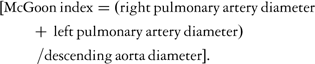
In addition, we calculated the McGoon indexReference Fogel, Donofrio and Ramaciotti9 for the branch pulmonary arteries. The formula for the McGoon index is as follows:
Measurements of the reconstructed aortic arch were made at the narrowest point of the proximal descending aorta (coarctation). This dimension was indexed against the dimension of the descending aorta, at the diaphragmatic level. In accordance with a common morphological classification in the literature, we classified a coarctation index of less than 0.7 as significant.Reference Lemler, Zellers and Harris10
Data analysis
Categorical variables, such as the type of Norwood Stage I operationReference Sano, Ishino and Kawada11 or the presence of a restrictive atrial septum, were expressed as proportions. Continuous variables, including the follow-up time or the patient's weight, were expressed as medians with inter-quartile ranges. The relationship between the type of operation and the number of narrowed areas in the circulation as defined above was explored using Fischer's exact test. The relationship between neo-aortic narrowing and the functional outcome measures (right ventricular ejection fraction and cardiac output) were explored using the Wilcoxon Rank Sum test for comparing medians. The relationship between the diameter of the neo-aorta and pulmonary arteries was explored using linear regression. Patient survival was investigated using Cox proportional hazard models, including all confirmed follow-up time for each patient, up to either the date of death or the most recent clinic appointment, with the last date as 1 January, 2009. The authors had full access to the data and take responsibility for its integrity. All authors have read and agreed to the manuscript as written.
Results
Patient inclusion
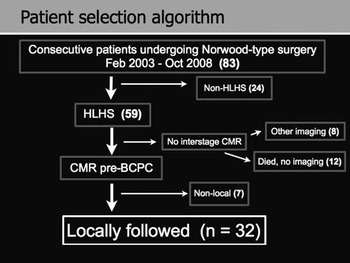
A total of 59 patients with typical hypoplastic left heart syndrome morphology underwent a Norwood-type surgical procedure at Great Ormond Street Hospital between 1 February, 2003 and 1 October, 2008. Of these patients, a total of 32 (54%) underwent elective cardiovascular magnetic resonance examination, before the Stage II procedure, and are included in this analysis. Of the 27 patients excluded from analysis, seven (12%) had completed further staged palliation at centres overseas and were excluded because their outcome was not definitely known; 12 (20%) patients died without undergoing magnetic resonance imaging; six of these patients (10%) died within 30 days of surgery; and a further 6 (10%) died later, between Stages I and II. Of the surviving patients, eight (14%), and one of the inter-stage death patients, underwent alternative imaging (such as cardiac computerised tomography or interventional cardiac catheterisation) for clinical reasons (Fig 1).
Patient selection algorithm.

Cohort demographics
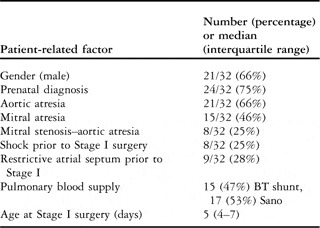
Table 1 shows the anatomical and surgical demographics of the cohort. During the study period, there was contemporary use of both shunt types for the Stage I procedure. In all, 15 (47%) patients underwent a classical Norwood procedure, with pulmonary blood supply through a right-sided modified Blalock–Taussig shunt and 17 (53%) patients had pulmonary blood supply via a right ventricle-pulmonary artery conduit.
Cohort anatomical and surgical demographics.

BT shunt = Blalock–Taussig shunt
Cohort clinical characteristics at cardiovascular magnetic resonance
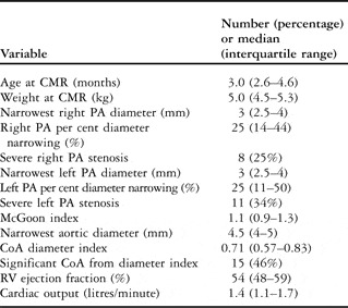
Table 2 shows the clinical characteristics of the cohort at the time of cardiovascular magnetic resonance. The median (inter-quartile range) age at cardiovascular magnetic resonance imaging was 92 (80–139) days and the median weight at cardiovascular magnetic resonance imaging was 5 (4.6–5.3) kilograms.
Cohort clinical characteristics at CMR.

CoA = coarctation defined by CoA index diameter greater than 0.7; CMR = cardiovascular magnetic resonance; PA = pulmonary artery; RV = right ventricular
No patient in this cohort had significant tricuspid valve regurgitation, as documented by serial echocardiography up to the time of the cardiovascular magnetic resonance scan. The cardiovascular magnetic resonance findings complied with this observation.
We did not include the pre-surgical right ventricular function reported from two-dimensional echocardiography in these data, as only qualitative estimates were available.
All patients were managed with standard medical therapy when indicated, including diuretics and afterload reduction. None of the patients received inotropic or invasive support at the time of the cardiovascular magnetic resonance imaging.
Relationships between morphologic and functional findings
Patients who had narrowing of one pulmonary artery were more likely to have narrowing in the contralateral pulmonary artery: regression coefficient 0.54 (95% confidence interval 0.17–0.92), p < 0.01. Narrowing of one or other pulmonary arteries was unrelated to aortic arch narrowing: left regression coefficient 4.34 (95% confidence interval −42.03 to 50.71), p = 0.84 and right regression coefficient −15.26 (95% confidence interval −55.34 to 24.81), p = 0.44.
There was some evidence that patients with a right ventricle-pulmonary artery conduit had a greater proportion of narrow right pulmonary arteries (p = 0.04); however, the proportion of narrow left pulmonary arteries and coarctation was similar between the two types of Norwood operation (p = 1.0 for both).
There was no evidence that the severity of aortic arch narrowing was related to either right ventricular ejection fraction or the cardiac output. When comparing the group of patients with a coarctation diameter index less than 0.7 against the group with coarctation index diameter greater than 0.7, there was no significant difference between the median right ventricular ejection fraction and cardiac output between the two groups (p = 0.41 and 0.29, respectively).
In this group, there was no statistical relationship between the cumulative number of areas of narrowing in the neo-aortic arch and pulmonary arteries found at cardiovascular magnetic resonance, and the right ventricular ejection fraction.
Interventions
In this cohort of 32 patients, five underwent specific intervention to the aortic arch and eight underwent a specific intervention to augment the left pulmonary artery, after the cardiovascular magnetic resonance scan and before the last follow-up date. In addition, some patients underwent pulmonary artery augmentation at the time of Stage II surgery, but these data were not uniformly recorded in surgical notes. In all cases, our Unit's multi-disciplinary forum made the complex decision to intervene on aorta or pulmonary arteries, and was based on the patients’ clinical condition, and a combination of cardiovascular magnetic resonance and echocardiographic features.
Influence of morphologic findings on survival
After considering post-operative (10%), inter-stage (10%), and late deaths, the actuarial survival at 1 year of age for the entire cohort was 63% (95% confidence interval 52–72%). The selected patients who underwent cardiovascular magnetic resonance protocol and were included in this study were followed up for a median of 19.2 (10.8–46.0) months, giving a total of 73.8 patient years of follow-up time, during which seven patients died.
The patient's age at cardiovascular magnetic resonance (in months) was unrelated to risk of death over time, expressed as a hazard ratio (hazard ratio 0.99, 95% confidence interval 0.98–1.01, p = 0.76).
The cardiac output expressed in litres per minute was unrelated to risk of death (hazard ratio 0.49, 95% confidence interval 0.09–2.67, p = 0.41).
The coarctation and McGoon indices both expressed as ratios were unrelated to risk of death expressed as a hazard ratio: coarctation index (hazard ratio 0.32, 95% confidence interval 0.01–25.76, p = 0.66) and McGoon index (hazard ratio 0.20, 95% confidence interval 0.02–1.99, p = 0.17).
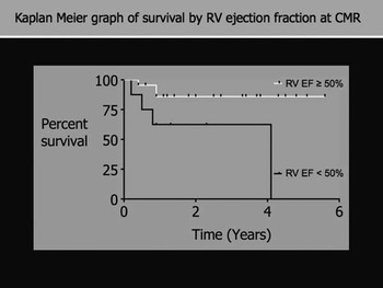
The risk of death was related to reduced right ventricular ejection fraction expressed as a percentage: hazard ratio 0.91, 95% confidence interval 0.85–0.98, p = 0.02. A right ventricular ejection fraction of 50% was the median index of global systolic function for a similar population in a previous study,Reference Muthurangu, Taylor and Hegde5 and thus we selected this value as a cutoff for further analysis. The Kaplan–Meier graph in Figure 2 shows the proportion of survivors over time with a right ventricular ejection fraction less than 50% at cardiovascular magnetic resonance and 50% or more at cardiovascular magnetic resonance, respectively.
The Kaplan–Meier graph shows the proportion of survivors over time on the basis of whether the right ventricular ejection fraction was above or below 50% at cardiovascular magnetic resonance. Nine patients were classified as having “low” right ventricular ejection fraction less than 50%, of whom four (44%) subsequently died, compared with 23 who were classified as having normal right ventricular ejection fraction equal to or greater than 50%, of whom three (13%) died.

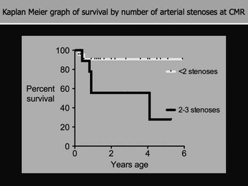
When assessing the effect of great vessel stenoses detected at cardiovascular magnetic resonance on survival over time, patients with single or multiple stenoses were each compared with those with no measured stenosis. There was no difference in the risk of death between patients with one or two-vessel stenoses; however, the cumulative number of areas of narrowing in the neo-aortic arch and pulmonary arteries found at cardiovascular magnetic resonance was strongly related to the risk of death with a hazard ratio 2.71 (95% confidence interval 1.14–6.44), p = 0.02. Figure 3 shows the proportion of survivors over time by the number of narrowed areas, in either pulmonary artery or aorta, at cardiovascular magnetic resonance imaging.
The Kaplan–Meier graph shows the proportion of survivors over time by the number of stenosed areas at cardiovascular magnetic resonance, in either the pulmonary arteries or aorta. Ten patients had no focal stenosis of which one (10%) later died, 12 patients had one stenosis of which one (8%) later died, eight patients had two stenoses of which three (38%) later died, and two patients had three stenoses both of which subsequently died.

Discussion
Right ventricular ejection fraction, cumulative stenoses, and survival
The outcome measure for this study was medium-term survival, with follow-up to a median age of 19.2 months. After considering post-operative, inter-stage, and late deaths, the actuarial survival of 63% for this cohort at 1 year of age was very similar to outcome elsewhere in the United Kingdom, for this population.3 Given the relatively large number of later deaths reported in infants who undergo staged, palliative surgery for hypoplastic left heart syndrome, scrutiny of factors related to outcome is crucial.
Our study shows that cardiovascular magnetic resonance-derived factors, including the measured right ventricular ejection fraction and the cumulative number of focal arterial stenoses, are strongly related to clinical outcome in these patients.
There is very good evidence to show that ventricular ejection fraction is accurately and reliably measured using cardiovascular magnetic resonance volumetric techniques, which use no geometric assumptions.Reference Denslow, Wiles and McKellar12, Reference Winter, Bernink and Groenink13 The measured ejection fraction is a simple, specific index of the change in global ventricular volume over the cardiac cycle. As such, the ejection fraction gives a load-dependent global index of systolic contractility. In patients with hypoplastic left heart syndrome, the ejection fraction of the functionally single, morphological right ventricle must represent the combined effect of the structural substrate of the myocardium, previous insults to the myocardium, such as shock or acidosis in the neonatal period, and the current loading on the myocardium, such as increased afterload from aortic arch stenosis. Owing to the fact that the study data contain information on a single magnetic resonance study before planning the stage two operation, we cannot comment on whether the right ventricular ejection fraction values changed over time.
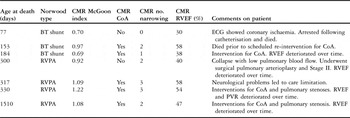
Focal pulmonary stenosis may lead to poor pulmonary flow, reduced saturations, and suboptimal oxygen delivery. Aortic coarctation increases the burden of afterload on the right ventricle. Our data suggest that the risk to outcome of focal stenoses in both aorta and pulmonary arteries is greater than focal stenosis in a single vessel alone. The influence on survival of subsequent specific intervention on the great vessels could not be assessed with this retrospective methodology; however, we present a review of the deceased patients in Table 3.
Review of deceased patients.

BT shunt = Blalock–Taussig Shunt; CMR = cardiovascular magnetic resonance; CoA = coarctation defined by CoA index diameter greater than 0.7; ECG = electrocardiogram; PVR = pulmonary vascular resistance; RVEF = right ventricular ejection fraction; RVPA = right ventricle to pulmonary artery conduit
Influence of coarctation
Using echocardiography and measuring the fractional area change, others have documented deterioration in right ventricular systolic function associated with recurrent aortic coarctation in infants with hypoplastic left heart syndrome, following the Norwood I operation.Reference Larrazabal, Tierney and Brown14
In our cross-sectional analysis, we did not identify a direct relationship between the coarctation index and the right ventricular ejection fraction, nor did we identify a significant relationship between the coarctation index and survival. The reasons for the lack of a relationship are unclear, including the reduced power of a small study, and the possibility that subsequent intervention on the arterial stenoses may have altered the natural history of the lesion.
Not all patients with aortic narrowing develop right ventricular dysfunction, and there are many other potential influences on right ventricular dysfunction, such as volume overload or hypoxaemia.
Defining coarctation
One distinct possibility is that our analysis oversimplified the definition of aortic coarctation for this population. We chose to use a simple, but sensitive, morphological classification of coarctation that is predominant in the current literature, the coarctation index.Reference Lemler, Zellers and Harris10 This index was first validated with two-dimensional angiographic data acquired during interventional catheterisation. To apply this index using cardiovascular magnetic resonance data, we reduced the isotropic, three-dimensional magnetic resonance angiographic data set to two dimensions, by averaging the orthogonal cross-sectional diameters of the vessels.
There is no clear consensus in the literature for the diagnosis or classification of recurrent aortic coarctation following the Norwood operation. As such, there is variation of incidence, which ranges from 11% to 37%, in published literature from different surgical centres.Reference Muthurangu, Taylor and Hegde5, Reference Larrazabal, Tierney and Brown14–Reference Hehir, Dominguez and Ballweg16
The clinical limitations inherent in relying on a single measurement of coarctation severity are well recognised. Moreover, the threshold and methods for intervention differ between centres. Methods utilising the three-dimensional angiographic and phase-contrast flow capabilities of magnetic resonance imaging to predict the coarctation gradient have been evaluated in cohorts with native and recurrent coarctation.Reference Nielson, Powell and Gauvreau17–Reference Simpson, Chung and Glass19 However, none of these cohorts included patients with a proximal aortopulmonary shunt. Following the Norwood I procedure, there are complex flow dynamics involving the aortic arch. A proximal low-resistance pathway (Blalock–Taussig shunt), variations in vessel geometry, irregular compliance properties of the reconstructed arch, and impaired ventricular function, all confound any simple analysis of stenosis.
Other factors
Other factors may contribute to a greater risk of inter-stage death. Pre-surgical and peri-surgical physiological parameters such as morphological type, intact atrial septum, lowest pH, episodes of shock, and the age at the time of surgery have been documented to influence outcome, and could confound the current data presented.Reference Hehir, Dominguez and Ballweg16, Reference Walsh, McCrindle and Dipchand20, Reference Gaynor, Mahle and Cohen21 Altman et alReference Altmann, Printz and Solowiejczyk22 found that the pre-surgical right ventricular systolic function influenced the intermediate and late survival for their patients. We have described these factors in our cohort but did not include them in the outcome analysis, as this was a selected group of patients undergoing cardiovascular magnetic resonance, rather than the entire population of hypoplastic left heart syndrome. We specifically did not include an initial, qualitative echocardiographic right ventricular functional assessment in our analysis. Echocardiographic and magnetic resonance assessment of right ventricular systolic function has been shown to have a poor correlation.Reference Muthurangu, Taylor and Hegde5, Reference Lim, Peeler and Matherne23
The cause and circumstances of death varied for each of the seven patients in our cohort, reflecting that in other cohorts.Reference Hehir, Dominguez and Ballweg16 Each patient underwent home monitoring and close outpatient follow-up in a similar manner to the Milwaukee group,Reference Ghanayem, Hoffman and Mussatto24 and not all deaths were directly attributable to myocardial failure at the time of death. Others have shown that the technical quality of the surgical procedures during the Norwood-type palliation has a strong correlation with outcome.Reference Bacha, Larrazabal and Pigula25 However, right ventricular ejection fraction, as measured with cardiovascular magnetic resonance, may be a sensitive marker for the general, functional reserve of these patients.
This study highlights the importance of measures to prevent and deal with focal arterial stenoses among patients with hypoplastic left heart syndrome. Moreover, the study reaffirms the knowledge that the preservation of right ventricular systolic function in these patients is critical for their survival.
Strengths and limitations
These data are retrospective and originate from a single centre, with the biases inherent in this type of evaluation. Our aim was to study the longer-term outcome in this group of hypoplastic left heart syndrome patients with a view to quality control in this challenging group.
Hypoplastic left heart syndrome is not a common condition and patients with other anatomic variants of single ventricle and aortic arch obstruction were excluded in order to make the cohort as homogeneous as possible. The original cohort of 59 patients with classic hypoplastic left heart syndrome was further reduced by exclusion of those who died early, who had further treatment overseas, or who did not follow the elective cardiovascular magnetic resonance protocol because of clinical concerns. This resulted in a small number, that is, 32 patients being included in the study. Therefore, the residual group is a selected population with the possibility for associated bias.
Conclusion
Cardiovascular magnetic resonance provides systematic and comprehensive evaluation of the circulation in infants with palliated hypoplastic left heart syndrome, allowing sensitive assessment of arterial stenoses and measurement of right ventricular systolic function. Measures to preserve right ventricular contractility and deal with important focal stenoses are paramount in patients undergoing palliative surgery for hypoplastic left heart syndrome.