INTRODUCTION
Taeniosis/cysticercosis due to Taenia solium is a major parasitic disease of global proportions that poses serious threats to human health and economy in the third world countries (Sciutto et al. Reference Sciutto, Fragoso, Fleury, Laclette, Sotelo, Aluja, Vargas and Larralde2000; Garcia et al. Reference Garcia, Del Brutto, Nash, White, Tsang and Gilman2005). The parasite's life cycle involves the pig as an intermediate host, harbouring larval stages called cysticerci and humans as the only known definitive host harbouring the adult tapeworm in the intestines. Humans can also acquire larval infection after accidental ingestion of human faeces contaminated with T. solium eggs, which evolves into human cysticercosis or neurocysticercosis (NCC) when the larvae lodge in the central nervous system (Flisser, Reference Flisser1994).
The nature of the immune response around the viable or degenerating cysticerci is vital in understanding the underlying pathology in NCC (Uddin et al. Reference Uddin, Gonzalez, Gilman, Garcia, Verastegui, Moore, Evans, Read and Friedland2006). The term ‘immune tolerance’ is used in cases of NCC where viable cysts without inflammation or surrounded by scanty inflammatory cells are observed (Carpio et al. Reference Carpio, Escobar and Hauser1998). Living parasites without inflammation are found more frequently in asymptomatic and immunodeficient patients whereas severe inflammation has only been seen in symptomatic patients (White et al. Reference White, Patrick and Diaz1995). The limited inflammatory reaction in NCC could be induced by parasite apoptosis that leads to clonal deletion of both thymocytes and mature T cells (Murphy et al. Reference Murphy, Heimberger and Loh1990; Lenardo, Reference Lenardo1991). Apoptosis is a type of programmed cell death, and an active process associated with distinctive biochemical and morphological changes that can be elicited by different triggers, including serum deprivation, and by extracellular stimuli acting through cell-surface receptors (Behnia et al. Reference Behnia, Robertson and Martin2000; Silva et al. Reference Silva, Sotoca, Johansson, Ludovico, Sansonetty, Silva, Peinado and Corte-Real2005).
Taenia solium cysticerci have been reported to employ various strategies to maintain a Th2 permissive environment (Villa and Kuhn, Reference Villa and Kuhn1996). Cysticerci of T. solium are known to secrete in vitro a factor that depresses human and murine lymphocyte proliferation (Tato et al. Reference Tato, Castro, Rodriguez, Soto, Arechavaleta and Molinari1995; Molinari et al. Reference Molinari, Mejia, White, Garrido, Borgonio, Baig and Tato2000) and that this factor impairs the humoral and cellular responses to metacestode antigens and inhibits the inflammatory reaction around metacestodes implanted subcutaneously in mice (Molinari et al. Reference Molinari, Tato, Rodriguez, Solano, Rubio and Sepulveda1998). The ability of the cysticerci to kill host lymphocytes could enhance parasite survival. It is therefore possible that the parasite may be directly affecting host lymphocytes. This may be through reducing the functional activity of the cells, which is advantageous to the parasite, and apoptosis rather than necrosis would reduce inflammation (O'Connell and Rogan, Reference O'Connell and Rogan2000). In this study, we report for the first time, apoptosis in the inflammatory reaction in porcine brain tissues fixed in formalin and paraffin embedded.
MATERIALS AND METHODS
Infected animals
A total of 35 pigs were used in this study. Thirty pigs naturally infected with T. solium were purchased from the Livestock market in Lusaka and 5 healthy T. solium-free pigs from a commercial piggery in Lusaka. Pigs that were found to be positive for T. solium cysticercosis on tongue examination met the selection criteria into the study.
Histopathological tissues and TUNEL staining
Pigs were euthanized using Euthasol® 20%, intravenously (200 mg/ml) (Produlab Pharma, Raamsdonksveer, Netherlands) to effect and immediately bled. Specimens containing cysticerci and their surrounding tissue were collected from the brain of all 35 pigs. The cysts and surrounding tissue were fixed in 10% formalin for 12–24 h and then transferred to 70% ethanol and thereafter paraffin embedded. Five μm thick sections (Londoño et al. Reference Londoño, Alvarez, Trujillo, Jaramillo and Restrepo2002) were mounted on positively charged slides (Fisher Scientific, Pittsburg, PA).
The terminal deoxynucleotidyl transferase-end labelling (TUNEL) staining was performed using the TdT-FragEL™ DNA Fragmentation Detection Kit (QIA33, Calbiochem/Oncogene, Denmark) according to the manufacturer's instructions. Briefly, slides were deparaffinized to TBS. The area immediately surrounding the specimen was dried. All washing steps were done 3 times in Tris-buffered saline (TBS) and incubations were done at room temperature unless specified. Then 100 μl of proteinase K diluted 1:200 in TBS were added to each specimen and incubated for 20 min, after which the slides were washed. To inactivate the endogenous peroxidases, 100 μl of 30% H2O2 were added on each specimen and incubated for 5 min. The slides were washed, following which they were incubated with 60 μl of equilibration buffer for 20 min. Fragment End Labeling (FragEL™) of DNA was done by addition of 60 μl of TdT-enzyme and slides incubated for 1 h 30 min at 37°C. The slides were washed and then incubated with stop buffer for 5 min before being washed again. Thereafter, the slides were incubated with a blocking buffer (50 μl per specimen) for 10 min, following which they were incubated with 60 μl of streptavidin-horseradish peroxidase (HRP) conjugate diluted at 1:50 in blocking buffer for 30 min. After washing, slides were incubated with 100 μl of diaminobenzidine (DAB) solution for 10 min. The resulting brown colour, after slides were washed, was counterstained with 0·3% methyl green for 3 min. To remove the excess dye, slides were washed in absolute ethanol 3 times, after which they were dried at 37°C and mounted with cover-slips for microscopical examination.
Staining for CD3+ lymphocytes
Tissue slides were deparaffinized to distilled water. All washing steps were performed twice in TBS, with each wash lasting for 5 min. Incubations were done at room temperature unless stated otherwise. Epitopes were retrieved by microwave oven heating (Taylor et al. Reference Taylor, Shi, Chen, Young, Yang and Cote1996; Pileri et al. Reference Pileri, Roncador, Ceccarelli, Piccioli, Briskomatis, Sabattini, Ascani, Santini, Piccaluga, Leone, Damiani, Ercolessi, Sandri, Pieri, Leoncini and Falini1997) at 700 W for 10 min in a buffer containing 0·01 M Tris-base, 0·0005 M ethylene glycol-bis(a-aminoethyl ether)-N,N,N,N-tetraacetic acid pH 9 ((EGTA) Sigma-Aldrich #E3889) and leaving the sections in the hot buffer for 15 min. The slides were then washed. Blocking of endogenous peroxidases was done by incubating slides with 100 μl of 0·6% H2O2 in TBS for 15 min. After washing, the slides were blocked for 30 min with 100 μl of 20% normal goat serum (DAKO X0907) diluted in TBS. Incubation with primary antibody CD3e-UNLB (SB 4510-01) diluted 1:1000 in TBS/0·1% BSA was done at 4°C overnight. The slides were then washed, after which they were incubated with EnVision/HRP (DAKO K4001) for 30 min. After washing, slides were incubated with 100 μl of DAB solution for 6 min, then washed and counterstained with Mayers hæmatoxylin for 10 sec. Slides were washed with tap water and then distilled water and finally mounted with glycerol-gelatine for microscopical examination.
Classification of cyst viability in brain granulomas
Cysticerci were classified as either viable or calcified (dead) according to their microscopical appearance. Viable cysticerci had well-limited thin cystic wall structures containing scolices characterized by little or no inflammatory infiltrate. Calcified cysticerci (late or advance granulomas) were those with caseous amorphous material with disintegrated cyst wall and without discernible parasite structures and heavy inflammatory infiltrate.
Co-localization of CD3+ and TUNEL-positive cells
In order to demonstrate co-localization of CD3+ cells and TUNEL-positive cells, 2 consecutive sections were cut from each paraffin block to be examined. Before the sections were mounted on slides, 1 from each pair was turned upside–down to ensure that the surfaces viewed at microscopy were 2 opposing surfaces. The sections were stained for CD3+ cells and TUNEL, respectively, as described above. The same areas with positive reaction were identified in each section from every pair using vessels and neurons as landmarks.
RESULTS
TUNEL and CD3 staining
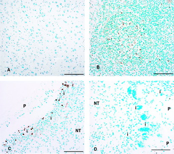
Of the 30 pig brains with cerebral cysticercosis, 27 had viable cysticerci only while the remaining 3 had both viable and calcified cysticerci. Calcified cysticerci were only observed on the periphery of the cerebral cortex and none were observed in the medulla. Five specimens from each stage of the infection (viable vs calcified) were further analysed by TUNEL staining. As expected, uninfected brain tissue was negative to the TUNEL staining (Fig. 1A). Porcine lymph node (Fig. 1B) was used as a positive control because in lymphoid organs, apoptosis occurs as a normal physiological process (Resendes et al. Reference Resendes, Majó, Segalés, Espadamala, Mateu, Chianini, Nofrarías and Domingo2004). However, light microscope examination of TUNEL-stained tissues containing cysticerci confirmed that apoptotic cells corresponded to small, rounded dark-brown stained cells with the characteristic condensation and fragmentation of nuclear chromatin and loss of cytoplasm (Fig. 1C). Apoptotic cells were usually found interspersed with normal cell types, and were mostly located in the inflammatory infiltrate. TUNEL-positive cells were only concentrated in inflammatory infiltrates adjacent to viable cysticerci. Late or advanced granulomas with disintegrated scolices (Fig. 1D) did not show TUNEL-positive cells.
TUNEL staining of porcine brain tissues naturally infected with Taenia solium cysticercosis. P, parasite; i, inflammatory infiltrate; NT, nervous tissue. (A) Normal nervous tissue, negative control. (B) Porcine lymph node showing tunnel-positive cells in germinal centre, positive control. (C) Early lesion. (D) Advanced lesion with dead cysticerci. Scale bar=100 μm.

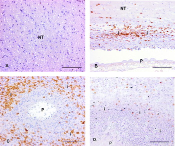
In normal brain tissue, no CD3+ cells were detected (Fig. 2A). Brain granulomas induced by T. solium cysticerci showed CD3+ cells in both early and advanced lesions (Fig. 2B and C). Many CD3+ cells associated with viable parasite were present in clusters in the inflammatory infiltrate adjacent to the parasite. However, the abundance of CD3+ lymphocytes was markedly increased in advanced lesions and apoptotic cells co-localized mainly with CD3+ lymphocytes (black spots) (Fig. 2D). The co-localization showed that almost all apoptotic cells were CD3 positive but not all CD3+ cells were apoptotic.
CD3 staining of porcine brain tissues from pigs naturally infected with Taenia solium cysticercosis. P, parasite; i, inflammatory infiltrate; NT, nervous tissue. (A) Normal nervous tissue, negative control. (B) Early granuloma. (C) Late granuloma. (D) CD3+ cells co-localization with TUNEL-positive cells. Scale bar=100 μm.

DISCUSSION
To our knowledge this is the first report of apoptosis in porcine brain tissues infected with T. solium cysticerci that have been fixed in formalin and paraffin embedded. We have shown that T. solium cysticerci trigger apoptosis or programmed cell death in porcine brain granulomas. Of interest was the observation that the TUNEL staining detected apoptotic cells only in inflammatory infiltrate associated with viable parasite. Apoptosis of CD3+ lymphocytes observed in areas adjacent to T. solium cysticerci suggests that these cells are constantly undergoing apoptosis and thus die as soon as they arrive at the site of infection.
Apoptotic cells were usually found interspersed with normal cell types, and were mostly located in the inflammatory infiltrate, indicating that the events of apoptosis probably and only affect a certain percentage of cells (including CD3+ lymphocytes) in the inflammatory reaction in early granulomas when the cysticerci are alive. These observations are consistent with other patterns of apoptosis described by Presas et al. (Reference Presas, Robert, Jiménez and Willms2005), in which apoptotic cells were interspersed with normal cells in differentiating tissues of adult T. solium and T. crassiceps strobilae.
Our study showed evidence of apoptosis in early granulomas when the parasite was alive, while advanced and late granulomas were devoid of apoptotic cells. Hence, in the early stage, the cysticercus is able to evade the host's immune response, but this is lost in the late stage when the cysticercus dies. This can be explained by the fact that the evading ability in disintegrating parasites is absent, and this provides a favourable condition to the host, and allows the establishment of cell immune responses leading to granuloma formation and eventual parasite removal.
Lee and Piedrahita (Reference Lee and Piedrahita2002) noted that apoptosis is an important aspect to be considered during host-parasite interactions given that bacteria, viruses and protozoa can regulate the death programmes of their hosts by activating, delaying or even preventing them. During host-parasite apoptosis, the rapid endocytosis of apoptotic bodies by phagocytic cells may prevent inflammation and excessive tissue damage (De Souza et al. Reference De Souza, Meuser-Batista, Batista, Duarte, Araújo-Jorge and Soeiro2008). Although it cannot be considered as the main type of cell death present in inflammatory reactions, parasite-induced apoptosis and its modulation by pro- and anti-apoptotic molecules (De Souza et al. Reference De Souza, Meuser-Batista, Batista, Duarte, Araújo-Jorge and Soeiro2008), may play a role in regulating the inflammatory response in early cysticercosis.
CD3 is a phenotypic marker that recognizes T cells. T cells are involved in cell-mediated immunity. They are essential in determining B cell antibody class switching, in the activation and growth of cytotoxic T cells, and in maximizing bactericidal activity of phagocytes such as macrophages (Koffeman et al. Reference Koffeman, Keogh, Klein, Prakken and Albani2007). It is probably this diversity in function that makes the T cell subgroup of lymphocytes a target for destruction by T. solium cysticerci. Whether CD3+ lymphocytes were the only cell types undergoing apoptosis in the inflammatory reaction could not be evaluated in this study as we did not search for other cell types. Nevertheless, our results suggest that in cysticercosis, the cysticerci have suppressive effects on the host immune responses through destruction of T cells.
The mechanism by which T. solium cysticerci induce apoptosis in CD3+ lymphocytes, as found in this study, and CD4+ T-cells as reported by Solano et al. (Reference Solano, Cortés, Copitin, Tato and Molinari2006) is still beyond the scope of many researchers of T. solium infections. Nevertheless, there are speculations that cysticerci may either excrete/secrete or express factors which can induce apoptosis directly, or induce cells to excrete/secrete, which in turn causes cells to apoptose (O'Connell and Rogan, Reference O'Connell and Rogan2000). However, in Trypanosoma cruzi infections, the interactions between Fas ligand and Fas receptor (Lopes et al. Reference Lopes, Nunes, Henriques-Pons, Giese, Morse, Davidson, Araujo-Jorge and DosReis1999), as well as nitric oxide (Martins et al. Reference Martins, Vieira, Cunha and Silva1999) has been linked to lymphocyte apoptosis induced by the parasite. Inhibition of caspase was reported to reduce lymphocyte apoptosis and improve host immune responses to infections caused by T. cruzi (Silva et al. Reference Silva, Guillermo, Ribeiro-Gomes, De Meis, Nunes, Senra, Soares, DosReis and Lopes2007). Similarly, analysis of parasite-induced apoptotic T cells in skin-stage schistosomula of Schistosoma mansoni infection showed increases in the expression of Fas, FasL, and the Fas-associated death domain (Chen et al. Reference Chen, Rao, He and Ramaswamy2002).
Our study carried out in porcine brain tissues has complemented the work conducted by Solano et al. (Reference Solano, Cortés, Copitin, Tato and Molinari2006) who cultured lymphocytes with T. solium metacestode products. Our conclusions are that, T. solium cysticerci appear to induce death of CD3+ T-lymphocytes in the inflammatory reactions and that whatever was causing cell death did not affect all cells. Our results further suggested that any apoptotic effects would act locally since not all cells were killed, and the neuronal cells immediately surrounding cysticerci were not apoptotic. Therefore, further research is needed to establish whether other cells besides T-lymphocytes are also targets for destruction by cysticerci in early cysticercosis as well as studies to assess whether cysteine protease is expressed by viable cysticerci in situ.
We are grateful to the DBL-Centre for Health Research and Development for financial support. The support from the School of Veterinary Medicine, University of Zambia is also acknowledged. Special thanks go to Lisbet Kiørboe, Hanne H. Møller, Betina Andersen and Dennis Brok of the Section of Pathology in the Department of Veterinary Pathobiology, Faculty of Life Sciences, University of Copenhagen, Denmark, for their technical assistance with immunohistochemistry and photography. We would also like to thank Messrs P. Zulu and B. Chulu of the School of Veterinary Medicine, University of Zambia for taking care of experimental animals and J. Kaumba, M. Masuku, M. Chembensofu and M. Kankomba for their assistance rendered during the dissection work. Drs Jørgen Kurtzhals and Lothar Wiese are thanked for assistance with apoptosis staining.