The term “potential Clostridium difficile excretor” (excretor) has been used to describe patients with diarrhea who have toxigenic C. difficile in stool but with no detectable free toxin.Reference Planche, Davies and Coen 1 , Reference Goldenberg 2 Practically, the samples from these patients test glutamate dehydrogenase (GDH) or polymerase chain reaction (PCR) positive but toxin enzyme immunoassay (EIA) or cytotoxin neutralization negative. They are distinguished clinically from asymptomatically colonized patients because they have active diarrhea and so are probably at risk of transmitting infection if they are not correctly identified and isolated.
Although the prevalence of asymptomatic colonization has been described in several studies, with rates ranging 6.6%–15%,Reference Eyre, Griffiths and Vaughan 3 – Reference Alasmari, Seiler, Hink, Burnham and Dubberke 5 the prevalence of excretors is less well described. Guerrero and colleaguesReference Guerrero, Chou, Jury, Nerandzic, Cadnum and Donskey 6 sampled the skin and environment of patients who tested positive by PCR. Of 132 patients studied, 32% had a negative toxin EIA, and the remaining patients were positive with both PCR and toxin EIA. Contamination rates of skin (both 57%) and the environment (29% vs 30%, respectively) were similar, regardless of toxin EIA status.
Kamboj and colleaguesReference Kamboj, Babady and Marsh 7 performed multiple-locus variable-number tandem repeat analysis on 42 isolates detected using PCR from a pediatric cancer hospital. Nineteen samples were cytotoxin neutralization negative. Although the risk of transmission was greater from cytotoxin neutralization–positive donors, there was clear evidence of transmission for those patients with samples that were negative with this assay.
We assessed environmental contamination in rooms of patients with C. difficile infection (CDI) (PCR positive-toxin EIA positive, PCR+/Toxin+) and excretors (PCR positive-toxin EIA negative, PCR+/Toxin−).
METHODS
The setting was a 1,100-bed academic hospital, with 180 single occupant rooms with private bathroom. Patients with diarrhea are placed in these rooms until at least 7 days following return to normal bowel habit or until discharge, dependent upon capacity. The study was classed as service evaluation with ethics board exemption; therefore, patient consent was not sought.
Clinicians are advised to investigate all cases of clinically significant diarrhea (2 or more episodes of liquid stool within 24 hours that is not obviously explained by an alternative cause, eg, laxatives) by testing for C. difficile. This is performed using GDH (Cdiff Chek-60 EIA; TechLab) followed by both EIA for toxins A/B (C. difficile Tox A/B II; TechLab) and PCR (GeneXpert; Cepheid). The combination of GDH plus toxin EIA has an estimated sensitivity of 81.8% compared with cytotoxin neutralization.Reference Planche, Davies and Coen 1 Samples not taking the shape of their container are rejected. All patients with positive results for either toxin EIA or PCR are reviewed by an infectious diseases and/or microbiology physician who makes recommendations on management, including specific anti–C. difficile therapy.
Environmental cleaning of all clinical areas is performed with a chlorine dioxide-containing product (Difficil-S; Clinimax) using microfiber cloths. Following discharge, the rooms of all patients with CDI undergo terminal cleaning consisting of standard cleaning as above followed by further disinfection with a hydrogen peroxide vaporized system (BioQuell). Nearby patient equipment is cleaned manually using peracetic acid disposable wipes (Clinell Sporicidal Wipes, GAMA Healthcare) before being placed in the room for exposure to hydrogen peroxide vapor. New disposable curtains are also used following this procedure. No changes to the environmental cleaning and disinfection protocols were made throughout the study period.
Samples were collected from consecutive patients from 1 to 3 days following the laboratory test and at times convenient to the scientist (9 AM–5 PM, Monday to Friday). Four standardized areas of the patient room were sampled: bed rails, bed controls/call buttons, toilet, and shower area. Samples were collected using cellulose sponges presoaked in neutralizing buffer (Whatman). Sponges were rubbed over a 10×10-cm area in 2 directions at right angles and placed in Robertson’s cooked meat broth supplemented with cycloserine, cefoxitin, lysozyme, and sodium taurocholate. Samples were incubated for 7 days anaerobically at 37°C as previously described.Reference Goldenberg, Patel, Tucker and French 8 Culture broth was subcultured onto agar containing cycloserine, cefoxitin, and fructose (Oxoid) and incubated for a further 2 days. Presumptive colonies were identified based on characteristic morphology confirmed with matrix-assisted laser desorption/ionization time-of-flight (MALDI-TOF) mass spectrometry (Bruker). All isolates underwent PCR ribotyping according to standard techniques.
Patient demographic characteristics (age, sex, community- or hospital-associated infection); markers of severity (peripheral white cell count, C-reactive protein, albumin, and creatinine), taking the most extreme value within 3 days of the laboratory test; and clinical outcomes (30-day all-cause mortality and length of stay) were measured. The PCR Ct value was used as a surrogate marker of bacterial load. Continuous variables were compared using unpaired t tests, and categorical variables were compared using the Fisher exact test.
RESULTS
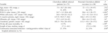
From June 1, 2012 through May 31, 2014, a total of 171 patients with positive C. difficile tests had environmental sampling performed within 3 days of the laboratory test result. Of these patients, 98 (57%) were GDH+/PCR+/toxin EIA- (excretors), and the remaining 73 patients were GDH+/PCR+/toxin EIA+ (active CDI). Summary characteristics of both groups are shown in Table 1.
Characteristics of Patients With PCR Positive and Toxin EIA Positive Results and Those With PCR Positive and Toxin EIA Negative Results

NOTE. EIA, enzyme immunoassay; PCR, polymerase chain reaction.
a Unpaired t test.
b Fisher exact test.
Patients in both groups were of a similar age and sex; however, those with CDI were more likely to be classified as community-associated (positive laboratory test within 3 days of hospital admission) than were the excretors (37% and 21%, respectively; P=.038).
Patients with CDI had a greater white blood cell count (16.6×10⁹) compared with that of excretors (13.3×10⁹); however, other markers of severity such as albumin and creatinine were similar. The PCR Ct values of patients with CDI were significantly lower than the excretor group (24.7 and 28.6, respectively; P<.001), suggesting a greater bacterial load in the CDI group. There were no differences in the clinical outcomes (30-day all-cause mortality and length of stay) between groups.
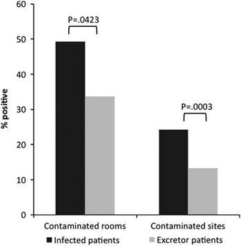
In the excretor group C. difficile was recovered from 33 (34%) of 98 patient rooms compared with 36 (49%) of 73 in the CDI group (P=.0423). In the excretor group 52/392 (13%) environmental sites were positive for C. difficile compared with 69/272 (25%) in the active CDI group (P=.0003). Environmental contamination results are shown in Figure 1. Isolates recovered from the environment matched the PCR ribotype of clinical isolates in 40 (77%) of 52 samples in the excretor group and 54 (78%) of 69 samples in the CDI group.
Environmental contamination rates for infected and excretor patients.

DISCUSSION
Although infected patients were significantly more likely to contaminate their immediate environment, excretors also disseminated spores widely. In this study C. difficile was isolated from the rooms of a third of patients. This is important since the hospital environment plays a significant role in transmission of C. difficile Reference Otter, Yezli and French 9 , Reference Mayfield, Leet, Miller and Mundy 10 and it has been shown that occupants of a room vacated by a previously colonized or infected patient are at increased risk of infection.Reference Shaughnessy, Micielli and Depestel 11
Limitations of the study include the omission of environmental sampling in “control rooms” that housed neither excretor nor CDI patients. Thus, it is difficult to estimate the background level of contamination with C. difficile; however, this may be important since approximately 25% of environmental isolates did not match the PCR ribotype of the patient isolates. Additionally, the laboratory method of detection (GDH followed by toxin EIA) has a sensitivity of 82%, which may result in some patients with CDI being misclassified as excretors.
Excretors may be an important reservoir of infection in the healthcare setting and this has relevant implications for onward transmission, particularly for hospitals whose laboratories are not using a testing strategy that can identify this patient group. We assumed our laboratory testing algorithm could correctly differentiate between patients with CDI and excretors; however, this may not be the case.Reference Planche, Davies and Coen 1 , Reference Goldenberg 2 The potential benefit of isolating these patients (and using other infection prevention interventions) warrants further investigation. Since we isolated both infected and excretor patients and used hydrogen peroxide vapor following discharge, it was not possible to measure potential transmission from excretors.
Acknowledgments
Financial support. None reported.
Potential conflicts of interest. J.A.O. reports that he is an employee of BioQuell. All other authors report no conflicts of interest relevant to this article.