Introduction
Kisspeptin, a polypeptide encoded by the Kiss1 gene containing 145 amino acids, is further processed in vivo into small peptides with different amino acids (kisspeptin54, kisspeptin14, kisspeptin13, kisspeptin10), and the structure and function of mammalian kisspeptin is conserved (Arnone and Oliveri, Reference Arnone and Oliveri2020; Beltramo et al., Reference Beltramo, Robert and Decourt2020; Clarke and Dhillo, Reference Clarke and Dhillo2016; Franssen and Tena-Sempere, Reference Franssen and Tena-Sempere2018; Tsukamura, Reference Tsukamura2022). In addition to its role in the central brain, Kiss1 and its receptor Kiss1r are widely expressed in peripheral tissues such as ovary, pancreas, liver, and adipose, suggesting that kisspeptin may be involved in regulating tissue and organ function in a paracrine or autocrine manner (Bhattacharya and Babwah, Reference Bhattacharya and Babwah2015; Cao et al., Reference Cao, Li, Jiang, Ling and Kuang2019; D’Occhio et al., Reference D’Occhio, Campanile and Baruselli2020). Numerous studies have identified the expression of Kiss1 and its receptors Kiss1r in ovaries including human (Owens et al., Reference Owens, Abbara, Lerner, O’Floinn, Christopoulos, Khanjani, Islam, Hardy, Hanyaloglu, Lavery, Dhillo and Franks2018), porcine (Xin et al., Reference Xin, Li, Zhong, Li, Wang, Zhang, Yuan, Li and Zhang2019), bovine (Guo et al., Reference Guo, Xu, Li, Liu, Zhao, Lu and Wang2022), domestic cat (Tanyapanyachon et al., Reference Tanyapanyachon, Amelkina and Chatdarong2018), and rabbit (Maranesi et al., Reference Maranesi, Petrucci, Leonardi, Bufalari, Parillo, Boiti and Zerani2019), which may be directly involved in the regulation of ovarian steroid hormone secretion, granulosa cell proliferation and apoptosis, and follicular development. Our previous studies have shown that continuous injection of kisspeptin can promote follicular development in non-breeding season of Tan sheep (unpublished data). Furthermore, some studies have demonstrated that kisspeptin treatment could regulate ovarian granulosa cell function in porcine (Fabová and Sirotkin, Reference Fabová and Sirotkin2021), feline (Loncová et al., Reference Loncová, Fabová and Sirotkin2023). However, another study has shown that kisspeptin treatment has no significant effect on ovarian granulosa cell function in bovine ovarian granulosa cells in vitro (Mattar et al., Reference Mattar, Cheewasopit, Samir and Knight2023). Until now, it is not clear whether Kiss1 and its receptor Kiss1r are expressed in ovarian granulosa cells of Tan sheep. It is not clear whether kisspeptin affects ovarian granulosa cell function in Tan sheep. Therefore, in this study, we first detected the expression of Kiss1/ Kiss1r in primary ovarian granulosa cells of Tan sheep, and then determined the effects of different concentrations of kisspeptin treatment on steroid hormones, granulosa cells proliferation and apoptosis by ELISA, Q-PCR, flow cytometric techniques. Further, we investigated the signaling pathway by which kisspeptin regulates ovarian granulosa cells function in Tan sheep. Our results will provide the basis for developing novel kisspeptin-mediated techniques to regulate reproduction in Tan sheep.
Materials and methods
Isolation and identification of primary ovarian granulosa cells
The ovaries of Tan sheep were obtained from Yongning Hongxiang slaughterhouse, Yinchuan, Ningxia. Approximately 150 fresh ovarian samples were collected from slaughtered female Tan sheep and transported to the laboratory within 1 h. After removing excess fat and connective tissue, the follicular fluid from 3–8cm size of follicle was aspirated into a centrifuge tube filled with DMEM/F12 culture medium (C3130-0500, Viva Cell, Shanghai, China) using a 10 mL disposable syringe (with a pillow diameter of 0.7 mm). The follicular fluid was then filtered through a 150-mesh cell sieve (XBGLS-60, Solarbio, Beijing, China) after pipetting. The cell filtrate was collected in another centrifuge tube and centrifuged at 1000 rpm for 8 min. The cells were inoculated into 6-well plates at 1 × 10⁶ cells/well after resuspending cell precipitate. The cells were incubated at 37°C in a 5% CO2 incubator. In addition, granulosa cells were also inoculated into 12-well plates at 1 × 10⁵ cells/well. The cells were rinsed with PBS (C3580-0500, Viva Cell, Shanghai, China) and then fixed in 4% paraformaldehyde (30525-89-4, Thermo Fisher Scientific, WI, USA) when the cells are fully adhered to the wall. Cells were incubated with a rabbit polyclonal antibody against FSHR (Bioss, #20658R), which, according to the manufacturer’s specifications, cross-reacts with sheep FSHR due to high sequence homology in the target epitope), at 4°C overnight after rinsing. Subsequently, cells were incubated with secondary antibody (FITC-labelled goat anti-rabbit IgG (H + L), ZB-0311, ZSGB-BIO, Beijing, China) for 2 h and then treated with 4’,6-diamidino-2-phenylindole (DAPI, C1005, Beyotime, Shanghai, China) for 10 minutes. Finally, cells were observed under a fluorescence microscope.
Detection of Kiss1/ Kiss1r gene in granulosa cells
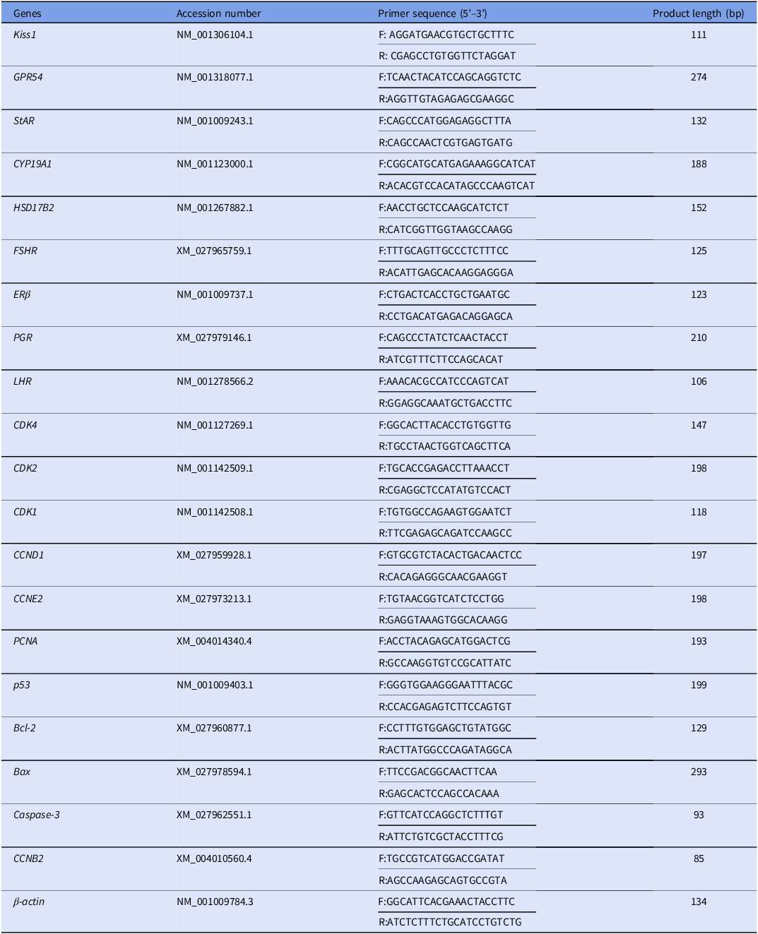
Ovarian granulosa cells of Tan sheep were inoculated into 6-well plates at 1 × 10⁶ cells/well. Total RNA was extracted from ovarian granulosa cells after 6 h, 12 h, 24 h, 48 h, 72 h, 96 h and 120 h of culture, respectively. At each time point, three replicates were collected for each treatment. The expression of Kiss1 and Kiss1r mRNA in granulosa cells was respectively detected by RT-PCR, and then amplified products were sequenced, respectively. PCR primers are shown in Table 1.
Sequences of PCR primers

Treatment of ovarian granulosa cells
Human-kisspeptin (Kisspeptin-10, 048-56, Phoenix Biotech, San Francisco, USA) was purchased from phoenix corporation. For hormone detection, cells were inoculated into culture plates and when the cell confluence reached 70%, serum-free DMEM/F12 culture medium containing 3%BSA (9048-46-8, Sigma-Aldrich, St. Louis, Missouri, United States), 1IU FSH (9002-68-0, Sigma-Aldrich, St. Louis, Missouri, United States), 1IU LH (39341-83-8, Sigma-Aldrich, St. Louis, Missouri, United States) and 10 nM dose of androstenedione (63-05-8, Glpbio, Montclair, CA) with different concentrations of kisspeptin (0 nM, 50 nM, 100 nM, 500 nM, 1000 nM) were added (Three replicate wells per treatment), and incubated for 24 h. The supernatant was collected, and the total RNA was extracted from the cells. Furthermore, cells were inoculated into culture plates and repeated with different concentrations of kisspeptin (0 nM, 50 nM, 100 nM, 500 nM, 1000 nM), and cell proliferation and apoptosis were detected separately. Finally, the cells were inoculated into culture plates until the cell confluence reached 70%, and then the cells were supplemented with 15 μM PKA inhibitor (SQ22536, MCE, New Jersey, USA), 50 μM PLC inhibitor (U-73122, MCE, New Jersey, USA), 20 μM PKC inhibitor (GO-6983, MCE, New Jersey, USA) and 10 μM Ca2+ inhibitor (thapsigargin, HY-13433, MCE, New Jersey, USA). The cells were then treated with kisspeptin after incubating for 90 min in serum-free medium. The supernatant was collected and total cellular RNA and total protein were extracted.
Hormone detection
Estrogen concentrations in cell supernatants were determined using the sheep estradiol (E2) ELISA kit (CSB-E13172Sh, CUSABIO, Wuhan, China), which has a sensitivity of 0.2 ng/ml and shows no cross-reactivity with other related proteins. Progesterone concentration was detected by sheep progesterone (P4) ELISA kit (CSB-E13176Sh, CUSABIO, Wuhan, China), which also demonstrates high specificity and sensitivity for sheep progesterone. The intra- and inter-assay coefficients of variation were less than 15% using four replicates per sample for both kits. Hormone testing is performed according to the kit instructions. The experiment was repeated three times.
Detection of cell proliferation
Ovarian granulosa cells of Tan sheep were inoculated into 96-well plates at 10,000 cells/well and serum-free culture medium containing different concentrations of kisspeptin (0 nM, 50 nM, 100 nM, 500 nM and 1000 nM) was added to incubate the cells for 12 h, 24 h and 48 h, respectively, with three replicate wells per treatment (n = 3 for each concentration and time point). A negative control group was treated with serum-free medium without kisspeptin (0 nM kisspeptin). Subsequently, 10 µL of CCK-8 was added to each well (Cd04, Dojindo, Kumamoto Prefecture, Japan), and the OD values were read at 450 nM wavelength after incubation.
Cell cycle and apoptosis assay
CCK-8 assays were performed using a CCK-8 kit (Cd04, Dojindo, Kumamoto Prefecture, Japan). In brief, cells in the log phase of growth were seeded in 96-well plates at a concentration of 1 × 10⁴ cells/well, with three replicate wells per treatment (n = 3 for each concentration). A negative control group was treated with serum-free medium without kisspeptin. Cell viability was assessed using the CCK-8 reagent, following the manufacturer’s protocol at the indicated time points (12, 24, and 48 h).
RT-PCR and q-PCR for detection of gene expression
Total cellular RNA was extracted from in vitro cultured ovarian granulosa cell of Tan sheep using TRIzol reagent (15596018CN, Invitrogen, Carlsbad, CA, USA) and reverse transcribed to cDNA using the PrimeScript RT reagent kit with gDNA Eraser (RRO47A, Takara, Beijing, China). RT-PCR amplification reaction conditions were as follows: 95°C, 3 min; 95°C, 15 s; 60°C, 15 s; 72°C, 45 s; 30 cycles, and the RT-PCR amplification products were detected by electrophoresis. For q-PCR, total RNA was extracted similarly, and 500 ng aliquots were reverse-transcribed. Q-PCR was performed with a TB Green Premix Ex TaqTM II (RR420A, Takara, Beijing, China) in a 20 µL reaction volume containing 10 µL of 2 × TB Green Premix Ex TaqII, 4 µL of cDNA, and 0.8 µM of each primer. All primers were validated with Primer Blast and synthesized by Sangon Biotech Company (Shanghai, China). Q-PCR was conducted in triplicates, using β-actin as the endogenous reference gene. The amplification conditions were as follows: 95°C for 30 s; 95°C for 5 s; 60°C for 30 s; 72°C for 20 s; for a total of 40 cycles.
Western blot
The RIPA lysis buffer (phosphatase inhibitor, PMSF, protease inhibitor, KGB5203-100, Nanjing KeyGen Biotech, Jiangsu, China) was used to extract cellular proteins. The BCA kit (KGP150, Nanjing KeyGen Biotech, Jiangsu, China) was used to determine the protein concentration. After adjusting the protein concentration, 6 × Protein Loading Buffer (P0015F, Beyotime, Shanghai, China) was added to mix with the proteins and boiled for 5 min. Then, each protein sample was separated by electrophoresis using 12% sodium dodecyl sulfate-polyacrylamide gel electrophoresis (MA0456, Meilunbio, Dalian, China), and then transferred to PVDF membrane (0.2 μm, ISEQ00010, Millipore, Massachusetts, United States). After incubation with Tris buffer (25 mM Tris and 150 mM NaCl, pH 7.6) containing 5% skimmed milk for 1 h, the membranes were incubated with primary antibodies against ERK 1/2 (1:1000, YT1625, Cell Signaling Technology, Boston, United States), p-ERK 1/2 (1:1000, YP0101, Cell Signaling Technology, Boston, United States), GAPDH (1:1000, YM3029, Immunoway, Texas, United States) overnight at 4℃. After rinsing with TBS (ST667-1L, Beyotime, Shanghai, China), the secondary antibody was added and incubated for 1 h. Immunoreactions were visualized using ECL (P0018S, Beyotime, Shanghai, China). WB images were captured on a Gel-Pro analyser version 4 (Media Cybernetics, Rockville, MD, USA), and bands were quantified using Image J software (version 1.51j8, National Institutes of Health, American).
Statistical analysis
The results are presented as the mean ± standard deviation (SD) and all statistical analyses were performed by SPSS 25.0 software (IBM Inc., Chicago, IL USA). Multiple comparisons among groups were performed using One-way ANOVA analysis and Duncan post hoc tests. P < 0.05 indicates significant differences. The same experiment was repeated three times. GraphPad Prism 8.0 software (version 8.0.2, San Diego, USA) was prepared the graphs.
Results
Immunofluorescence identification of primary ovarian granulosa cells
The isolated ovarian granulosa cells of Tan sheep were completely adherent to the wall after 24 h of culture, and the cell confluence reached about 85% at 96 h. Immunofluorescence identification revealed that FSHR-positive cells showed green fluorescence in the cytoplasm (Figure 1B1) and blue fluorescence in the nucleus (Figure 1B2), and the purity of FSHR-positive cells by cell counting reached more than 95%, indicating that the isolated cultured cells were ovarian granulosa cells.
Immunofluorescence identification of ovarian granulosa cells from Tan sheep. (A1–A2) Negative control (NC) group stained only with the secondary antibody (FITC-conjugated, green) and DAPI (blue), showing negligible non-specific binding. (A) Overlapping images of A and B; (B1) Cells exhibiting positive staining for follicle-stimulating hormone receptor (FSHR) (green, FITC channel). (B2) Nuclei of the same field of view counterstained with DAPI (blue). (B) Overlapping images of A1 and B1.

Effects of kisspeptin treatment on steroid hormone secretion and related gene expression in Tan sheep ovarian granulosa cells. (A) The effect of kisspeptin treatment at different concentrations on estrogen and progesterone secretion. (B) The effect of kisspeptin treatment at different concentrations on the expression of steroidogenesis-related genes. Different letters between bars indicate significant differences (p < 0.05).

Tan sheep ovarian granulosa cells expressed Kiss1/Kiss1r
The Kiss1 and its Kiss1r were expressed in ovarian granulosa cells of Tan sheep cultured for 0, 6, 12, 24, 48, 72, 96 and 120 h. The amplification products of Kiss1 and Kiss1r were 111 bp (Fig. S.A) and 274 bp (Fig. S.B), respectively, which were the same size as that of the target fragments. Furthermore, the nucleotide sequences of the amplification product were100% identical to the gene sequence of Kiss1 and Kiss1r. This indicated that Kiss1 and Kiss1r were presented in the ovarian granulosa cells of Tan sheep.
Kisspeptin promotes E2 and P4 secretion in ovarian granulosa cells of Tan sheep
Both 100 nM and 500 nM doses of kisspeptin significantly increased progesterone secretion (P < 0.05), whereas 1000 nM dose of kisspeptin significantly decreased progesterone secretion in ovarian granulosa cells (P < 0.05, Figure 2A). Furthermore, 500 nM dose of kisspeptin treatment also significantly stimulated estrogen secretion (P < 0.05), whereas other concentrations of kisspeptin had no significant effect on estrogen secretion in Tan sheep ovarian granulosa cells (Figure 2A). The average coefficient of variation (CV) for the ELISA experiments of progesterone and estrogen, which were 11.22% and 5.37%, respectively. In addition, 500 nM dose of kisspeptin treatment was able to significantly up-regulate the expression of steroidogenesis-related genes including StAR, HSD17B2, CPY19A1, FSHR, LHR, ERβ, and PGR (P < 0.05, Figure 2B). This suggests that kisspeptin promotes granulosa cell estrogen and progesterone secretion by up-regulating the expression of steroidogenesis-related genes in ovarian granulosa cells of Tan sheep.
Kisspeptin promotes granulosa cell proliferation
500 nM and 1000 nM doses of kisspeptin significantly promoted the proliferation of ovarian granulosa cells at 12 h post-treatment (P < 0.05). When granulosa cells were treated for 24 h, 50 nM, 500 nM and 1000 nM doses of kisspeptin significantly promoted the proliferation of granulosa cells (P < 0.05), among which 500 nM doses of kisspeptin significantly promoted the proliferation of granulosa cells (P < 0.01, Figure 3). Treatment with different concentrations of kisspeptin significantly promoted the rate of granulosa cells entering S phase (P < 0.05, Figure 4). Meanwhile, 500 nM doses of kisspeptin significantly up-regulated the expression of cell proliferation-related genes including CCNB2, CCNE2, CDK1, CDK2, CDK4 and PCNA (P < 0.05, Figure 5). This suggests that specific concentrations of kisspeptin may promote the proliferation of ovarian granulosa cells in Tan sheep.
Effect of kisspeptin treatment at different concentrations on granulosa cell proliferation, ‘*’indicates significant difference (p < 0.05).

Effect of kisspeptin treatment at different concentrations on cell cycle. (A) Control; (B) 50 nM kisspeptin treatment; (C) 100 nM kisspeptin treatment; (D) 500 nM kisspeptin treatment; (E) 1000 nM kisspeptin treatment; (F) The rate of granulosa cells entering S phase after kisspeptin treatment, ‘*’indicates significant difference (p < 0.05).

Effect of kisspeptin treatment at different concentrations the expression of cell proliferation-related genes. Completely different letters between bars indicate significant differences (p < 0.05).

Kisspeptin inhibits granulosa cell apoptosis
500 nM doses of kisspeptin significantly inhibited granulosa cell apoptosis (P < 0.05, Figure 6F). Furthermore, 100 nM, 500 nM and 1000 nM doses of kisspeptin significantly up-regulated the expression of Bcl-2 and down-regulated the expression of caspase-3 and p53 (P < 0.05, Figure 6J). Moreover, 500 nM and 1000 nM doses of kisspeptin also significantly down-regulated the expression of Bax. These results suggest that kisspeptin may inhibit the apoptosis of ovarian granulosa cells in Tan sheep.
Effect of kisspeptin treatment on granulosa cell apoptosis. Effect of kisspeptin treatment at different concentrations on cell cycle. (A) Control; (B) 50 nM kisspeptin treatment; (C) 100 nM kisspeptin treatment; (D) 500 nM kisspeptin treatment; (E) 1000 nM kisspeptin treatment; (F) The rate of granulosa cell apoptosis after kisspeptin treatment, ‘*’indicates significant difference (p < 0.05); J: Effect of kisspeptin on apoptosis-related gene expression, completely different letters between bars indicate significant differences (p < 0.05).

Kisspeptin promotes steroid hormone secretion through the MAPK/ERK pathway
Progesterone and estradiol concentrations were significantly lower in the group co-treated with PKC inhibitor, PLC inhibitor and Ca2+ inhibitor compared to the 500 nM doses of kisspeptin-treated group (P < 0.05, Figure 7A). However, co-treatment of kisspeptin with PKA inhibitor had no significant effect on secretion of both progesterone and estradiol compared to the kisspeptin-treated group. In addition, co-treatment of kisspeptin with PKC inhibitors, PLC inhibitors and Ca2+ inhibitors significantly down-regulated the expression of StAR, CYP19A1, and HSD17B2, (P < 0.05, Figure 7B). In contrast, co-treatment of kisspeptin with PKA inhibitor did not significantly change the expression of CYP19A1 and HSD17B2, although it decreased the expression of StAR. Additionally, 500 nM dose of kisspeptin treatment significantly increased the expression of p-ERK1/2 protein, whereas co-treatment of kisspeptin with PLC and PKC inhibitors significantly down-regulated the expression of p-ERK1/2 protein (P < 0.05, Figure 7C). This suggests that kisspeptin promotes steroid hormone secretion in ovarian granulosa cells of Tan sheep by activating the MAPK/ERK signaling pathway.
Kisspeptin promotes steroid hormone secretion through the MAPK/ERK pathway. (A) Effect of co-treatment with signaling pathway inhibitors and 500 nM kisspeptin on estrogen and progesterone secretion in Tan sheep ovarian granulosa cells; (B) Effect of co-treatment with signaling pathway inhibitors and 500 nM kisspeptin on steroidogenesis-related genes in Tan sheep ovarian granulosa cells; (C) Effect of co-treatment with signaling pathway inhibitors and 500 nM kisspeptin on the expression of p-ERK protein in Tan sheep ovarian granulosa cells. Different letters between bars indicate significant differences (p < 0.05).

Discussion
Kisspeptin injection can induce oestrus and follicles development in non-breeding season of Tan sheep (unpublished data). Granulosa cells are closely related to follicular development (Martinez et al., Reference Martinez, Rizos, Rodriguez-Martinez and Funahashi2023). However, it is not clear whether kisspeptin directly regulates steroid hormone secretion, cell proliferation and apoptosis in ovarian granulosa cells of Tan sheep. In this study, we have shown for the first time that kisspeptin can directly promote cell proliferation, inhibit apoptosis and stimulate steroid hormone secretion via the MAPK-ERK signaling pathway in ovarian granulosa cells of Tan sheep. Our findings may provide strong evidence that kisspeptin directly regulates follicular development in a paracrine or autocrine manner in the ovary of Tan sheep.
Firstly, RT-PCR analysis revealed that Kiss1and Kiss1r were expressed in ovarian granulosa cells of Tan sheep. Previous studies have found that Kiss1/KISS1R is expressed in ovarian granulosa cells of human (García-Ortega et al., Reference García-Ortega, Pinto, Fernández-Sánchez, Prados, Cejudo-Román, Almeida, Hernández, Romero, Tena-Sempere and Candenas2014), pig (Basini et al., Reference Basini, Grasselli, Bussolati, Ciccimarra, Maranesi, Bufalari, Parillo and Zerani2018), domestic cat (Tanyapanyachon et al., Reference Tanyapanyachon, Amelkina and Chatdarong2018), and buffalo (Rajin et al., Reference Rajin, Patra, Sheikh, Singh, Mishra, Karikalan, Singh, Kumar, Gaur and Krishnaswamy2022). However, it is unclear whether Kiss1and Kiss1r are expressed in ovarian granulosa cells of goat and sheep. The study clearly showed for the first time that Kiss1and Kiss1r are expressed in ovarian granulosa cells of Tan sheep. Our finding may provide direct evidence that kisspeptin is involved in the regulation of ovarian function in Tan sheep.
Next, we investigated the effect of different concentrations of kisspeptin on ovarian granulosa cell function in Tan sheep. Our study found that kisspeptin10 treatment significantly promoted steroid hormone secretion in ovarian granulosa cells of Tan sheep, and 500 nM dose of kisspeptin treatment significantly up-regulated the expression of steroidogenesis-related genes including StAR, HSD17B2, CPY19A1, FSHR, LHR, ERβ and PGR. Previous studies have shown that kisspeptin10 treatment significantly increased progesterone secretion in chicken ovarian granulosa cells (Xiao et al., Reference Xiao, Ni, Huang, Wu, Grossmann and Zhao2011). Furthermore, kisspeptin10 treatment significantly promoted progesterone secretion and up-regulated StAR expression in ovarian granulosa cells of cattle (Guo et al., Reference Guo, Xu, Li, Liu, Zhao, Lu and Wang2022). Similarly, Kiss1 overexpression increased steroid hormone levels and up-regulated CYP19A1 expression in ovarian granulosa cells of porcine 10. Our results were consistent with those described above. This suggests that kisspeptin may promote steroid hormone secretion by up-regulating the expression of steroidogenesis-related genes in ovarian granulosa cells of Tan sheep. However, there appears to be a species difference in regulating ovarian function by kisspeptin. Recent study found that kisspeptin treatment did not stimulate steroid hormone secretion in bovine ovarian granulosa cells in vitro (Mattar et al., Reference Mattar, Cheewasopit, Samir and Knight2023). In addition, we found that kisspeptin did not have a dose-dependent effect on the secretion of steroid hormones from the ovarian granulosa cells of Tan sheep. An earlier study found that kisspeptin increased the expression of StAR, CPY19A1 and HSD3B2 in vivo, but kisspeptin treatment had no significant effect on the expression of key steroidogenesis genes in vitro (Owens et al., Reference Owens, Abbara, Lerner, O’Floinn, Christopoulos, Khanjani, Islam, Hardy, Hanyaloglu, Lavery, Dhillo and Franks2018). We speculated that this may be related to the dose of kisspeptin. Previous studies have shown that high-dose kisspeptin treatment inhibits steroid hormone secretion in ovarian granulosa cells (Fabová et al., Reference Fabová, Loncová, Mlyn Ek and Sirotkin2022). Similarly, our results also found that high-dose kisspeptin (1000 nM) treatment instead significantly reduced progesterone secretion in ovarian granulosa cells of Tan sheep. This suggests that kisspeptin may regulate ovarian granulosa function in a non-dose-dependent manner. However, we did not perform a systematic, quantitative analysis of morphological changes during granulosa cells luteinization, future studies should incorporate morphological analysis to directly investigate the potential luteinizing effects of kisspeptin. Additionally, we also recognized that a detailed time-course experiment would be necessary to elucidate such dynamic changes and to fully address whether kisspeptin initially promotes estradiol synthesis before potentially promoting a transition towards a more progesterogenic phenotype. Thus, the temporal dynamics of kisspeptin’s effects on E₂ and P₄ synthesis remain to be determined in future studies.
More importantly, as shown in this study, kisspeptin may regulate steroid hormone secretion through the MAPK-ERK pathway in ovarian granulosa cells of Tan sheep. The MAPK-ERK signaling pathway is involved in the regulation of many biological processes, including steroid hormone synthesis, proliferation and apoptosis (Park, Reference Park2023). In primate pituitary cells, it has been shown that kisspeptin increases LH and GH secretion by activating PLC, PKC, Ca2+, but not PKA signaling pathway (Luque et al., Reference Luque, Córdoba-Chacón, Gahete, Navarro, Tena-Sempere, Kineman and Castaño2011). However, the signaling pathway by which kisspeptin stimulates steroid hormone secretion in ovarian granulosa cells of Tan sheep is unclear. In present study, we found that the level of steroid hormone secretion and the expression of key enzyme genes regulating steroidogenesis were significantly decreased after treatment with MAPK-ERK pathway inhibitors including PKC inhibitors, PLC inhibitors and Ca2+ inhibitors. In contrast, co-treatment of PKA inhibitors with kisspeptin had no significant effect on steroid hormone secretion in ovarian granulosa cells. Furthermore, western blotting assay demonstrated that kisspeptin promoted steroid hormone secretion via the ERK pathway in ovarian granulosa cells of Tan sheep. This suggests that kisspeptin regulates steroid hormone secretion through the MAPK-ERK signaling pathway in ovarian granulosa cells of Tan sheep. Recently, 100 nM dose of kisspeptin treatment stimulated steroid hormone secretion by activating the ERK1/2 signaling pathway in luteal cells of the buffalo (Thejaswini et al., Reference Thejaswini, Patra, Sharma, Raza, Sasidharan, Karikalan, Dubal, Ghosh, Gaur, Singh and Krishnaswamy2024). Thus, kisspeptin may regulate steroid hormone secretion through the MAPK-ERK pathway in ovarian granulosa cells of Tan sheep.
Interestingly, kisspeptin may promote granulosa cell proliferation in Tan sheep. Although numerous studies have shown that kisspeptin inhibits cancer cell proliferation (Alkafaji et al., Reference Alkafaji, Raji, Rahman, Zekiy, Adili, Jalili, Hojjatipour, Cid-Arregui, Shomali, Tarzi, Tamjidifar, Heshmati, Marofi, Akbari, Hasanzadeh, Deljavanghodrati, Jarahian and Sandoghchian Shotorbani2021; Ciaramella et al., Reference Ciaramella, Della Corte, Ciardiello and Morgillo2018; Li et al., Reference Li, Cao, Wang, Fu, Cheng, Ma, Zhang, Guo, Kan and Liu2020a). However, kisspeptin may play different role in normal tissues, regulating the biological functions of organs and tissues. It has been shown that kisspeptin promotes the proliferation of bovine mammary epithelial cells through the AKT/mTOR/ERK1/2 pathway in vitro (Li et al., Reference Li, Cao, Wang, Fu, Cheng, Ma, Zhang, Guo, Kan and Liu2020b). In human ovarian granulosa cells, low-dose kisspeptin treatment up-regulated the expression of PCNA and cyclinB1 and promoted granulosa cell proliferation, and inhibited granulosa cell apoptosis. Similarly, overexpression of Kiss1 promoted granulosa cell proliferation and inhibited apoptosis by activating PI3K/AKT and ERK signaling pathways in ovarian granulosa cell lines (Sun et al., Reference Sun, Zhang, Sun, Sun, Wang and Ma2023). Our study also found that 500 nM dose of kisspeptin treatment significantly up-regulated the expression of proliferation-related genes and down-regulated the expression of apoptosis-related genes, thereby promoting the proliferation and inhibiting the apoptosis of ovarian granulosa cells in Tan sheep. This is the first time that kisspeptin has been found to promote the proliferation and inhibit the apoptosis of ovarian granulosa cells in sheep. Our results further suggest that kisspeptin may be involved in regulating follicular development in a paracrine and autocrine manner by directly modulating ovarian granulosa cell function in Tan sheep. However, one study found that kisspeptin10 treatment induced apoptosis and inhibited proliferation in bovine ovarian granulosa cells (Liu et al., Reference Liu, Xu, Yuan, Dong, Wang and Lu2017). More recently, 100 nM dose of kisspeptin was able to significantly up-regulate the expression of BCL-2 (5.0-fold) and PCNA (94.9-fold) in bubaline ovarian granulosa cells, which in turn promoted granulosa cell proliferation and inhibited cell apoptosis (Sharma et al., Reference Sharma, Patra, Puttanarsappa, Hitesh, Sahu, Mathesh, Dubal, Ghosh, Gaur, Das, Singh and Krishnaswamy2024). Thus, there appears to be a degree of species variability in regulating the functions of ovarian granulosa cell by kisspeptin, but this needs to be further investigated.
We have not yet confirmed the signaling pathway through which kisspeptin promotes granulosa cell proliferation and suppresses cell apoptosis. However, many studies have demonstrated that the MAPK-ERK signaling pathway plays a role in regulating cell proliferation, apoptosis and steroid hormone secretion (Park et al., Reference Park2023; Wu et al., Reference Wu, Xiao, Pi, Zhang, Pan, Pu, Liang, Shen and Du2019; Yang et al., Reference Yang, Yu, Peng, Chang and Chen2023). Therefore, in addition to regulating the secretion of steroid hormones in the ovarian granulosa cells of Tan sheep, we speculate that the MAPK-ERK signaling pathway also regulates the proliferation and apoptosis of these cells (as summarized in Figure 8). The binding of kisspeptin to its receptor Kiss1r activates a downstream cascade involving PLC, PKC, and intracellular Ca2+, leading to the phosphorylation of ERK1/2. This activation serves as a central node, hierarchically coordinating multiple cellular processes: 1) Steroidogenesis: p-ERK1/2 upregulates the expression of key steroidogenic enzymes (StAR, CYP19A1, HSD17B2) and receptors (FSHR, LHR), thereby enhancing the production of estradiol and progesterone; 2) Proliferation: ERK activation may promote the transition of cells into the S phase of the cell cycle by upregulating cyclins (CCNB2, CCNE2) and CDKs (CDK1, CDK2, CDK4), while simultaneously increasing the expression of the proliferation marker PCNA; 3) Apoptosis: ERK activation may also lead to up-regulation of the apoptosis suppressor gene Bcl-2 and down-regulation of apoptosis-promoting gene (Bax, caspase-3, p53), which in turn regulate granulosa cell proliferation and apoptosis. These pathways are not isolated but are highly interconnected. For instance, the enhanced steroidogenic capacity provides a favourable microenvironment for cell growth, while the suppression of apoptosis ensures the survival of proliferating granulosa cells, collectively contributing to follicular development.
Kisspeptin may regulate steroidogenesis, cell proliferation and apoptosis in ovarian granulosa cells of Tan sheep through MAPK-ERK signaling pathway.

This study has several limitations. Firstly, the experiments were conducted in vitro using isolated granulosa cells, which lack the complex follicular microenvironment and the interplay with theca cells and oocytes. Secondly, the concentrations of kisspeptin used were pharmacological; further studies are required to determine its physiological relevance and optimal effective dose in vivo. Thirdly, the application of MAPK-ERK signaling pathway inhibitors and activators is needed in the future to further confirm the mechanism of MAPK-ERK signaling pathway in regulating the proliferation and apoptosis of ovarian granulosa cells in Tan sheep. Furthermore, the potential crosstalk between the MAPK/ERK pathway and other signaling pathways (e.g., PI3K/AKT) should be investigated in future. Despite these limitations, our findings present significant novelty. This is the first comprehensive study to confirm the expression and direct functional role of the kisspeptin/Kiss1r system in ovarian granulosa cells of Tan sheep. We provide mechanistic evidence that kisspeptin concurrently stimulates steroidogenesis primarily via the MAPK/ERK pathway. These results contribute substantially to reproductive biology by elucidating a local regulatory mechanism within the ovine ovary, expanding our understanding of kisspeptin’s actions beyond the hypothalamic-pituitary axis. From a practical standpoint, our research suggests that kisspeptin might be developed as a potential therapeutic agent to improve ovarian function and fecundity in Tan sheep. For instance, exogenous kisspeptin administration could be strategized to enhance follicular development and ovulation rates, particularly during the non-breeding season, thereby improving reproductive efficiency and genetic management in this valuable breed.
In conclusion, we found for the first time that Kiss1 and Kiss1r are presented in ovarian granulosa cells of Tan sheep, 500 nM dose of kisspeptin significantly stimulates the secretion of estrogen and progesterone through the MAPK-ERK signaling pathway (Figure. 8). Additionally, kisspeptin promotes the cell proliferation and inhibits cell apoptosis in ovarian granulosa cells of Tan sheep. Our findings provide strong evidence that kisspeptin is directly involved in the regulation of ovarian function through paracrine or autocrine manner, which will provide the basis for the development of novel kisspeptin-mediated techniques to regulate reproduction in Tan sheep.
Credit authorship contribution statement
Wei Song: Original draft, Data curation. Tianshu Dai: Writing- review & editing, Formal analysis. Di Yang: Software. Shuang Li: Resources, Formal analysis. Ming Cao: Supervision, Investigation. Xingang Dan: Resources, Project administration.
Competing interests
The authors have nothing to declare.
Funding information
This research was funded by The National Natural Science Foundation of China (31760665, 32460835).