Introduction
Nasal septal deviation is the most commonly seen deformity of the nose, and can be either congenital or acquired.Reference Gray 1 There is no standard definition of nasal septal deviation, but it is usually defined as deviation of the bony and cartilaginous septum from the midline.
Patients with nasal septal deviation complaining of nasal obstruction have varying degrees of concomitant hypertrophy of the middle turbinate and the inferior turbinate on the contralateral side to the nasal septal deviation.Reference Demir, Asil, Güven, Kayabaşoğlu and Yılmaz 2 , Reference Egeli, Demirci, Yazýcý and Harputluoglu 3 This compensatory mechanism is characterised by an increase in both mucosal and bony structures, as the hypertrophy protects the more patent passage from excess airflow, which has drying and crusting effects on nasal mucous membranes.Reference Demir, Asil, Güven, Kayabaşoğlu and Yılmaz 2 – Reference Berger, Hammel, Berger, Avraham and Ophir 4 Accordingly, most physicians may prefer turbinate surgery in combination with septoplasty in these patients. However, middle turbinate management is more controversial than inferior turbinate management, lacking definitive consensus. Although there are many studies indicating the outcomes of nasal surgery, studies that evaluate the effect of septoplasty on middle turbinate hypertrophy have not previously been reported.
This study aimed to investigate whether there was an effect of septoplasty on middle turbinate hypertrophy using computed tomography (CT) in patients with nasal septal deviation.
Materials and methods
This retrospective study was approved by the Sakarya University Ethical Committee and consent was obtained from the patients. We reviewed the paranasal sinus CT scans of patients with nasal septal deviation identified from the radiology department database at the Training and Research Hospital of the University of Sakarya.
A total of 985 patients who had undergone septoplasty between November 2011 and January 2014 were included in the study. After reviewing their medical records, patients were excluded from the study if: they had undergone previous nasal surgery; they had caudal nasal septal deviation, nasal polyposis or other abnormalities besides nasal septal deviation; or they had a history of tobacco use. Patients with findings of allergy, and those who had received systemic or topical medication for upper respiratory tract disease within two weeks before CT, were also excluded.
We performed CT for clinical reasons such as headache, facial trauma and a reduced sense of smell, at least one year after septoplasty. All post-operative images were taken both in the pre-operative angular position of the head and at the shooting time of pre-operative images to take account of the nasal cycle. Patients did not use topical or systemic medications for at least two weeks before post-operative CT examination, and did not have nasal surgery after septoplasty. Patients with findings of sinonasal pathology were excluded from this study after post-operative CT examination. A14-year-old child was included in the analysis. This patient suffered from facial pain as a result of trauma and needed evaluation by CT two years after septoplasty. After ensuring that this patient had no sinonasal pathology, the child was referred to the neurosurgery clinic for future examination. As a result, the study group included 40 evaluable patients.
In the current study, all patients were evaluated using the same CT scanner (Toshiba Medical Systems, Otawara, Japan) at a tube voltage of 12 kVp and 230 mA, both pre- and post-operatively. We compared the pre-operative images with the post-operative images using continuous scanning from anterior to posterior, and using 3 mm slices in the coronal plane.
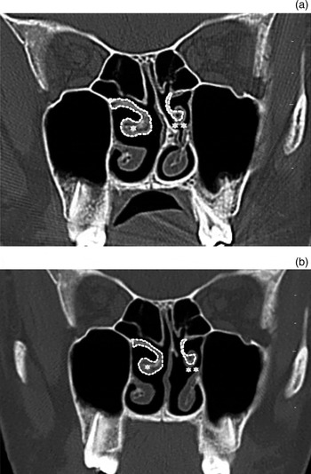
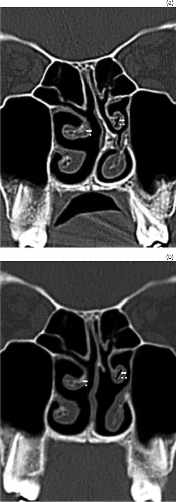
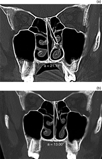
The boundaries of the middle turbinate and conchal bone were outlined on the bone window CT images, and the overall cross-sectional area was measured bilaterally (Figure 1). The corresponding areas were called the middle turbinate total area and the middle turbinate bone area, respectively. The value of the middle turbinate mucosal area was determined by subtracting the middle turbinate bone area from the middle turbinate total area. Thicknesses of the medial mucosa, bone and lateral mucosa were measured on both sides (Figure 2). The angle of the nasal septal deviation was defined as the angle between the line from the crista galli to the pre-maxilla and a second line from the crista galli to the most prominent point (Figure 3). All images were magnified to allow for exact measurements. All measurements were obtained from the CT section of the widest middle part of the middle turbinate and were taken by a radiologist.
Coronal computed tomography scans showing the boundary of the middle turbinate (asterisk) and bone outlined (double asterisk) on bone window before septoplasty (a) and after septoplasty (b).

Coronal computed tomography scans showing measurements of medial mucosa (asterisk), lateral mucosa (double asterisk) and bone structure (black line) before septoplasty (a) and after septoplasty (b).

Coronal computed tomography scans showing the angle of septal deviation (white lines) before septoplasty (a) and after septoplasty (b). a = angle

Statistical analysis
To calculate the necessary sample size, the effect size was calculated as 0.5, given the lack of information available from previous studies. The sample size was calculated as 34, assuming an effect size of 0.5, type I error (alpha) = 0.05, and type II error (beta) = 0.20, with power = 80 per cent. Power analysis was performed using the G*Power statistical power analysis program (version 3.1.9).Reference Faul, Erdfelder, Lang and Buchner 5 We opted to include 40 patients in our study.
Categorical data are reported in terms of numbers and percentages. The Kolmogorov–Smirnov test was used to check the normality of the continuous data. The paired sample t-test was used to compare the parametric continuous data between the concave side group and the convex side group, and between pre- and post-operative periods. The Wilcoxon rank sum test was used to compare non-parametric continuous data between the concave side group and the convex side group, and between pre- and post-operative periods. Spearman's correlation coefficients were calculated to determine the correlation between the angle of deviation and other parameters. Continuous data are presented as means ± standard deviations (SDs). P values less than 0.05 were considered statistically significant. Analyses were performed using SPSS Statistics software, version 22.0 (IBM, Armonk, New York, USA).
Results
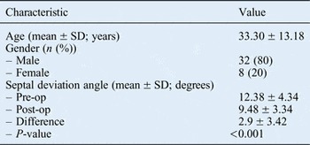
Participants' ages ranged between 14 and 66 years (mean ± SD, 33.30 ± 13.18 years). Thirty-two participants (80 per cent) were male and 8 (20 per cent) were female.
Nasal septal deviation was to the left side in 20 cases and to the right side in 20 cases. The angle of deviation ranged between 4.83 and 19.94 degrees (mean ± SD, 12.38 ± 4.34 degrees) pre-operatively, and between 4.36 and 16.39 degrees (mean ± SD, 9.48 ± 3.34 degrees) post-operatively. The post-operative angle of deviation had decreased significantly (p < 0.001) (Table I).
Distributions of patient characteristics

SD = standard deviation; pre-op = pre-operative; post-op = post-operative
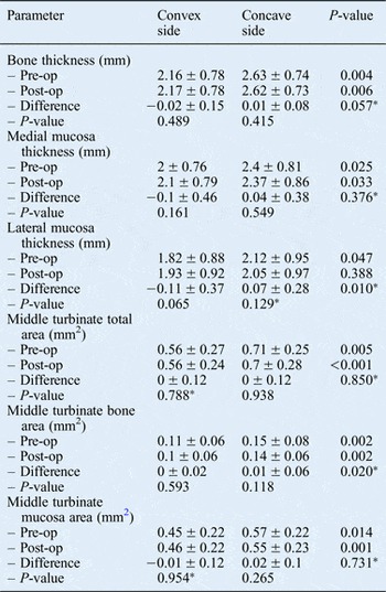
Measurements of bone thickness, medial mucosa thickness and lateral mucosa thickness were statistically analysed. There were no significant differences observed between the pre- and post-operative values for either the concave or convex sides. However, the values for the concave side were significantly higher than those for the convex side, both pre- and post-operatively, except for the post-operative value of the lateral mucosa thickness. The results for the middle turbinate bone area, middle turbinate total area and middle turbinate mucosal area values were similar (Table II).
Comparisons of thickness and area between convex and concave sides of septum

Data are shown as mean ± standard deviation. *Wilcoxon rank sum test. Pre-op = pre-operative; post-op = post-operative
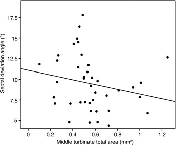
A correlation was performed to assess the relationships between the post-operative angle of deviation and other parameters following septoplasty. A significant correlation was found between the convex side of the middle turbinate total area and the angle of deviation (p = 0.033) (Figure 4).
Correlations between post-operative angle of the deviation and middle turbinate total area on the convex side of the septum (r = −0.338, p = 0.033).

Discussion
The middle turbinate is the medial component of the osteomeatal complex. It is an important anatomical structure located on the lateral nasal wall. Although the concha bullosa is the most frequently encountered anatomical structure in the middle turbinate, other variations may be determined by endonasal and radiological examinations. These variations include a paradoxical, secondary or accessory middle turbinate, and agenesis or hypertrophy of the middle turbinate.Reference Demir, Asil, Güven, Kayabaşoğlu and Yılmaz 2 , Reference Cho, Park, Chung, Hong, Kwon and Kim 6 , Reference Aydil and Ozçelik 7 Turbinate growth can occur congenitally or secondarily to conditions such as tobacco use, recreational drug use, pregnancy, hyperthyroidism, allergic rhinitis and nasal septal deviation.Reference Egeli, Demirci, Yazýcý and Harputluoglu 3
Permanent hypertrophy of the middle turbinate, which occupies the expansive space of the contralateral nasal cavity, is often associated with nasal septal deviation being toward one side. This is frequently considered to be compensatory to the deflection of the nasal septum. An alternative explanation relates to primary unilateral growth of the turbinate bone, which can be genetic or originate from an early-life trauma.Reference Berger, Hammel, Berger, Avraham and Ophir 4 This turbinate bone may produce pressure on the growing nasal septum during childhood and adolescence, and eventually cause it to angle toward the other side of the nose. Although observations of remarkable bone growth compared to mucosal growth contribute reliability to the second, alternative theory, Aslan et al. indicated that unilateral growth of bone was compensatory and not congenital.Reference Aslan, Uzun, Ugur, Uckurt, Sagun and Yazicilar 8 In that study, cross-sectional areas of bone and soft tissue components were significantly increased in the inferior turbinate of adults with nasal septal deviation, but such a difference was not found between paediatric patients with and without nasal septal deviation.Reference Aslan, Uzun, Ugur, Uckurt, Sagun and Yazicilar 8
In our earlier study, we demonstrated that the middle turbinate on the concave side of the septum was significantly wider than the middle turbinate on the convex side of the septum in patients with nasal septal deviation.Reference Demir, Asil, Güven, Kayabaşoğlu and Yılmaz 2 Although septoplasty was performed successfully, we did not observe a significant change of the middle turbinate bilaterally in the current study. That is, even after one year, compensatory hypertrophy of the middle turbinate continued in the patients who had undergone septoplasty.
Our findings are in contrast to the results of Kim et al.Reference Kim, Park, Kim, Kang, Park and Han 9 Their study showed that septoplasty significantly decreased both the thickness of the medial mucosa of the inferior turbinate and the mean dimensions of the inferior turbinate. In addition, septoplasty significantly increased both the thickness of the medial mucosa of the inferior turbinate and the mean dimensions of the inferior turbinate on the deviated side.Reference Kim, Park, Kim, Kang, Park and Han 9 In another study, although the medial mucosa and cross-sectional area of the inferior turbinate on the concave side of the septum were significantly decreased by septoplasty, a significant change in the dimensions of the inferior turbinate was not observed on the convex side of the septum.Reference Xu, Gao, He, Zhang, Ma and Thou 10
In the current study, all post-operative measurements of the middle turbinate on the concave side became smaller; however, these findings were not statistically significant. This result could be related to the location and relative amount of erectile tissue in the nose. In fact, the greatest proportion of erectile tissue seems to be located in the inferior turbinate.Reference Ng, Ramsey and Corey 11 Therefore, the inferior turbinate may demonstrate a much greater change than the middle turbinate, by expanding and shrinking with the change of air volume in the nose.
The structure and dimensions of the middle turbinate play crucial roles in the maintenance of normal nasal physiology, including the deflection of inspired air superiorly toward the olfactory epithelium, the heating and humidification of inspired air, and the lamination of air flow. Moreover, secretions produced by the nasal and paranasal sinus mucosal lining are transported posteriorly along the surfaces of the middle turbinate and inferior turbinate to reach the nasopharynx.Reference Aydil and Ozçelik 7 Thus, enlargement of the middle turbinate may cause sinus dysfunction and may interfere with respiration. Nevertheless, Cho et al. showed that there was no statistical relationship between nasal septal deviation or middle turbinate variations and rhinosinusitis.Reference Cho, Park, Chung, Hong, Kwon and Kim 6
In the present study, although pre-operative measurements involving both mucosa and bone thickness did not correlate with the angle of deviation, after surgery we detected a significant negative correlation between the angle of deviation and middle turbinate total area on the convex side of the septum. As the angle of septum deviation decreased, the middle turbinate total area on the deviated side increased in our study. According to Orhan et al., a positive correlation was found between the measurements for deviation angle and inferior turbinate bone thickness in the compensatory group.Reference Orhan, Aydın, Ormeci and Yılmaz 12 In another study, Grymer et al. claimed that mucosal hypertrophy of the inferior turbinate is not dependent on the degree of septal deviation, but this is not the case for the middle turbinate.Reference Grymer, Illum and Hilberg 13
A hypertrophic inferior turbinate can be managed with different therapeutic procedures.Reference Akoğlu, Karazincir, Balci, Okuyucu, Sumbas and Dağli 14 Many authors have suggested that successful management of nasal obstruction requires a reduction of the inferior turbinate at the time of septoplasty.Reference Pollock and Rohrich 15 , Reference Martinez, Nissen, Stock and Tesmer 16 However, according to Kim et al., preserving the medial mucosa of the inferior turbinate may be useful because septal surgery may decrease the medial mucosa thickness.Reference Kim, Park, Kim, Kang, Park and Han 9
-
• Middle turbinate anatomical variations include paradoxical, secondary or accessory middle turbinate, and agenesis or hypertrophy
-
• Middle turbinate compensatory hypertrophy involves both mucosal and bony structures
-
• Septoplasty did not result in significant decreases in middle turbinate mucosa or bone thickness after one year
-
• There was a significant negative correlation between septum angle and middle turbinate total area on the deviated side
Management of middle turbinate hypertrophy is more controversial than that for inferior turbinate hypertrophy. Middle turbinate hypertrophy may cause middle turbinate syndrome or sinus dysfunction. Middle turbinate syndromes were described by Morgenstein and Krieger as being due to contact between the middle turbinate and the nasal septum, and/or due to contact between the middle turbinate and other lateral nasal wall structures.Reference Morgenstein and Krieger 17 However, many surgeons may prefer to perform middle turbinate resection, reporting the possible development of frontal or ethmoidal-maxillary rhinosinusitis in patients with chronic rhinosinusitis.Reference Choby, Hobson and Lee 18 Further investigations are needed to determine whether middle turbinate hypertrophy obstructs patency of the osteomeatal complex.
Conclusion
In this study, although compensatory hypertrophic middle turbinate dimensions remained unchanged for one year after septoplasty, as the angle of septum deviation decreased, the middle turbinate total area on the deviated side increased.