Introduction
Wolff–Parkinson–White syndrome is defined as documented supraventricular tachycardia or symptoms consistent with supraventricular tachycardia in a patient with ventricular pre-excitation during sinus rhythm. Reference Page, Joglar and Caldwell1 The Wolff–Parkinson–White pattern is defined as abnormal electrocardiogram findings with pre-excitation. The Wolff–Parkinson–White pattern is characterised by the presence of an accessory electrical conduction pathway that bypasses the atrioventricular node, directly connecting the atria to the ventricles, and it affects 0.1–0.3% of the general population. Reference Page, Joglar and Caldwell1 Its characteristic electrocardiogram features include a shortened PR interval, slurred QRS upstroke (delta wave), and prolonged QRS duration.
A Japanese nationwide school-based electrocardiogram screening programme for heart diseases was established by law in 1994 for 1st, 7th, and 10th graders (aged 6, 12, and 15 years, respectively), as well as for 4th graders in some regions. Reference Yoshinaga, Kucho, Nishibatake, Ogata and Nomura2 The Wolff–Parkinson–White pattern was among the several findings frequently detected in school-based cardiovascular screening programmes. Most patients with the Wolff–Parkinson–White pattern are asymptomatic, although tachycardia can occur. The risk of sudden death due to rapid ventricular response and subsequent induction of ventricular fibrillation, particularly in patients with atrial fibrillation or atrial flutter, is approximately 0.13% per year, with initial symptoms including syncope or sudden death. Reference Obeyesekere, Leong-Sit and Massel3,Reference Olen, Baysa, Rossi, Kanter and Fishberger4 Additionally, the rates of sudden death and supraventricular tachycardia are higher in children than in adults. Reference Obeyesekere, Leong-Sit and Massel3 Moreover, a previous study reported that 40% of patients aged ≤ 21 years who experienced a fatal arrhythmic event (e.g., sudden death, near-miss sudden death, shortest pre-excited RR interval ≤ 250 ms, or haemodynamic compromise in clinical atrial fibrillation) were asymptomatic at the onset of the event. Reference Etheridge, Escudero and Blaufox5 Therefore, it is difficult to estimate the risks for patients with Wolff–Parkinson–White syndrome based on history alone, necessitating appropriate diagnosis and management.
Delta waves can also be observed with fasciculoventricular pathways. The fasciculoventricular pathway does not form a re-entry pathway as it solely involves a bundle branch in the ventricles, and only shows pre-excitation without tachycardia, as observed in Wolff–Parkinson–White syndrome. Consequently, fasciculoventricular pathway is not associated with a risk of tachycardia or sudden death and requires no management. Reference Gormel and Yasar6
In Japan, once a Wolff–Parkinson–White pattern electrocardiogram is identified by school-based electrocardiogram screening, even in the absence of palpitations or other prodromal symptoms, the patient undergoes outpatient follow-up. Fasciculoventricular pathway is considered a rare condition, accounting for an estimated 1.2–5.1% of all cases of pre-excitation syndrome. Reference Sternick, Gerken, Vrandecic and Wellens7,Reference Ratnasamy, Khan, Wolff and Young8 This proportion has been reported in patients with palpitations and other symptoms who underwent electrophysiological testing, suggesting the possibility of many more fasciculoventricular pathway cases among asymptomatic patients with delta waves who are monitored for the Wolff–Parkinson–White pattern.
As electrophysiological testing is an invasive procedure, a less invasive approach is required for distinguishing Wolff–Parkinson–White syndrome from fasciculoventricular pathways. As adenosine is unavailable in Japan, adenosine triphosphate is used instead; the equivalent dose of adenosine triphosphate is 1.9-fold higher than that of adenosine. Adenosine triphosphate briefly inhibits atrioventricular nodal conduction and has proven useful in differentiating Wolff–Parkinson–White syndrome from fasciculoventricular pathways. Reference Suzuki, Nakamura, Yoshida, Yoshida and Shintaku9 Following the rapid infusion of adenosine triphosphate during sinus rhythm, atrioventricular block, along with changes in the PR interval and QRS waveform, can be used to identify fasciculoventricular pathways, which do not involve a tachycardia circuit or cause sudden death. Unfortunately, previous studies focusing on using adenosine for differentiation are lacking. In the 2016 Pediatric and Congenital Electrophysiology Society/Heart Rhythm Society expert consensus on paediatric ablation therapy, fasciculoventricular pathway was included for the first time in the Class III guidelines for ablation therapy for Wolff–Parkinson–White syndrome (ablation is not recommended), highlighting the increasing importance of differentiating the diagnosis of Wolff–Parkinson–White syndrome from fasciculoventricular pathway. Reference Philip Saul and Kanter10 Therefore, in this study, we aimed to evaluate the efficacy and safety of adenosine administration in differentiating Wolff–Parkinson–White syndrome from fasciculoventricular pathway during a school-based electrocardiogram screening programme for heart diseases.
Materials and method
In this retrospective study, we analysed 127 children with Wolff–Parkinson–White patterns who were referred to Saitama Children’s Medical Center in Saitama, Japan, between April 2008 and March 2022. The children were identified through school-based cardiovascular screening programmes using electrocardiogram findings indicative of the Wolff–Parkinson–White pattern. We included children whose 12-lead electrocardiogram showed delta waves.
All children had been managed at our hospital for several years, had no tachycardia events, and were in good health. Children with a history of asthma, anti-arrhythmic medication use, cardiac structural abnormalities, or who did not undergo echocardiography or did not consent to participate in this test were excluded. Holter electrocardiogram was performed simultaneously with the adenosine administration test, whereas most treadmill tests were performed before the adenosine administration test.
The study was approved by the Ethics Committee of Saitama Children’s Medical Center (approval number 2023-03-008) and was conducted in accordance with the principles of the 1975 Declaration of Helsinki, as revised in 2013. Written informed consent for the adenosine triphosphate administration test was obtained from all participants or their parents.
We observed changes in the PR interval and QRS waveform by suppressing atrioventricular conduction via rapid intravenous adenosine triphosphate infusion during sinus rhythm under continuous electrocardiogram monitoring in the NASA and CM5 leads. The adenosine triphosphate dose was initiated at 0.2 mg/kg and subsequently increased to 0.5 mg/kg, as needed (maximum dose, 20 mg/dose), with a dosing interval of approximately 3 min. Given the short half-life of adenosine triphosphate, 20 mL of saline was rapidly injected intravenously. Regarding the interpretation of waveform changes, speculated Wolff–Parkinson–White syndrome was diagnosed when the PR interval did not change or the QRS waveform changed (Figure 1a). In contrast, fasciculoventricular pathway was diagnosed when the QRS dropped out due to an atrioventricular conduction block or when the QRS waveform did not change with a prolonged PR interval (Figure 1b). Cases in which these changes did not occur were considered indeterminate. Temporary symptoms, including bradycardia, chest pain, discomfort, and nausea due to the adenosine administration, that did not require treatment were not considered side effects. An automatic analyser (Fukuda Denshi, Tokyo, Japan) was used for electrocardiogram analysis, and a paediatric cardiologist performed the adenosine administration test. The procedures were performed on an outpatient basis and did not require inpatient management.
Changes in ECG findings induced by adenosine administration in delta wave ECGs. (a) ECG of a 9-year-old girl. In response to the adenosine administration, the QRS waveform becomes wider, while the PR interval remains unchanged. sWPW is diagnosed based on this finding. (b) 1) ECG of a 13-year-old boy. A complete AV block occurs without a change in the QRS waveform in response to the adenosine administration test. FVP is diagnosed based on this finding. 2) ECG of a 9-year-old girl. PR lengthening with no QRS change would help to diagnose FVP. AV = atrioventricular, ECG = electrocardiogram, FVP = fasciculoventricular pathway, sWPW = speculated Wolff–Parkinson–White syndrome, WPW = Wolff–Parkinson–White syndrome.

All statistical analyses were performed using IBM SPSS Statistics for Windows, version 24 (IBM, Armonk, NY, USA) and STATA 16.1 (Stata Corp, College Station, TX, USA). Statistical significance was set at p < 0.05. Categorical and continuous variables were compared using Fisher’s exact test and the Mann–Whitney U test, respectively. Continuous and categorical variables are expressed as medians (ranges) and numbers (percentages), respectively. Logistic regression analysis was used to identify independent risk factors for speculated Wolff–Parkinson–White. Variables were selected as potential confounding factors according to clinical knowledge or previous reports. Reference Suzuki, Nakamura, Yoshida, Yoshida and Shintaku9,Reference Sumitomo, Baba and Doi11
Results
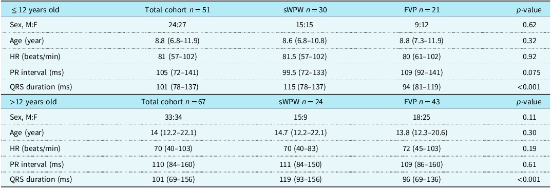
This study included 127 children (65 males and 62 females), with a median age of 12.8 (6.8–22.1) years, resting heart rate of 75 (40–103) beats/min, PR interval of 109 (72–160) ms, and QRS duration of 101 (69–157) ms (Table 1). The adenosine administration test revealed fasciculoventricular pathway, speculated Wolff–Parkinson–White, and indeterminate findings in 64 (50.4%), 54 (42.5%), and 9 (7.1%) children, respectively. The most frequent indeterminate cases were suppression of automaticity at the sinus node (n = 5 children), no electrocardiogram changes (n = 3 children), and ST changes (n = 1 child). The speculated Wolff–Parkinson–White and fasciculoventricular pathway groups were separately compared for children aged ≤ 12 years (51 children) and >12 years (67 children), respectively, in accordance with the diagnostic criteria for Wolff–Parkinson–White syndrome. The results showed a significant difference only in QRS duration, while no significant differences were noted in sex, age, resting heart rate, and PR interval (Table 2).
Patient characteristics

Note: FVP = fasciculoventricular pathway, HR = heart rate, M:F = male-to-female ratio, sWPW = speculated Wolff–Parkinson–White syndrome.
* PR interval and QRS duration were measured using a 12-lead electrocardiogram automatic analyser (Fukuda Denshi, Tokyo, Japan).
†Data are presented as median (range).
Patient characteristics (≤ 12 and >12 years old)

Note: FVP = fasciculoventricular pathway, HR = heart rate, M:F = male-to-female ratio, sWPW = speculated Wolff–Parkinson–White syndrome.
Notably, most children with fasciculoventricular pathway had a QRS duration ≤ 120 ms, with only one child showing >120 ms, which was complicated by bundle branch block. Additionally, >60% of children with a QRS duration ≤ 120 ms were diagnosed with fasciculoventricular pathway (Figure 2). A QRS duration of 120 ms was valuable for distinguishing between speculated Wolff–Parkinson–White and fasciculoventricular pathway. Notably, speculated Wolff–Parkinson–White was significantly more common in the QRS duration >120 ms group than in the QRS duration ≤ 120 ms group (75% vs. 33%, p < 0.001; Supplementary Table S1). Children with the Wolff–Parkinson–White pattern were divided into type A and B categories based on the direction of the dominant QRS deflection in lead V1 on electrocardiogram. Children in the type A category had R/S ratios of >1, whereas children in the type B category had R/S ratios of <1 in lead V1. Type A is highly likely to be speculated Wolff–Parkinson–White. However, for type B, distinguishing between speculated Wolff–Parkinson–White and fasciculoventricular pathway using only the 12-lead electrocardiogram was challenging (Table 3, Figure 3). Among the Type A cases, 2 out of 30 were identified as fasciculoventricular pathway (Figure 3).
Proportion of children with sWPW and FVP according to the adenosine administration test for QRS durations ≤ 120 ms and >120 ms on ECG. More than 60% of children with QRS duration widths ≤ 120 ms referred to our department with WPW pattern were diagnosed with FVP. ECG = electrocardiogram, FVP = fasciculoventricular pathway, sWPW = speculated Wolff–Parkinson–White syndrome, WPW = Wolff–Parkinson–White syndrome.

Proportion of children with sWPW and FVP according to type A and type B categories. Most children with type A category were diagnosed with sWPW. ECG = electrocardiogram, FVP = fasciculoventricular pathway, sWPW = speculated Wolff–Parkinson–White syndrome, WPW = Wolff–Parkinson–White syndrome.

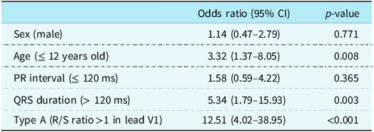
Multivariate logistic regression analysis with odds ratios (95% CI) of various sWPW predictors

Note: CI = confidence interval, sWPW = speculated Wolff–Parkinson–White syndrome.
Multivariate logistic regression analysis revealed age ≤ 12 years (odds ratio: 3.32; 95% confidence interval: 1.37 to 8.05; p = 0.008), QRS duration >120 ms (odds ratio: 5.34; 95% confidence interval: 1.79 to 15.93; p = 0.003), and type A category (R/S ratios >1 in lead V1) (odds ratio: 12.51; 95% confidence interval: 4.02 to 38.95; p < 0.001) as independent risk factors for speculated Wolff–Parkinson–White. PR interval and sex were not considered risk factors for speculated Wolff–Parkinson–White (Table 3).
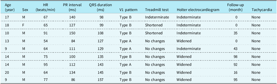
Table 4 presents the clinical characteristics, additional test results (treadmill test and Holter electrocardiogram monitoring), presence of tachycardia, and duration of observation of the nine indeterminate cases. The median follow-up duration was 35 (0–98) months, during which none of the children experienced tachycardia. None of the children underwent electrophysiological testing. Furthermore, two children with and six children without disappearance of the delta waves during treadmill testing had a QRS duration of ≤ 120 and >120 ms, respectively. One case with indeterminate findings could not be adequately examined because the examination was interrupted due to fatigue. Among the six children with QRS duration of >120 ms, five exhibited a change in the QRS waveform when premature atrial contraction appeared on Holter electrocardiogram.
Patients for whom adenosine administration yielded nondiagnostic results

F = female, HR = heart rate, M = male, PAC = premature atrial contraction. Treadmill test = Indicating changes in the QRS waveform; Holter electrocardiogram: indicating changes in the QRS waveform in the presence of PAC; evaluation was not possible when PAC was absent. Type A patients had R/S ratios >1 in lead V1, and type B patients had R/S ratios 1 in lead V1.
Adverse events attributed to adenosine triphosphate therapy were not reported in any child. The adenosine triphosphate doses administered at the time of speculated Wolff–Parkinson–White or fasciculoventricular pathway diagnosis were 0.2 mg/kg (81 children, 68.6%; adenosine at approximately 0.1 mg/kg), 0.3 mg/kg (27 children, 22.9%; adenosine at approximately 0.15 mg/kg), 0.4 mg/kg (six children, 5.1%; adenosine at approximately 0.2 mg/kg), and 0.5 mg/kg or 20 mg (four children, 3.4%; adenosine at approximately 0.25 mg/kg).
Discussion
This study investigated the efficacy of the adenosine administration test in differentiating between Wolff–Parkinson–White syndrome and fasciculoventricular pathway. We found that the adenosine administration was effective in identifying children at risk of sudden death and tachycardia, thereby reducing unnecessary management. To the best of our knowledge, this is the largest study to demonstrate the efficacy and safety of adenosine administration for distinguishing Wolff–Parkinson–White syndrome from fasciculoventricular pathway.
According to the Japanese school-based cardiovascular screening guidelines, children with a Wolff–Parkinson–White pattern without a history of tachycardia should be followed up every 1–3 years without exercise restriction. Reference Sumitomo, Baba and Doi11 Children with fasciculoventricular pathway exhibiting delta waves are frequently subjected to similar treatment, typically leading to over-management. In the Japanese guidelines for Heart Disease Screening in Schools (the Japanese Circulation Society 2016 and the Japanese Society of Pediatric Cardiology and Cardiac Surgery 2016), the diagnostic criteria for the Wolff–Parkinson–White pattern have two definitions based on age: one for middle- and high school-aged children and another for primary school-aged children. Here, we conducted a sub-analysis with 12 years of age as the cut-off. In middle- and high school-aged children, prolonged QRS duration (≥120 ms), short PR interval (<120 ms), and ventricular activation time >60 ms are included, whereas the diagnostic criteria in primary school-aged children include prolonged QRS duration (≥100 ms), short PR interval (<100 ms), and ventricular activation time >50 ms. Reference Sumitomo, Baba and Doi11
Delta wave amplitudes >5 mm, measured as the maximum height of the delta wave in the first 40 ms of the most pre-excited frontal limb lead QRS, have a specificity of 96% for the atrioventricular accessory pathway. In contrast, delta wave amplitudes <2 mm have a specificity of 96% for fasciculoventricular pathway. Reference O’Leary, Dewitt, Mah, Gauvreau, Walsh and Bezzerides12 However, the Wolff–Parkinson–White pattern is often managed based on the presence of delta waves rather than the PR/QRS duration in a clinical setting.
In our study, we observed that at least 50% of children with Wolff–Parkinson–White pattern were diagnosed with fasciculoventricular pathway, suggesting the possibility of over-management. Previous research has indicated that the Wolff–Parkinson–White pattern is diagnosed in approximately 0.1% of school-based cardiovascular screening programmes in Japan Reference Hosaki13 and in 0.1–0.3% of the general population. Reference Page, Joglar and Caldwell1 Although the epidemiological rate of fasciculoventricular pathway remains unknown, our study findings indicate the possibility of many cases of fasciculoventricular pathway among those considered to have a Wolff–Parkinson–White pattern.
Holter electrocardiogram can occasionally be used to identify the Wolff–Parkinson–White pattern. The presence of atrial extrasystoles can lead to a widened QRS waveform, facilitating the diagnosis of speculated Wolff–Parkinson–White. This widening is induced by changes in atrioventricular conduction in atrial extrasystoles, as the QRS waveform in speculated Wolff–Parkinson–White is constructed from waves passing through the atrioventricular node and accessory conduction pathway, with accessory conduction persisting even during atrial extrasystoles. In contrast, this phenomenon was not observed in fasciculoventricular pathway cases, as the QRS waveform was exclusively generated from the wave passing through the atrioventricular node. Diagnosing fasciculoventricular pathway through Holter monitoring is challenging. However, comparing electrocardiogram waveforms recorded during daytime activity with those captured during sleep can aid in diagnosing Wolff–Parkinson–White, especially when the QRS waveform widens during deep sleep.
While the treadmill test is often used to differentiate between speculated Wolff–Parkinson–White and fasciculoventricular pathway, distinguishing them remains challenging. Evaluating younger children using the treadmill test is particularly difficult because they cannot follow instructions or reach their maximum heart rate owing to physical limitations.
Our study analysed 127 children, which is a larger sample size than those in previous studies Reference Suzuki, Nakamura, Yoshida, Yoshida and Shintaku9,Reference Follansbee, Beerman, Wu, Bertolet and Arora14 involving patients who underwent adenosine administration. The test was proven to be useful in identifying patients at risk of sudden death and tachycardia and in reducing unnecessary routine examinations.
Our study revealed that the adenosine triphosphate dose was 0.2–0.3 mg/kg (adenosine at approximately 0.1–0.15 mg/kg) in approximately 90% of the cases. Based on our study, which indicates that 31.4% of cases cannot be diagnosed with a single dose of adenosine triphosphate at 0.2 mg/kg, administering multiple doses may be necessary, potentially causing discomfort. Therefore, to minimise the number of adenosine triphosphate doses, setting the initial dose at 0.3 mg/kg is better than 0.2 mg/kg.
Children with type A category (R/S ratios >1 in lead V1) were more likely to present with speculated Wolff–Parkinson–White, which is consistent with the findings of a previous report. Reference Suzuki, Nakamura, Yoshida, Yoshida and Shintaku9 When considering age, the PR interval and QRS duration are prolonged in bradycardia, with these parameters tending to be more prolonged in older children. Despite this confounding factor, a QRS duration of >120 ms in younger patients indicates an increased risk. In the multivariate analysis, the study population was classified as “speculated Wolff–Parkinson–White.” Moreover, although selection bias exists, we aimed to differentiate patients with fasciculoventricular pathways that exist in a population managed for Wolff–Parkinson–White pattern. The study results are consistent with those of previous reports, which indicated that children with type A and QRS duration >120 ms are more likely to have a true Wolff–Parkinson–White pattern. Reference Suzuki, Nakamura, Yoshida, Yoshida and Shintaku9,Reference Sumitomo, Baba and Doi11 Furthermore, these findings indicate that the adenosine administration is a useful test for identifying patients with a QRS duration ≤ 120 ms who are not at high risk for speculated Wolff–Parkinson–White.
Children with speculated Wolff–Parkinson–White who have not experienced tachycardia typically undergo outpatient follow-up every several years, which is similar to the management of children with fasciculoventricular pathway with delta waves. In this study, approximately 50% of the children with Wolff–Parkinson–White pattern were classified as having fasciculoventricular pathway. The adenosine level was considered useful for identifying patients at risk of sudden death and tachycardia and reducing unnecessary management. Moreover, distinguishing speculated Wolff–Parkinson–White from fasciculoventricular pathway helps prevent future health morbidities that could occur in cases of uncertainty. For example, for children with electrocardiogram findings suggestive of speculated Wolff–Parkinson–White who have an unclear diagnosis and are scheduled for noncardiac interventions (e.g., knee surgery), treatment could be delayed until the uncertainty is resolved. In contrast, if a diagnosis of fasciculoventricular pathway had been established in such children, the delay in care would have been prevented.
This study is limited by its dependence on the precision of data extracted from a single centre. Therefore, further large-scale multicentre studies are warranted to validate the results of our study. The absence of adverse events suggests that the adenosine administration test is excellent in terms of simplicity and safety.
In conclusion, this study demonstrated the efficacy of the adenosine administration test in distinguishing between speculated Wolff–Parkinson–White and fasciculoventricular pathway, as it involves no risk of sudden death and tachycardia and thus can reduce unnecessary management. Furthermore, the adenosine administration test can be considered useful with respect to its safety and simplicity in children with delta waves.
Supplementary material
The supplementary material for this article can be found at https://doi.org/10.1017/S104795112403659X.
Data statement
The datasets used and analysed during the current study are available from the corresponding author upon reasonable request.
Acknowledgements
None.
Author contribution
Kentaro Kogawa: Conceptualisation, Data curation, Formal analysis, Investigation, Methodology, Project administration, Supervision, Validation, Visualisation, and Writing – original draft, Writing – review & editing; Reiji Ito: Writing – review & editing, Supervision; Daishi Hirano: Formal analysis, Supervision; Kenji Hoshino: Supervision.
Financial support
This research received no specific grant from any funding agency, commercial, or not-for-profit sectors.
Competing interests
None.
Ethical standards
The authors assert that all procedures contributing to this work comply with the Helsinki Declaration of 1975, as revised in 2008, and have been approved by the Ethics Committee of Saitama Children’s Medical Center (approval number 2023-03-008).
Patient consent
The authors confirm that patient consent forms were obtained for this article.