Introduction
Zoisite, Ca2Al3(SiO4)(Si2O7)O(OH), is an orthorhombic sorosilicate crystallising in the Pnma space group (Liebscher et al., Reference Liebscher, Gottschalk and Franz2002; Dörsam et al., Reference Dörsam, Liebscher, Wunder, Franz and Gottschalk2007). Zoisite is a polymorphic modification of clinozoisite and its structure (Fig. 1) is similar to that of the epidote supergroup but is formed by only one type of octahedral chain oriented in the b-axis direction (Fig. 2) (Liebscher et al., Reference Liebscher, Gottschalk and Franz2002; Armbruster et al., Reference Armbruster, Bonazzi, Akasaka, Bermanec, Chopin, Gieré, Heuss-Assbichler, Liebscher, Menchetti, Pan and Pasero2006). The structure of zoisite consists of seven cationic positions, of which two are surrounded by 7- to 9-fold coordination polyhedra (A1; A2), two by octahedra (M1,2; M3), three by tetrahedra (T1; T2; T3), and ten anionic positions, the O10 position being occupied by an OH group. Substitutions of Fe3+, Cr3+, Mn2+ and V3+ ions replacing Al3+ in the structure are commonly present (Dörsam et al., Reference Dörsam, Liebscher, Wunder, Franz and Gottschalk2007).
Visualisation of the zoisite structure in a view perpendicular to b for the sample studied. Yellow = Ca; blue = octahedra, M; and red = tetrahedra, T. Drawn using Crystal Impact Diamond (version 3.2k).

The chains of M1,2O6 (blue) octahedra parallel to b with attached M3O6 (green) octahedra. Drawn using Crystal Impact Diamond (version 3.2k).

Zoisite was first described as ‘saualpite’, named after the Saualpe type locality in Carinthia, Austria. The name zoisite was given in 1805 by A. G. Werner, in honour of Siegmund Zois, Baron von Edelstein (1747–1819), an Austrian mineral collector, from whom Werner obtained the holotype of a new mineral from Saualpe (Gaines et al., Reference Gaines, Skinner, Foord, Mason and Rosenzweig1998). Zoisite occurs mainly in metamorphic and hydrothermally transformed rocks, and in pegmatites which were melted from eclogites (Franz and Smelik, Reference Franz and Smelik1995). Zoisite is a mineral that includes several colour varieties. The most sought-after variety of zoisite is tanzanite, whose blue-violet colour is caused by the presence of vanadium. Tanzanite was first identified by George Kruchiuk in 1962, who obtained several samples of alleged blue sapphire from the Merelani area of Tanzania (Dirlam et al., Reference Dirlam, Misiorowski, Tozer, Stark and Bassett1992). At present, yellow, brown, green, blue–green and pink zoisites are mined on the site; these are often subjected to heat treatment to change the colour to exclusively blue (Zancanella, Reference Zancanella2004).
The goal of this work is to determine the possible structural position of vanadium in the zoisite var. tanzanite structure. A fragment of blue tanzanite from the Merelani area, Tanzania (from the personal collection of Peter Bačík) was investigated by a multi-analytical chemical, structural and spectroscopic approach. Direct determination using structural refinement has limited application due to the small amount of vanadium in tanzanite. Therefore, we applied optical absorption spectroscopy with its interpretation using crystal field Superposition Model calculations and structural refinement accompanied by Bond-Valence Model calculations and considered the possible influence of vanadium and other substituents on the zoisite structure.
Geological settings
The Merelani area is located in the western part of the Lelatema antiform ~65 km from the city of Arusha in Tanzania (Malisa, Reference Malisa1998, Reference Malisa2004) and consists of granulite complexes of the Pan-African Mozambican zone (Muhongo and Lenoir, Reference Muhongo and Lenoir1994). Merelani is a world-famous area of minerals of gemmological quality, also called the ‘Gemstone Belt of East Africa’. Here, it is possible to find garnets (tsavorite, spessartine and rhodolite), ruby, sapphire, tanzanite, kyanite, diopside and many other minerals (Malisa, Reference Malisa2004; Le Goff et al., Reference Le Goff, Deschamps and Guerrot2010; Feneyrol et al., Reference Feneyrol, Giuliani, Ohnenstetter, Fallick, Martelat, Monié, Dubessy, Rollion-Bard, Le Goff, Malisa, Rakotondrazafy, Pardieu, Kahn, Ichang'i, Venance, Voarintsoa, Ranatsenho, Simonet, Omito, Nyamai and Saul2013; Harris et al., Reference Harris, Hlongwane, Gule and Scheepers2014). Notably, in addition to blue–violet V-bearing tanzanite, the Merelani area is also the unique occurrence of light blue V-bearing axinite-(Mg) (Jobbins et al., Reference Jobbins, Tresham A and Young1975; Andreozzi et al., Reference Andreozzi, Lucchesi and Graziani2000). The site extends along the Lelatema fault system, which consists of Proterozoic metasediments, graphitic gneisses, dolomitic marbles and shales. The rocks went through a metamorphic peak from 7.7 to 9.1 kbar and 600–740°C at 640 Ma, while the Lelatema fracture system formed during deformation at 560 Ma (Appel et al., Reference Appel, Möller and Schenk1998; Muhongo et al., Reference Muhongo, Tuisku and Mtoni1999; Hauzenberger et al., Reference Hauzenberger, Bauernhofer, Hoinkes, Wallbrecher and Mathu2004, Reference Hauzenberger, Sommer, Fritz, Bauerhofer, Kröner, Hoinkes, Wallbrecher and Thöni2007; Malisa, Reference Malisa2004; Le Goff et al., Reference Le Goff, Deschamps and Guerrot2010). After the Pan-African tectonothermal event, there was a hydrothermal dissolution of rocks rich in Ca, Mg, CO2, and SO3 and also enriched in V, U, Sr, Zn and heavy rare earth elements. Subsequently, fluids with these elements got into local fractures and fissures, where they reacted with the bedrock layers. This reaction resulted in the formation of tanzanite and other zoisite varieties, green grossular (var. tsavorite), diopside, quartz, graphite and calcite (Bocchio et al., Reference Bocchio, Adamo, Bordoni, Caucia and Diella2012).
Sample description and possible treatment
The crystal studied was a heat-treated fragment of uniformly blue colour up to 1 cm in size with inclusions of flaky graphite crystals. It is important to consider that blue tanzanites are artificially treated from original yellow, brown, green, blue–green and pink zoisites mined at Merelani, Tanzania. The temperature range of this treatment is from 350 to 700°C (Zancanella, Reference Zancanella2004). Dichroism (actually trichroism, but with similar blue and bluish-green directions) observed in the sample studied indicates that this crystal was artificially heat-treated. Natural high-quality blue–violet tanzanite samples are very rare, most of the bright blue tanzanite in the market has undergone heat treatment (Yang et al., Reference Yang, Ye, Liu, Lu, Liu, Gu and Gurzhiy2021).
Analytical methods
Chemical composition determination
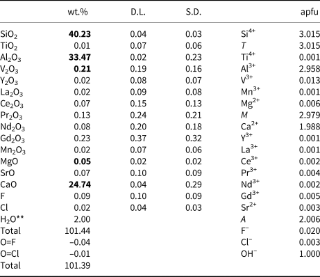
The composition of zoisite studied was established using a Cameca SX100 electron microprobe analysis (EMPA) in wavelength-dispersion mode at the Masaryk University (Brno, Czech Republic), Department of Geological Sciences, under the following conditions: accelerating voltage = 15 kV, beam current = 20 nA and beam diameter = 3 μm. The samples were analysed using the following standards (all Kα lines): hematite (Fe); Mn2SiO4 (Mn); TiO (Ti); wollastonite (Ca); andalusite (Al); Mg2SiO4 (Mg); titanite (Si); chromite (Cr); gahnite (Zn); vanadinite (V); Ni2SiO4 (Ni) and Co (Co). The measured element detection limits ranged from 150 to 700 ppm. The tanzanite crystal-chemical formula (Table 1) was calculated on the basis of 8 cations per formula unit, the OH content was calculated assuming OH = 1 apfu.
Electron probe microanalysis and ionic proportions of V-zoisite (tanzanite) from Merelani, Tanzania.*

* Only contents of oxides highlighted in bold were convincingly above their detection limits, however the presence of other elements is very likely according to LA-ICP-MS.
** Calculated assuming OH = 1 apfu.
D.L. detection limit; S.D. –standard deviation
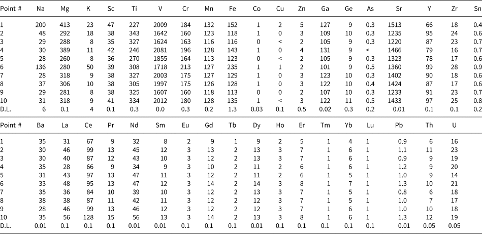
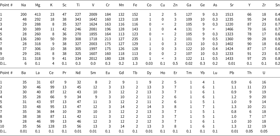
Instrumentation for laser ablation inductively coupled plasma mass spectrometry (LA-ICP-MS) at the Department of Chemistry, Masaryk University, Brno, Czech Republic consists of the laser ablation system Analyte G2+ (CETAC Technologies) emitting laser radiation of λ = 213 nm, and the quadrupole ICP-MS spectrometer Agilent 7900 (Agilent Technologies, Japan). The sample was enclosed in two-volume ablation cell Helex2. The ablated material was carried out from the cell by He flows of 0.600 and 0.300 l/min. Argon was admixed (1.0 l/min) into the helium flow before ICP-MS. NIST 610 standard was used for the optimisation of ICP-MS parameters with respect to maximal S/N ratio, oxide formation <0.2% (ThO+/Th), and U+/Th+ <1.1%. The laser ablation of the sample was done under these optimised parameters: laser beam spot of 100 μm diameter, repetition rate of 20 Hz and laser beam fluence of 6.0 J/cm2. Each spot was drilled for 60 s, and the pause between the ablation of individual spots was 30 s. Glass reference material NIST610 and normalisation on Al content was used for the quantification purpose. Respective detection limits for each element are included in Table 2.
The trace-elements contents (in ppm) of V-zoisite (tanzanite) from Merelani measured with LA-ICP-MS.
D.L. – detection limit.
Crystal structure refinement
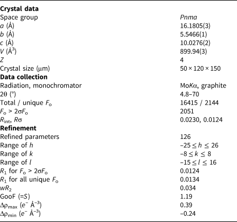
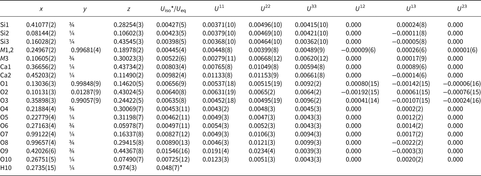
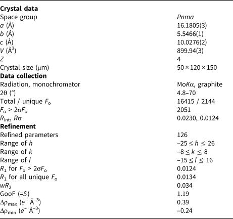
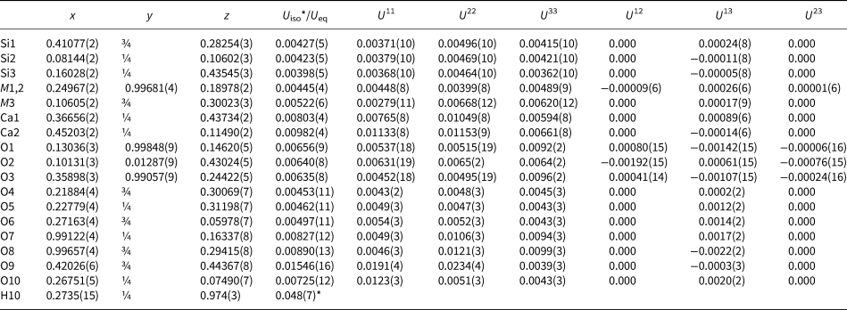
A platy fragment extracted from the tanzanite sample was studied using single-crystal X-ray diffraction. The measurement was done at the Centre for Higher Order Structure Elucidation (C-HORSE) at the University of British Columbia, Canada, using a Bruker X8 APEX II diffractometer with graphite-monochromated MoKα radiation. Crystal data and details of the structure refinement are listed in Table 3. The structure was solved using SHELXT (Sheldrick, Reference Sheldrick2015) and symmetry-transposed to the earlier published zoisite structure model (Alvaro et al., Reference Alvaro, Angel and Cámara2012). The CrysAlis (Oxford Diffraction Ltd.) and SHELXL (Sheldrick, Reference Sheldrick2015) program packages were used for data reduction and structure refinement, respectively, using neutral scattering factors and anomalous dispersion corrections for cation atoms and ionised species for oxygen (O2–; Azavant and Lichanot, Reference Azavant and Lichanot1993). Atoms at all cation sites (Ca at Ca1 and Ca2; Al at M1,2 and M3; and Si at Si1, Si2 and Si3) were set to vary freely (tested both individually and simultaneously) however their deviations from full occupancies were negligible; therefore, full occupancies of all sites were used for final refinement. The position of the H(10) hydrogen atom was located on the residual electron-density map. The structure was refined in space group Pnma and converged to a final R1 index of 1.24% for 2051 reflections with F o2 > 2σ(F o2) and 126 refined parameters. The refined atomic coordinates and displacement parameters are listed in Table 4 and selected geometric parameters in Table 5. The crystallographic information file has been deposited with the Principal Editor of Mineralogical Magazine and is available as Supplementary material (see below).
Crystal and refinement data for V-zoisite (tanzanite) from Merelani.
Fractional atomic coordinates and isotropic or equivalent isotropic displacement parameters (Å2) for V-zoisite (tanzanite) from Merelani.
* Isotropic displacement parameter (Å2).
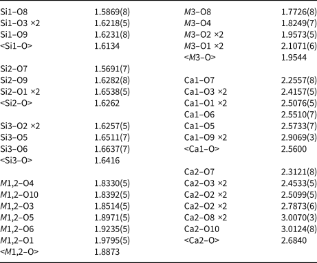
Selected geometric parameters (Å) for V-zoisite (tanzanite) from Merelani.

Polarised optical absorption spectroscopy
Polarised optical absorption spectra of the tanzanite samples investigated were recorded on double-sided polished crystal slabs in the spectral range 36000–3400 cm–1, i.e. covering the near ultraviolet (UV), the visible (Vis) and the near-infrared (NIR) ranges of the electromagnetic spectrum. Slabs were prepared in two different orientations, i.e. (010) and (001), both in two different thicknesses (the former slab at 9.03 mm, then thinned to 2.05 mm, the latter one at 6.05 and 2.20 mm), on the one hand to allow polarised measurements with the electric light vector parallel to the three crystallographic axes, on the other hand allowing recording of strongly different intensities of various absorption features with reliable S/N ratios. As the relevant literature on the mutual assignment of pleochroic colours, optical axes, crystal morphology and crystallographic axes for untreated as well as for heat-treated tanzanites is highly inconsistent (see respective comments by Deer et al., Reference Deer, Howie and Zussman1986), the slab orientations and cell-axes directions were checked by X-ray diffraction, referring to the generally consistent assignment of a ≈ 16, b ≈ 5.5 and c ≈ 10 Å. The spectroscopic measurements were performed in the sample chamber of a Bruker Vertex 80 FTIR spectrometer, using a calcite Glan-prism polariser and appropriate combinations of light sources (Tungsten or Xenon lamp), beam splitters (CaF2-NIR or CaF2-ViS/UV), and detectors (InGaAs-, Si- or GaP-diodes) to cover the desired spectral range, at measuring spots of 1–2 mm in diameter (the specific measuring spots were selected under a stereomicroscope, avoiding inclusions and cracks in the two sample slabs as far as possible). Hence, each full spectrum is combined from three partial spectral regions (36000–20000 cm–1: spectral resolution 40 cm–1, averaged from 512 scans; 20000–11000 cm–1: resolution 20 cm–1, 128 scans; 11000–3400 cm–1: resolution 10 cm–1, 64 scans), if necessary merged from respective measurements at different slab thicknesses. The subspectra were aligned in absorbance for a perfect match and calculated to linear absorption coefficient α (cm–1). Observed transition energies for the crystal field calculations were extracted from the spectra by visual inspection.
Crystal field Superposition Model calculations
Crystal field (CF) calculations were performed in the framework of the semiempirical Superposition Model (SPM) of crystal fields, originally developed by Newman (Reference Newman1971) to separate the geometrical and physical information contained in CF parameters, taking into account the exact geometry of the coordination polyhedra in the respective phases. The SPM is based on the assumption that the CF can be expressed as the sum of axially symmetric contributions of all i nearest-neighbour ligands of the transition metal cation. The CF parameters Bkq (in Wybourne notation) are then obtained from:

where $\bar{B}_k$ are the ‘intrinsic’ parameters (related to a reference metal–ligand distance R 0), tk are the power-law exponent parameters, both for each rank k of the crystal field, Ri are the individual metal–ligand distances and Kkq(Θi,Φi) are the coordination factors calculated from the angular polar coordinates of the ligands. For details and comprehensive reviews on the SPM refer to Newman (Reference Newman1971), Newman and Ng (Reference Newman and Ng1989, Reference Newman and Ng2000), Rudowicz et al. (Reference Rudowicz, Gnutek and Açikgöz2019), and (with geoscientific focus) Andrut et al. (Reference Andrut, Wildner and Rudowicz2004).
The actual CF calculations were done using the HCFLDN2 module of the computer program package by Y.Y. Yeung (Rudowicz et al., Reference Rudowicz, Yeung, Du and Chang1992; Chang et al., Reference Chang, Rudowicz and Yeung1994; Yang et al., Reference Yang, Hao, Rudowicz and Yeung2004), which includes imaginary CF terms and is thus applicable to arbitrary low symmetries of all 3d N electron systems. A suite of supplementary programs (Wildner, unpublished) was used to manage the input and output of the HCFLDN2 program, in particular (i) for the transformation of atomic to polyhedral polar coordinates; (ii) for the systematic variation of intrinsic and power-law SPM parameters, as well as of the Racah parameters B and C; (iii) for the SPM calculation itself, giving the actual values for the Bkq parameters of the CF; (iv) for the corresponding communication with a slightly modified version of the HCFLDN2 program (Yeung, pers. comm.); and (v) for the interpretation and evaluation of the HCFLDN2 output results in terms of a reliability index for the agreement of calculated and observed transition energies.
According to the low point symmetries 1 (M1,2) and m (M3) of the potentially V (or other transition metal) -bearing AlO6 polyhedra in zoisite, symmetrically unrestricted SPM calculations were performed; however, to reduce the number of variables (accompanied by reduced CPU time) and to improve the transferability of intrinsic $\bar{B}_k$ parameters, the power-law exponent parameters tk were fixed at their ideal electrostatic values of t 4 = 5 and t 2 = 3. The reference metal–ligand distance R 0 for V3+ was set to 2.01 Å, the sum of the ionic radii (Shannon, Reference Shannon1976) of octahedrally coordinated V3+, 0.64 Å, plus 1.37 Å for O2– in three-to fourfold coordination and equalling the overall mean bond length in V3+O6 polyhedra extracted by (Schindler et al., Reference Schindler, Hawthorne and Baur2000). Cubically averaged Dqcub values were calculated from the Bkq parameters via the rotational invariant s 4 (Leavitt, Reference Leavitt1982).
Bond-Valence Model calculations
Bond-length and bond-valence calculations are based on the following equations:


where dij is the bond length (in Å) between the two given ions, the bond valence (ν ij) measures the bond strength (in vu – valence units), R 0 is the length of a single bond (for which ν ij = 1 vu), and b is the universal parameter for each bond (Brown, Reference Brown2006). The R 0 and b values for each cation from the list of Gagné and Hawthorne (Reference Gagné and Hawthorne2015) were used, as this list provides the most current and consistent data on the bonding parameters. For more details, see Bačík and Fridrichová (Reference Bačík and Fridrichová2019). Bond lengths were calculated only for the most common major, minor and trace elements occurring in zoisite, although a similar approach can be used for any chemical element.
Results
Chemical composition
The composition of the Merelani tanzanite based on the EMPA is close to pure zoisite (Table 1). Among the octahedrally coordinated cations, the most abundant is Al with limited substitutions of V and Mg. The amounts of other cations were below their detection limits. At the A sites, the observed amounts of Ca below 2 atoms per formula unit (apfu) allow for some substitution of Sr and rare earth elements, however, their contents are very near their respective detection limits. Tetrahedrally-coordinated sites are only occupied by Si.
The LA-ICP-MS analyses of the sample studied show that V and Sr are the most abundant among trace elements exceeding 1000 ppm. The contents of Mg, Ti, Cr, Mn, Fe and Ga vary between 100–450 ppm; those of Na, K, Sc, Y, Zr, Ba, La, Ce, Pr, Nd, Sm, Gd, Dy, Th and U are usually below 100 ppm; and the remaining elements are only slightly above their respective detection limits (Table 2). Moreover, the measurements from 10 points in the line profile show no significant variations in any of the measured elements and therefore, no significant zoning of the crystal studied.
Crystal structure refinement
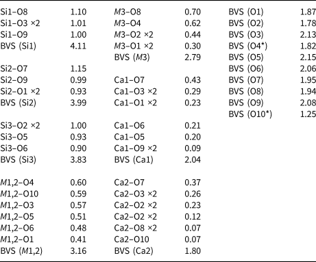
The structure of V-zoisite from Merelani agrees with the previously reported data (e.g. Alvaro et al., Reference Alvaro, Angel and Cámara2012). The zoisite structure is characterised by chains of edge-sharing M1,2 octahedra parallel to [010], decorated by M3 octahedrally coordinated sites that share edges with two M1,2 octahedra. Typically, the M1,2 octahedra are occupied by Al3+ whereas M3 can be occupied by both Al3+ and Fe3+ (Tsang and Ghose, Reference Tsang and Ghose1971b; Alvaro et al., Reference Alvaro, Angel and Cámara2012). The octahedral chains are linked by isolated SiO4 tetrahedra (T3) in the c direction and by Si2O7 groups (T1 and T2) in the a and c directions; cavities in this framework of octahedra and tetrahedra contain two sevenfold- to ninefold-coordinated sites (A1 and A2) occupied by Ca. Hydrogen is bonded to oxygen O10 bonded to two M1,2 cations (Franz and Liebscher, Reference Franz, Liebscher, Liebscher and Franz2004). The bond-valence analysis of the refined structure shows discrepancies between calculated bond valences and formal cation charges (Table 6) at all sites but T2 and A1.
Bond valences (vu) for V-zoisite (tanzanite) from Merelani (BVS – bond-valence sum)

* Hydrogen bond donor (O10) and acceptor (O4)
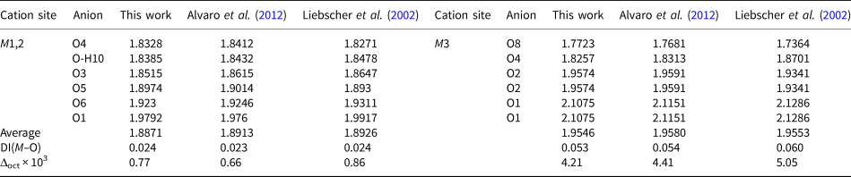
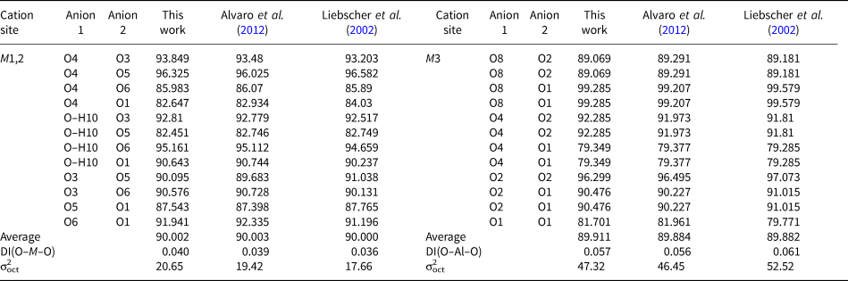
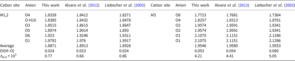
The average bond length of the M1,2O6 octahedron is below 1.90 Å; M3O6 has slightly longer bonds with an average of ca. 1.96 Å (Table 7). The M1,2O6 octahedron also has a smaller bond-length distortion than M3O6. Its distortion parameter DI(Al–O) is 0.024, whereas that of M3O6 is more than twice higher (Table 7). The difference in Δoct is even higher, in M1,2O6 it is 0.77 and in M3O6 it is as much as 4.21. Similarly, the M3O6 octahedron is stronger bond-angle distorted than M1,2O6; both DI (O–M–O) and σoct2 parameters of M3O6 are higher: 0.057 vs. 0.040 and 47.33 vs. 20.67 (Table 8), respectively.
Empirical bond lengths (in Å) at the octahedral sites of the sample studied compared to published data and calculated octahedral bond-length distortions.
Distortion parameters: Bond-length distortion ${\rm DI}\;( {M\ndash O} ) = \left({\mathop \sum \limits_{i = 1}^6 \vert {d_i-d_m} \vert } \right)/( {6 \times d_m} )$
Quadratic elongation $\Delta _{oct} = {1 \over 6}\mathop \sum \limits_{i = 1}^6 [ {( {d_i-d_m} ) /d_m} ] ^2$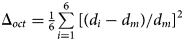
Empirical bond angles (°) at the octahedral sites of the sample studied compared to published data and calculated octahedral bond-angle distortions.

Distortion parameters: Bond-angle distortion $\rm DI{\rm \;}( {O\ndash{\it M} \ndash O} ) {\rm \;} = \left({\mathop \sum \limits_{i = 1}^{12} \vert {\rm \alpha_i-\rm\alpha_m} \vert } \right)/( {12 \times \rm\alpha_m} )$
Octahedral angle variance $\rm\sigma _{\rm{oct}}^2 = {1 \over {11}}\mathop \sum \limits_{\it i \rm{= 1}}^{12} ( {\rm\alpha_i-90} ) ^2$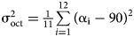
Optical absorption spectra
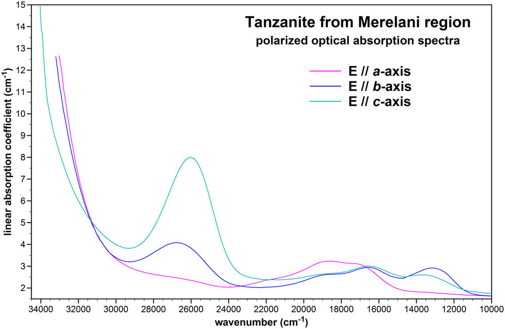
The polarised optical absorption spectra of the investigated tanzanite sample are shown in Fig. 3. Four major regions of absorptions can be distinguished: (1) the NIR region ≤ 6500 cm–1, containing combination and overtone modes of fundamental vibrations that are not the focus of the present study (and not included in Fig. 3); (2) the NIR and Vis region between ~12000–23000 cm–1, containing various CF absorption bands with maxima around ~13500 and 16500–19000 cm–1, also including faint remains of the band at ~22000 cm–1 (in polarisation ‖ the a-axis) that is responsible for the colour-change of tanzanites upon heat-treatment; (3) an absorption band system centred around ~26500 cm–1, i.e. close to the Vis-Near-UV boundary and thus still influencing the colour and pleochroism of tanzanites; and (4) the UV absorption edge, strongly ascending between 33000–34000 cm–1, perhaps showing a faint structure in polarisation ‖ the c axis at ~34000 cm–1. The bands or band components have half-widths typical for spin-allowed d–d CF-transitions, whereas there are no spectral features that could be attributed to respective field-independent (i.e. sharp) spin-forbidden transitions. The trichroism of the investigated tanzanite referring to the crystallographic axes, i.e. purple ‖ a, blue ‖ b, and bluish green ‖ c, can be correlated easily with the respective absorption behaviour within the visible spectral range between ~14000 and 25000 cm–1.
Polarised optical absorption spectra of tanzanite from the Merelani area, Tanzania.

Bond-valence and bond-length calculation
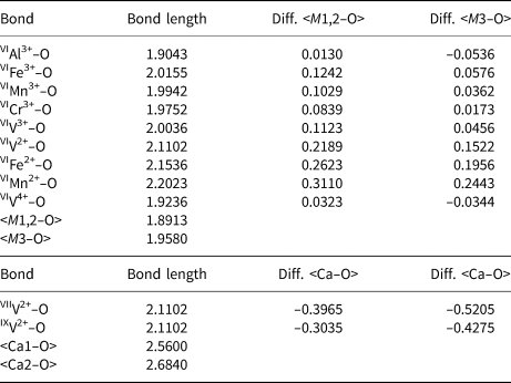
Ideal bond lengths for typical octahedral cations in zoisite were calculated from the ideal bond valences of 0.5 vu for trivalent and 0.33 vu for divalent cations (Table 9). The Al–O bond has the shortest ideal bond length; other trivalent cations form slightly longer bonds, the largest difference (0.11 Å) was observed for the Fe3+–O bond. For comparison, the difference in ideal bond lengths of divalent cations compared to the Al–O bond is >0.25 Å. Interestingly, the V4+–O bond length is <0.02 Å larger than the Al–O bond.
Calculated ideal bond lengths (in Å) for selected cations in vi, vii and ix coordination, empirical average bond length and their difference (diff.) for each octahedral site in zoisite.

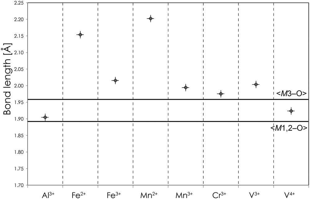
This also makes V4+–O the only bond (except Al–O) which is shorter than the average bond length of the M3O6 octahedron – the larger of the two octahedra in the zoisite structure (Fig. 4). Bonds of trivalent cations (except Al) are longer than <M3–O> though the difference is not larger than 0.05 Å, which is less than ca. 2.5% of the bond length. The difference of Fe2+–O and Mn2+–O bond lengths to <M3–O> is 0.20 and 0.25 Å, respectively. This is equal to 10–13% of the <M3–O> bond length. The difference to <M1,2–O> is even larger, more than 16% in case of the Mn2+–O bond.
Calculated bond lengths for selected cations (symbols) compared to empirical average bond lengths for the octahedral sites in zoisite (full horizontal lines).

As some works (Ghose and Tsang, Reference Ghose and Tsang1971; Tsang and Ghose, Reference Tsang and Ghose1971a) suggest the presence of V2+ as a possible chromophore in tanzanite, its ideal bond lengths were calculated in the octahedral, 7- and 9-fold coordination (Table 9). The calculation revealed that V2+–O bonds in the octahedral coordination are significantly longer than average bond length of both octahedra in zoisite. In contrast, if we assume higher coordination, V2+–O bonds are shorter than Ca–O bonds at both possible sites in zoisite with an even larger difference. Consequently, it is possible to conclude that the presence of any V2+ in tanzanite is highly unlikely.
Discussion
Estimation of the V position in the tanzanite structure in such low concentrations is based on several necessary assumptions. Firstly, it is well established that in zoisite, V occupies octahedrally-coordinated sites with an assumed preference of the M3 site (Ghose and Tsang, Reference Ghose and Tsang1971; Tsang and Ghose, Reference Tsang and Ghose1971a, Reference Tsang and Ghose1971b). Then, the oxidation state of V is not straightforward. In the sample studied, the presence of any yellow–green shade typical for untreated tanzanite (Yang et al., Reference Yang, Ye, Liu, Lu, Liu, Gu and Gurzhiy2021) was not found and has probably been shifted to bluish green, hence we assume the heat treatment of the sample. This change in pleochroism was proposed to be the result of V3+ to V4+ oxidation or Ti3+ to Ti4+ oxidation (Schmetzer and Bank, Reference Schmetzer and Bank1978; Pluthametwisute et al., Reference Pluthametwisute, Wanthanachaisaeng, Saiyasombat and Sutthirat2020). This would indicate that a significant part of the V should be four-valent, while some V could remain in a trivalent state. Consequently, the position and oxidation state of V in the tanzanite structure was studied and derived from the optical spectra accompanied by CF calculations and the structure by the bond-valence calculations.
Crystal field Superposition Model calculations
The chemical analysis (Table 1) shows that V (1854 ppm) is by far the major transition element and hence will be decisive for the crystal field absorption spectra, with possible minor influence of Ti (301 ppm). Theoretically, vanadium as a V2+ or V3+ cation (with d 3 and d 2 d-orbital configuration, respectively), i.e. cations with a spectroscopic F ground term plus one spin-allowed excited P-term, could be responsible for the observed complex spectral envelope, though V2+ is neither compatible with available structural sites in zoisite, nor expected to be stable enough in oxidic environments (Faye and Nickel, Reference Faye and Nickel1971) or upon heat-treatment. Hence V3+, substituting for aluminium in one or both available Al-sites, very probably dominates the absorption spectra. However, in the present case, V4+ and Ti3+, both with d 1 configuration may also contribute to some extent.
Previous detailed crystal-field analyses of V3+ in tanzanite (Faye and Nickel, Reference Faye and Nickel1971; Tsang and Ghose, Reference Tsang and Ghose1971a) assumed that “the entire d-d spectrum of Tanzanian zoisite is in the range ~13000 to 27000 cm–1”, and consequently assigned the band systems centred around 13500, 18000 and 27000 cm–1 to electronic transitions from the 3T1(F) ground state of V3+ to the excited 3T2(F), 3T1(P) and 3A2(F) levels, respectively. Whereas Faye and Nickel (Reference Faye and Nickel1971) concluded that V3+ only occupies the M3 site and hence derived Dq ≈ 1400 cm–1 and Racah B <630 cm–1, Tsang and Ghose (Reference Tsang and Ghose1971a) proposed that V3+ occupies both Al-sites randomly, resulting in Dq values of 1850 cm–1 at the M1,2 site and 1400 cm–1 at M3 (Racah B values were not determined). As part of a comprehensive review on the spectroscopy of epidote-group minerals, Liebscher (Reference Liebscher, Liebscher and Franz2004) gives a detailed summary of the previous studies on the optical absorption spectra of tanzanites.
However, a detailed inspection of the present optical absorption (Fig. 3) combined with crystal chemical arguments and considerations reveals that both the above-mentioned previous interpretations have to be dismissed, at least in part, for the following reasons: (1a) the intense absorption band around 26500 cm–1 (Fig. 3), previously assigned as 3A2(F), definitely shows a minor but unambiguous splitting with maxima around ~26100 (E ‖ c) and 26700 cm–1 (E ‖ b) – this means that it may not be assigned to a non-degenerate A-state which does not split, whatever the local symmetry may be (spin-orbit splitting can also be ignored in case of V3+); (1b) assuming a distribution of V3+ on both Al-sites with clearly different Dq-values (as Tsang and Ghose Reference Tsang and Ghose1971a did) would result in a much larger difference of the two respective 3A2(F) levels than the mere ~600 cm–1 found in the present study; (1c) the 3T1(F)→ 3A2(F) transition in d 2 systems corresponds to a forbidden ‘two-electron jump’ in the classical CF view and is hence expected to be much weaker than the other spin-allowed transitions; in contrast, the band around 26500 cm–1 is comparatively intense, especially in polarisation ‖ c, where it is by far the most intense band (Fig. 3).
(2a) Assigning the band around 13500 cm–1 to the first spin-allowed band 3T1(F)→ 3T2(F) of V3+ at the M3 site (Faye and Nickel, Reference Faye and Nickel1971; Tsang and Ghose, Reference Tsang and Ghose1971a) leads to an extremely low Dq value of 1350 cm–1, compared to usual values for V3+ in oxygen-based compounds (as compiled, e.g. by Wildner et al., Reference Wildner, Andrut, Rudowicz, Beran and Libowitzk2004) ranging between Dq ≈ 1700–1850 cm–1; (2b) on the contrary, considering the fact that both MO6 polyhedra in tanzanite have average bond lengths (Table 5) well below the typical octahedral V3+–O distance of 2.01 Å (Schindler et al., Reference Schindler, Hawthorne and Baur2000), typical or even elevated Dq values are to be expected rather than low ones.
Though the splitting of the band at ~26500 cm–1 (points 1a,b,c argued above) has not been observed before, the latter two critical points (2a,b) have already been raised by Schmetzer and Bank (Reference Schmetzer and Bank1978) who propose that the band around 13500 cm–1 (with partial contribution at ~16000 cm–1) may instead be attributed to V4+. From these revised assignments of the earlier spectra (Faye and Nickel, Reference Faye and Nickel1971; Tsang and Ghose, Reference Tsang and Ghose1971a), Schmetzer (Reference Schmetzer1982) derived Dq = 1917 cm–1 and Racah B = 633 cm–1 for V3+ in tanzanite.
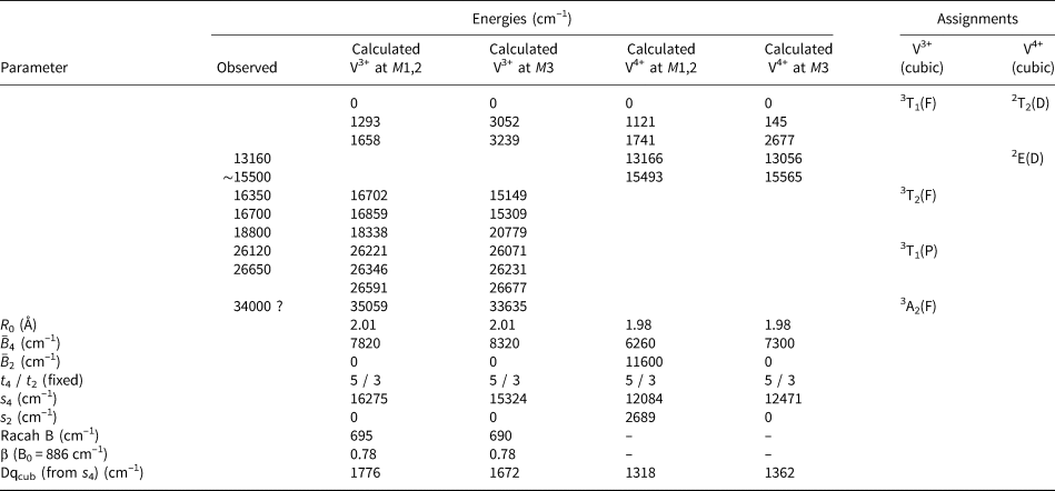
For the present study, we adopt the main features of the interpretation of Schmetzer (Reference Schmetzer1982) and perform semi-empirical CF calculations based on our polarised tanzanite absorption spectra (Fig. 3). The observed transition energies and the energy levels calculated for V3+ as well as V4+ at both available M-sites are summarised in Table 10, together with respective level assignments (for cubic symmetry) and resulting SPM-CF parameters, rotational invariants, Dqcub and (where applicable) Racah B parameters; due to the absence of noticeable spin-forbidden transitions in the tanzanite spectra, Racah C could not be determined.
Observed and calculated transition energies, level assignments for cubic symmetry (with parental terms in parentheses), and obtained crystal field and Racah B parameters for V3+ and V4+ at both M sites in tanzanite.

As Table 10 shows, the SPM-CF calculations for V3+ yield good agreements of calculated and observed transition energies for both M sites, but especially so for V3+ at the M1,2 site, thus hinting to a preference of the latter site. The resulting parameters, Dqcub = 1776 cm–1 and Racah B = 695 cm–1 comply well with expectations for V3+ in oxygen-based structures. However, in calculations for both M sites the second-rank parameter $\bar{B}_2$ was refined to $\bar{B}_2$
 = 0 cm–1, in gross contradiction to expectations. Generally, $\bar{B}_2$
 >> $\bar{B}_2$
 is expected from theory (Newman and Ng, Reference Newman and Ng1989), however in case of 3d N transition ions this sequence is most often not observed, and sometimes $\bar{B}_2$
 < $\bar{B}_4$
 is found (Andrut et al., Reference Andrut, Wildner and Rudowicz2004; Wildner et al., Reference Wildner, Beran and Koller2013). The uncommon values of $\bar{B}_2$
 = 0 might also be biased to some extent by the fixation of the power-law exponents tk to reduce the number of refined parameters, however additional calculations reveal that in the tanzanite spectra the main factor governing $\bar{B}_2$
 is the small splitting of the 3T1(P) level at ~26500 cm–1. In spite of the good agreement of observed and calculated energy levels, a meaningful interpretation of the observed polarisation behaviour in terms of symmetry selection rules is not feasible in view of the low symmetries as well as the irregular distortions of both M sites, especially regarding the M1,2-site (point symmetry 1) preferred by V3+.
Concerning the results for V4+, the limited number of bands attributable to this d 1-configurated cation and the partial overlapping with the lowest-energy V3+-absorptions do not allow assignation to its preferred M site or to extract any further details; the averaged Dqcub value from both sites is 1340 cm–1 (Table 10). Similarly, a reliable assessment of the V3+:V4+ ratio in the tanzanite sample investigated from the absorption spectra is not possible; therefore, the high intensity of the 3T1(P) absorption of V3+ at ~26500 cm–1, compared to the lowest energy band at ~13100 cm–1, exclusively caused by V4+, indicates that V3+ dominates over V4+. Finally, as to the origin of the very weak band found around ~21500 cm–1 (in E ‖ a), we propose that it represents the remains of a respective intense absorption band typically occurring in untreated tanzanite samples, and tentatively assign it to residual traces of Ti3+, not completely oxidised to Ti4+ during heat-treatment.
Bond-Valence Model calculations
The Bond-Valence Model calculation based on the sample structure studied revealed discrepancies between the bond-valence sum and the ideal charge of the site. Bond-valence sum (BVS) calculations at mixed occupancy sites show apparent violations of the Valence Sum Rule (Bosi, Reference Bosi2014). Deviations of the weighted BVS from the weighted sum of formal cation charges results from the difference between the atomic valences and bond-valance parameters (both R 0 and b) of involved cations at the same crystallographic site. The apparent failure of the Valence Sum Rule is expected even for regular and unstrained polyhedra and should be corrected for accurate bond-strain analyses in crystal structures (Bosi, Reference Bosi2014). Such BVS distribution asymmetry between Si1 and Si3, and among Al sites was observed in zoisite (Liebscher et al., Reference Liebscher, Gottschalk and Franz2002) and is analogous to that observed in allanite-group minerals (e.g. Škoda et al., Reference Škoda, Cempírek, Filip, Novák, Veselovský and Čtvrtlík2012). This results from different local arrangements of individual crystallographic sites.
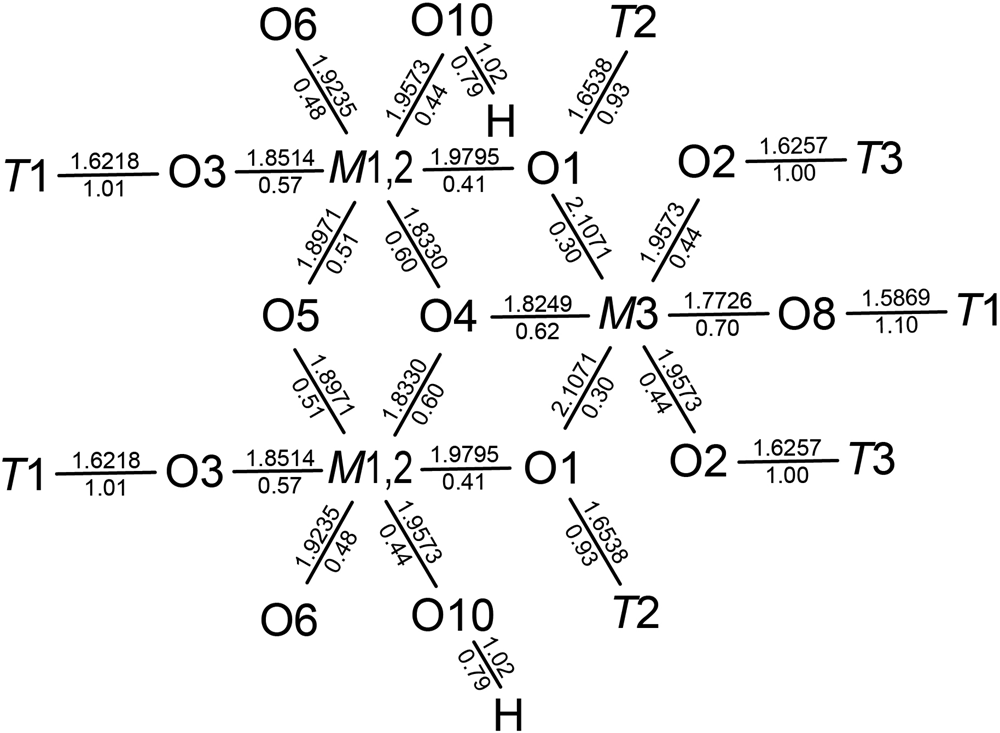
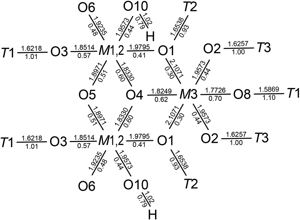
There are two separate octahedral sites in the zoisite structure – the M1,2 site forming the chains parallel to b and M3 attached to the chain of M1,2O6 octahedra. They differ in the size, bond lengths, bond angles (Fig. 5) and also bond-length and bond-angle distortions (Tables 7, 8). Use of angle variance (σoct2) and quadratic elongation (Δoct) as a measure of distortion was first proposed by (Robinson et al., Reference Robinson, Gibbs and Ribbe1971). The distortion indices (DI), originally defined by Baur (Reference Baur1974) for tetrahedra, were adapted for octahedra by (Wildner, Reference Wildner1992).
Bond-topological graphs for M1,2 and M3 sites and their first and second coordination sphere. The number above the line is the bond length, the number below the line is the bond valence in the studied zoisite.
The bond-length and bond-angle distortions of the zoisite investigated can be compared with published structures. To avoid any influence from other cations, pure synthetic zoisite was selected (Liebscher et al., Reference Liebscher, Gottschalk and Franz2002). The second structure for comparison is natural zoisite from Merelani with a similar composition to the sample studied but supposedly without heat treatment (Alvaro et al., Reference Alvaro, Angel and Cámara2012). In the M1,2O6 octahedron, the value of Δoct in the sample studied is between the two reference samples. The value of DI(M–O) for the sample studied is similar to synthetic zoisite (Liebscher et al., Reference Liebscher, Gottschalk and Franz2002) and larger than that of untreated zoisite from Merelani (Alvaro et al., Reference Alvaro, Angel and Cámara2012). However, both values of angular distortion, DI(O–M–O) and σoct2, of the sample studied are the largest in the compared set, whereas the smallest value was found in synthetic zoisite (Liebscher et al., Reference Liebscher, Gottschalk and Franz2002). The situation is different at the M3 site. The sample studied has the smallest bond-length distortion, both in terms of Δoct and DI(M–O). The angular distortion of the sample studied is in-between the reference samples, slightly higher than in untreated V-bearing zoisite and smaller than in synthetic zoisite.
On the basis of the geometry of both sites and bond-length calculations, it is possible to divide octahedral cations according to their site preference. It is almost certain that divalent cations including Fe2+ and Mn2+, if present, prefer the larger and more distorted M3 site. The ideal bond lengths of trivalent cations are closer to the average bond lengths (Fig. 4, Table 9). They differ by 0.084–0.124 Å from <M1,2–O> and 0.017–0.058 Å from <M3–O> bond lengths. Therefore, it can be assumed that they would prefer the larger octahedral site, but this is not definite. Only V4+ has the calculated bond length in the interval between <M1,2–O> and <M3–O>. Therefore, it is possible that it can occupy both sites. There are no bond-length constraints for that.
As indicated by the bond-length distortion values, the M3O6 octahedron is more distorted, the shortest bond is 1.77 Å, the longest pair exceeds 2.10 Å. The difference in metrics of the M1,2O6 octahedron is smaller, 1.83 vs. 1.98 Å (Table 7). Consequently, the differences in bond valences are larger in the M3O6 octahedron (0.70 vs. 0.30 vu) than in M1,2O6 (0.41 vs. 0.60 vu). Moreover, the M1,2 site is slightly overbonded with BVS of 3.03 vu and M3 is slightly underbonded (BVS = 2.78 vu). This increases the strength of the M1,2O6 octahedral chains.
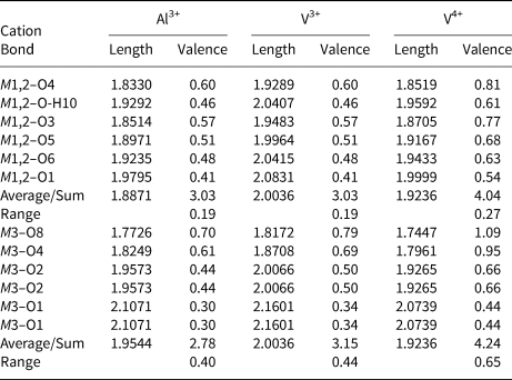
If we assume that in the original tanzanite sample, V was trivalent, it should prefer the M3 site based strictly on the calculated average bond lengths (Table 9). However, as both octahedra in zoisite are significantly distorted, it is appropriate to calculate bond valences according to the actual octahedral metrics. Original proportions of individual bond lengths in Al3+-bearing octahedra can be used for the calculation of theoretical bond lengths and valences in V3+- and V4+-bearing octahedra (Table 11).
Calculation of individual bond valences (in vu) and lengths (in Å) in both zoisite octahedra occupied by Al3+, V3+ and V4+. The Al3+–O bond lengths are empirical from the single-crystal structure refinement, individual V3+–O and V4+–O bond lengths are in the same proportions as Al3+–O.

If a similar geometry of both octahedra after the substitution is assumed, we can calculate bond valences for each bond in both octahedra. Trivalent V at M1,2 retains its BVS compared to Al3+, if the calculated ideal bond valence is used for the BVS. In contrast, V increases the BVS at the M3O6 octahedron. With the same BVS, the longer V3+–O would induce extension of M1,2–O4 and M1,2–O6 bonds shared by two neighbouring edge-sharing M1,2O6 octahedra and that would require bond-angle distortion of V3+-bearing or neighbouring octahedra. Considering bond lengths, the M3 site is a more natural choice for V3+, because the larger volume and smaller number of shared edges with other octahedra (M1,2 – three shared edges, M3 – two shared edges) allows greater variability in accommodation of larger cations.
However, if the Bond-Valence Model is considered, a different view can be established. Crystal structures prefer the smallest possible distortions resulting in the smallest energetical requirements for the structural stability. Therefore, the distribution of bond valences for each bond of each ion should be as even as possible. The substitution of ions with the same charge should also not produce too large a difference in the BVS for the specific site. Consequently, if we consider the bond-valence distribution for each bond, V3+ at M1,2 has bond valences in the range 0.41–0.60 vu (difference of 0.19 vu), which is very similar to Al at this site. However, V3+ at the M3 site has a significantly larger variation of bond valences (0.34–0.78 vu, i.e. 0.44 vu difference), which is larger than Al (0.40 vu difference) at the M3 site. Moreover, the BVS of V3+ at M3 is significantly larger (3.15 vu) than M 3Al3+ (2.78 vu). This may indicate the bond-valence distribution requirements result in the preference of the M1,2 site for V3+.
A similar consideration can be done with V4+. Logically, the BVS should be ~4, which is the nominal charge of V4+. In both octahedra, the BVS is >4 vu, if the calculated ideal bond valence is used as the BVS. However, the higher BVS value is observed at the M3 site of 4.24 vu, which is quite significantly overbonded. Moreover, the M3–O8 bond with V4+ in M3 has more than 1 vu (1.08 vu). As already mentioned, the M3–O8 bond is constrained by the Si–O8 bond in the neighbouring tetrahedron. The Si–O8 bond in the zoisite studied is 1.5870 Å with a bond valence of 1.08 vu. If its valence was 0.92 vu, which is the subtracted difference of 2 – 1.08 vu, the resulting bond length would have to be >1.65 Å. To obtain such a structural change in the tetrahedron seems unlikely. Moreover, there was no significant shortening of the M3–O8 bond observed in the sample studied, it is very similar to untreated zoisite from Merelani. Consequently, V4+ at M3 site would increase the M3O6 distortion and result in lower structural stability. In contrast, the presence of V4+ at the M1,2 site does not require significant changes in the octahedral geometry except slight isotropic expansion and seems to be more stable in terms of individual bond valences. This is in accordance with the observed structural data. The studied zoisite has the lowest M3O6 bond-length distortion among compared zoisites and its M1,2O6 bond-length distortion is within the range of the two reference structures (Tables 7 and 8).
The preference of V4+ at the M1,2 site is supported by the bond-valence distribution; the range of the individual valences is 0.34–0.78 vu (difference of 0.44 vu), which is significantly smaller than 0.44–1.09 vu, i.e. 0.65 vu difference at the M3 site. This is similar to V3+ and also suggests that V4+ at the M1,2 site would be more stable from the electrostatic perspective.
The possible presence of V at M1,2 also makes sense, if charge-balancing of V4+ for Al3+ substitution is considered. The cation at M1,2 is coordinated by the O10 anion, to which H10 is usually bonded. Therefore, excess in charge due to a four-valent cation can be easily charge-balanced by the deprotonation of O10. This would also influence the bond lengths, mostly M1,2–O10 which shortens due to the increase in bond valence after the release of H. In fact, the sample studied has the shortest M1,2–O10 bond. This deprotonation should also increase the angular distortion of this octahedron, which was actually observed in the sample studied.
Conclusion
The position of V in the structure of studied heat-treated zoisite var. tanzanite was not possible to determine from the structural refinement. Therefore, advanced theoretical interpretations of structural and optical absorption spectroscopic data were applied. Crystal field Superposition Model calculations from the optical spectra indicated that V3+ prefers occupying the M1,2 site, however determination of the V4+ preference from the present data was not possible. Bond-Valence Model calculations revealed no unambiguous preference for V3+, although the simple bond-length calculation suggests the M3-site preference, however it is quite straightforward that the M1,2 site is more suitable for V4+. However, if the possible octahedral distortion is considered, the M1,2O6 octahedron is subject to a smaller distortion if occupied by V3+ than the M3O6 octahedron. Consequently, considering results of the crystal field Superposition Model and Bond-Valence Model calculations, we propose that both V3+ and V4+ prefer the M1,2 site.
Acknowledgements
The authors thank Andreas Wagner (Vienna) for the careful preparation of the tanzanite crystal slabs for the polarised optical absorption measurements. This work was supported by the Slovak Research and Development Agency under contract No. APVV-18-0065, and VEGA Agency VEGA-1/0137/20 grant. Finally, we thank reviewers and editors for their constructive suggestions for improving the quality of our work.
Supplementary material
The supplementary material for this article can be found at https://doi.org/10.1180/mgm.2023.48.
Competing interests
The authors declare none.