Direct communication between the right pulmonary artery and the left atrium is a rare congenital cardiac malformation, initially reported by Friedlich et alReference Friedlich, Bing and Blount 1 . Since then, >100 patients have been diagnosed with right pulmonary artery–left atrial fistula. Such patients usually present with central cyanosis with clubbing of fingers and exertional dyspnoea and uncommonly with complications of systemic embolismReference Kavarana, Devaney, Torres and Cotts 2 or cerebral abscess.Reference Kawano, Hirano, Ikeno, Fuwa and Uchino 3 Surgical repair or transcatheter closure with a variety of devices has been performed with good results.Reference Margaryan, Arcieri, Cantinotti and Murzi 4 – Reference Ding, Xu, Liu, Yan and Qin 7 Spontaneous closure of a right pulmonary artery–left atrial fistula has also been reported by Bachani et alReference Bachani, Pinto and Dalvi 8 . In this study, we report the successful transcatheter closure and surgical repair of right pulmonary artery–left atrial fistula in three cases.
Case 1: transcatheter closure
A 5-year-old girl was referred for evaluation of cyanosis and clubbing of fingers. Chest radiography showed abnormal vascular shadow adjacent to the right heart (Fig 1a). The two-dimensional transthoracic echocardiogram (TTE) showed only a small patent duct arteriosus (1–2 mm). Selective pulmonary artery angiogram (Fig 1b) was performed to profile the right pulmonary artery–left atrium communication, and the narrowest diameter measured was 10 mm. Pulmonary artery pressure was 21/10 (14) mmHg. A 16/18 mm Duct occluder (Lifetech Scientific, Shenzhen, China) was selected to close the right pulmonary artery–left atrial fistula. Again selective right pulmonary artery angiogram showed a well-positioned device and no residual shunt (Fig 1c). Pulmonary artery pressure increased to 29/14 (19) mmHg. There were no procedure-related complications. Multidetector (MD) CT angiography showed the position of the device and no other accompanied malformations (Fig 1d and e). The child was observed for 48 hours before discharge, and was asymptomatic with significant improvement of cyanosis at her 20-month follow-up.
( a ) Chest X-ray showing an abnormal right hilar shadow, no cardiomegaly, and normal pulmonary blood flow. ( b ) Pulmonary artery angiogram showing a direct communication between the right pulmonary artery and the left atrium. ( c ) Postoperative pulmonary artery angiogram showing complete closure of the fistula with no residual shunt. ( d and e ) Contrast-enhanced CT presents the closure outcome with no device displacement and thrombus formation.

Cases 2 and 3: surgical repair
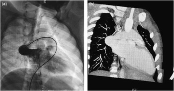
A 9-year-old boy presented with cyanosis and clubbing of fingers. He underwent right cardiac catheterisation and selective right pulmonary arteriography, which showed a large right pulmonary artery–left atrial fistula (20 mm) (Fig 2a), also confirmed by Multidetector CT (Fig 2b). His pulmonary arterial pressure was 22/3 (10) mmHg. Considering the large size and as there was no obvious anatomical narrow portion, the operator gave up on transcatheter closure of the fistula, and the patient was transferred to the department of cardiac surgery and surgical ligation of the lesion was performed. After 26 months of follow-up, the patient had normal level of activity with no cyanosis and dyspnoea on exertion. The other patient was an 11-year-old girl who was referred for cyanosis and dyspnoea. Chest radiography showed no evident abnormalities in the right lower lung region. Transthoracic echocardiogram showed communication between the right pulmonary artery and the left atrium with a slightly enlarged left ventricle (39 mm). Contrast echocardiography revealed early dense opacification of the left atrium and the left ventricle with microbubbles within two cardiac cycles. CT and three-dimensional volume-rendered CT imaging confirmed the large fistulous communication (16 mm) between the right pulmonary artery and the left atrium. Finally, the patient received surgical repair and with no symptoms and uncomfortable complaints at the 38-month follow-up.
( a ) Pulmonary arteriogram shows a large right pulmonary artery–left atrial fistula. ( b ) CT presents a large shunt between the right pulmonary artery and the left atrium (oblique view).

Discussion
Right pulmonary artery–left atrial fistula is a rare cardiovascular anomaly with right-to-left shunt. No definite embryological explanation has been described for right pulmonary artery–left atrial communication. Some have hypothesised that this anomaly may result from a communication between the right pulmonary artery and a primitive pulmonary vein and later the pulmonary vein would become incorporated into the wall of the left atrium, resulting into a fistulous connection between the right pulmonary artery and the left atrium.Reference Vadlamudi, Verma, Christopher, Penumatsa and Koneti 6 It often presents with central cyanosis, clubbing of fingers, exertional dyspnoea, or even systemic embolism, infective endocarditis, and cerebral abscess. Therefore, when a patient presents with these symptoms, pulmonary arteriovenous fistula should be considered. In our case, all three patients complained of cyanosis since early childhood, and all three patients had a delay in diagnosis.
Generally, chest radiography and transthoracic echocardiogram cannot confirm this diagnosis. Contrast echocardiography can detect the extent of the right-to-left shunt. CT can offer a better picture of the condition to confirm the diagnosis. Cardiac catheterisation with selective angiography is considered to be the most effective procedure for confirming this anomaly and can accurately describe the anatomical details. In our case, the abnormality was not detected on transthoracic echocardiogram and was diagnosed with CT or selective pulmonary artery angiography. In cases 1 and 2, angiography was performed to confirm the direct communication between the proximal right lower pulmonary artery and the left atrium.
Surgical closure is a traditional treatment for fistulas, as in cases 2 and 3, especially large ones; however, transcatheter closure of the fistula can also be a good option in most cases. Some authors have reported using an Amplatzer duct occluder,Reference Vadlamudi, Verma, Christopher, Penumatsa and Koneti 6 Amplatzer septal occluder,Reference Ergul, Nisli and Aydogan 5 muscular ventricular septal occluder,Reference Ding, Xu, Liu, Yan and Qin 7 or coilsReference Slack, Jedeikin and Jones 9 to treat fistulas. Rarity of the disease, a certain rate of missed diagnosis, morphology of the fistula, and fear of embolisation directly into the left atrium are some of the possible reasons for the rarity of interventional treatment. Generally, device choice depends on the size of the vessel, the anatomy of the narrowing, and also the experience of operators. The retrograde deployment from the left atrial side is a method using a duct occluder device in the presence of an atrial septal defect or patent foramen ovaleReference Ece, Uner, Cuce and Balli 10 or by trans-septal puncture. In the absence of an atrial septal defect or patent foramen ovale, however, double-disc devices may be used such as ventricular septal occluder, Amplatzer septal occluder,Reference Ece, Uner, Cuce and Balli 10 or vascular plug device. Coils are only suitable for right pulmonary artery–left atrial fistulas with a narrow neck, and not for large ones. Duct occluders with a larger disc on the distal end are not ideally suited for closing fistulas from the pulmonary arterial approach because of the possibility of displacement and systemic embolism. In case 1, we chose a Duct occluder and implanted the device from the pulmonary arterial site and obtained good results; however, this is not recommended, because it cannot be released as distally as possible to prevent distal right pulmonary artery obstruction, and needs larger devices. In conclusion, transcatheter closure of right pulmonary artery–left atrial fistula appears to be a good alternative choice.
Acknowledgement
None.
Financial Support
This study was supported by grants from the Peking Union Medical College Graduate Innovation Fund (2015-1002-01-14).
Conflicts of Interest
None.
Ethical Standards
This study was conducted in accordance with the Declaration of Helsinki (World Medical Assembly) and its amendments, and was approved by the Ethics Committee of Fuwai Hospital.