Case report
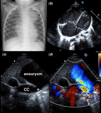
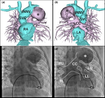
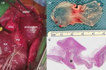
A 1-year-old girl was referred to our institution complained of cyanosis. Upon admission, her oxygen saturation was 85%. Chest X-ray showed an abnormal mass shadow in the left upper lung field (Fig 1A). An echocardiogram revealed significant enlargement of the right heart with a large atrial septal defect (Fig 1B). All pulmonary veins were determined to be connected to a common chamber, which drained into the left innominate vein through the extremely dilated vertical vein (Fig 1C, D). Furthermore, enhanced CT revealed total anomalous pulmonary venous connection with aneurysmal formation of the vertical vein (size 45 × 35 mm, Fig 2C, D). The area between the common chamber and aneurysm was narrowed with a diameter of 8 mm. Cardiac catheterisation confirmed a slight elevation of pulmonary artery pressure of 28/12(21) mmHg (Fig 2A, B). The pressure gradient through the narrowest site was 5 mmHg. Calculated pulmonary-to-systemic blood flow ratio was 2.7, and pulmonary vascular resistance was 1.4 wood units/m2. Surgical intervention was performed by posterior approach including resection of the aneurysm (Fig 3A, B). Her post-operative course was uneventful. Pathological findings revealed normal layers of thinned venous walls with partially thickened endothelium (Fig 3C).
A: Abnormal shadow in the left upper lung field depicted in chest X-ray. B: Four chamber view TTE indicating dilatation of the right heart and a large ASD. C/D: Suprasternal TTE view showing the connection between the common chamber (CC) and aneurysm along with the narrowing site (*), and color Doppler. ASD: atrial septal defect, TTE: transthoracic echocardiogram.

A/B: RV angiography illustrating four PVs drain into the common chamber which further drains into the aneurysm (*). C/D: CT angiography tridimensional reconstruction of PVs, aneurysm (*), innominate vein, and atrium on anterior view(C) and posterior view (D). PV: pulmonary vein, CC: common chamber, RU: right upper pulmonary vein, RL: right lower pulmonary vein, LU: left upper pulmonary vein, LL: left lower pulmonary vein, RA: right atrium, LA: left atrium, INNV: innominate vein, SVC: superior vena cava.

A: Intraoperative appearance with a median sternotomy approach demonstrating the aneurysm. B: Resected specimen of the venous aneurysm which has thinned vascular wall. C: Pathological mapping stained by Elastica van Gieson. Normal layers of venous walls with partially thickened endothelium (*). AAo: ascending aorta, PA: pulmonary artery, RA: right atrium, INNV: innominate vein.

Aneurysm of the vertical vein is extremely rare, with few cases reported in children younger than 10 years of age. This is the youngest patient reported to have a vertical vein aneurysm, and her age at diagnosis suggests that a vertical vein aneurysm can develop antenatally or within the first year of birth. Phadke et al. reported a vertical vein aneurysm with a narrowing site between the left bronchus and the left pulmonary artery and speculated that the possible mechanism for the aneurysm was post-stenotic dilatation. Reference Phadke, Mate and Kerkar1 In this case, the left bronchus was distant from the stenotic area and was not producing an extrinsic obstruction to the vertical vein. The degree of endothelial thickness may support the presence of elevated shear stress in the aneurysm.
Supplementary material
To view supplementary material for this article, please visit https://doi.org/10.1017/S1047951123000719
Financial support
This research received no specific grant from any funding agency, commercial, or not-for-profit sectors.
Conflict of interest
None.
Ethical standards
Informed consent was obtained from the patient’s guardian for publication of this case study.