The lower jaws of gnathostomes are complex, variable, and character-rich structures that often fossilise in remarkably good condition, thereby providing an important source of data for phylogenetic, biomechanical, palaeoecological, and macroevolutionary analyses (Ahlberg & Clack Reference Ahlberg and Clack1998; Ruta & Bolt Reference Ruta and Bolt2008; Anderson & Friedman Reference Anderson and Friedman2012; Anderson et al. Reference Anderson, Friedman and Ruta2013; Neenan et al. Reference Neenan, Ruta, Clack and Rayfield2014; Hill et al. Reference Hill, Puttick, Stubbs, Rayfield and Donoghue2018; Sakamoto et al. Reference Sakamoto, Ruta and Venditti2019; Deakin et al. Reference Deakin, Anderson, den Boer, Smith, Hill, Rücklin, Donoghue and Rayfield2022; So & Mann Reference So and Mann2024). In recent years, studies of lower jaws have been invigorated by newly collected, newly recognised, and newly prepared specimens, particularly among stem-group gnathostomes (Anderson Reference Anderson2010; Anderson et al. Reference Anderson, Friedman, Brazeau and Rayfield2011; Anderson & Westneat Reference Anderson and Westneat2012; Zhu et al. Reference Zhu, Yu, Ahlberg, Choo, Lu, Qiao, Qu, Zhao, Jia, Blom and Zhu2013; Choo et al. Reference Choo, Zhu, Zhao, Jia and Zhu2014; Dupret et al. Reference Dupret, Sanchez, Goujet, Tafforeau and Ahlberg2014), tetrapodomorph fish (Zhu & Yu Reference Zhu, Yu, Arratia, Wilson and Cloutier2004; Porro et al. Reference Porro, Rayfield and Clack2015a; Lebedev & Clément Reference Lebedev, Clément, Ruta, Ahlberg and Smithson2019), and Devonian, Carboniferous, and Mesozoic early tetrapods (Ahlberg & Clack Reference Ahlberg and Clack1998, Reference Ahlberg and Clack2020; Daeschler Reference Daeschler2000; Bolt & Lombard Reference Bolt and Lombard2001, Reference Bolt and Lombard2006; Clack & Ahlberg Reference Clack, Ahlberg, Arratia, Wilson and Cloutier2004; Ahlberg et al. Reference Ahlberg, Luksevics and Lebedev1994, Reference Ahlberg, Friedman and Blom2005; Lombard & Bolt Reference Lombard, Bolt, Carrano, Gaudin, Blob and Wible2006; Ruta & Bolt Reference Ruta and Bolt2008; Clack Reference Clack2012; Clack et al. Reference Clack, Ahlberg, Blom and Finney2012, Reference Clack, Porro and Bennett2018; Anderson et al. Reference Anderson, Friedman and Ruta2013; Clément & Lebedev Reference Clément and Lebedev2014; Sookias et al. Reference Sookias, Böhmer and Clack2014; Porro et al. Reference Porro, Rayfield and Clack2015b; Chen et al. Reference Chen, Alavi, Brazeau, Blom, Millward and Ahlberg2018; Rawson et al. Reference Rawson, Porro, Martin-Silverstone and Rayfield2021; So & Mann Reference So and Mann2024). However, our knowledge of lower jaws in late Palaeozoic early tetrapods, especially from the Early Permian, has not kept pace. This statement may appear counterintuitive, given that many Permian taxa have been known for quite some time and that several of them have been monographed in considerable detail (e.g., Eryops megacephalus; Sawin Reference Sawin1941). In spite of this, our observations suggest that the lower jaws of some well described and common taxa differ from their published accounts, and that certain features have been consistently poorly documented or have received cursory treatment (e.g., configuration of sutures in the symphyseal region; structural details of the adductor fossa; location and proportions of foramina). To fill these gaps, we initiated comparative investigations into the structure and variation of the lower jaw (Ruta & Bolt Reference Ruta and Bolt2008), particularly in temnospondyls, the most diverse and longest ranging group of early tetrapods (Schoch & Milner Reference Schoch and Milner2000, Reference Schoch and Milner2014).
The present contribution focusses on a common taxon that has been known since the second half of the 19th Century, namely the aquatic dvinosaur temnospondyl Trimerorhachis. The type species of this genus, T. insignis Cope Reference Cope1878, is one of the most abundant Early Permian vertebrates (Sander Reference Sander1989) and is represented by extensive collections in several museums around the world. Until recently, the only comprehensive description of Trimerorhachis was that of Case (Reference Case1935), which is still widely cited as the primary source for T. insignis and has provided the basis for most of our current understanding of this species. In recent decades, various papers have dealt with specific aspects of the anatomy of Trimerorhachis, with focus on its stapes (Lombard & Bolt Reference Lombard, Bolt, Fritzsch, Ryan, Wilczynski, Hetherington and Walkowiak1988), braincase (Schoch Reference Schoch1999), postcranial skeleton (Pawley Reference Pawley2007), and skull (Milner & Schoch Reference Milner and Schoch2013). Milner & Schoch (Reference Milner and Schoch2013) re-examined the taxonomy and stratigraphy of this genus and summarised the relevant literature to date. Their work is particularly useful in the present context, because it informs attributions of the material described and figured in this paper.
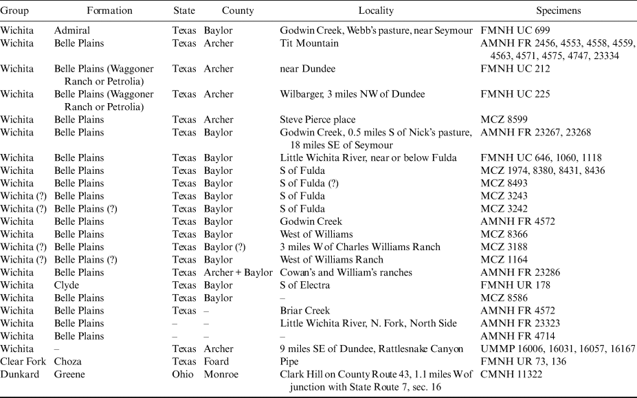
From October 2004 to March 2006, we carried out observations on numerous lower jaw rami of Trimerorhachis from various museum collections in North America (Table 1). Mechanical preparation of this material revealed new information that helped us amend and complement former descriptions to a substantial degree. The main conclusion from our initial survey was that published works on the lower jaw of Trimerorhachis are not entirely adequate and sometimes provide inexplicably contradictory accounts of its morphology. Not only is the lower jaw variable, including in specimens of comparable size and/or from the same locality and horizon, but it also exhibits traits that, so far, have been overlooked or addressed superficially. Milner & Schoch (Reference Milner and Schoch2013) briefly redescribed the lower jaw of Trimerorhachis (fully acknowledging our work in progress), clarified numerous aspects of the shape and proportions of its constituent elements, and presented a reconstruction. Major differences between their reconstruction and ours concern details of individual bones, the shape and distribution of foramina (especially on the dorsal, medial, and symphyseal surfaces of the ramus), the proportions and position of major openings (e.g., Meckelian fenestrae), the course of sutures, and the pattern of dentition on the dentary and coronoids.
Stratigraphic attribution and geographic provenance of specimens examined in this study.

Sequence of anatomical illustrations
The aims of this paper are to describe in detail the lower jaw of Trimerorhachis, including its individual and, where applicable, ontogenetic variation, and produce a new, improved reconstruction. Figures 1–4 illustrate the general proportions and variation of the jaw ramus. Figure 5 presents a new jaw reconstruction in lateral, dorsal, and medial aspects. Figures 6–9 show the anterior one-third to anterior half of the ramus, while Figures 10–12 show details of its symphyseal region and precoronoid fossa. For comparisons with the symphyseal region of tetrapodomorphs, we illustrate the anterior extremity of a right jaw ramus of the elpistostegalian Panderichthys rhombolepis in the Museum für Naturkunde, Berlin (specimen MB. f. 17548; Fig. 13), a line drawing of which featured in Ahlberg and Clack (Reference Ahlberg and Clack1998). Figures 14–17 show the middle and posterior one-thirds of the ramus and the three coronoids. The sutural patterns at the posterior extremity of the ramus are reproduced in lateral (Figs 18, 19) and medial views (Figs 20–22). Figures 23–26 display the adductor fossa and adductor process. Two rami of Trimerorhachis rogersi figured by Olson (Reference Olson1955, fig. 104) appear in Figures 27, 28. Figure 29 shows the adductor fossa of a left jaw ramus of the dvinosaur Acroplous vorax in the University of Kansas Museum of Natural History (KUVP 28352; figured by Coldiron Reference Coldiron1978, fig. 1), in which mechanical preparation has revealed an adductor process.
Institutional abbreviations
AMNH, American Museum of Natural History, New York (FR initials preceding AMNH catalogue numbers refer to the fossil fish and reptile collections in that institution); CMNH, Cleveland Museum of Natural History, Cleveland, Ohio; FMNH, Field Museum of Natural History, Chicago, Illinois (UC and UR initials preceding FMNH catalogue numbers refer to specimens formerly housed at the Walker Museum, University of Chicago, and subsequently transferred to the Field Museum, or collected by University of Chicago expeditions); KUVP, University of Kansas Museum of Natural History, Vertebrate Paleontology, Lawrence, Kansas; MB, Museum für Naturkunde, Berlin; MCZ, Museum of Comparative Zoology, Harvard University, Cambridge, Massachusetts; UMMP, University of Michigan Museum of Paleontology, Ann Arbor, Michigan.
1. Brief timeline of research
1.1. Historical background
Milner & Schoch's (Reference Milner and Schoch2013) contribution makes it unnecessary to provide a detailed account of the taxonomy, stratigraphy, and history of research on Trimerorhachis. However, some considerations that are relevant to the present paper are warranted. Cope (Reference Cope1878) originally assigned the binomen Trimerorhachis insignis to several specimens from the Tit Mountain locality, Archer County, Texas (Olson Reference Olson1955). In terms of current stratigraphic nomenclature (Hentz Reference Hentz1988), this locality belongs to the Petrolia Formation (Hook Reference Hook and Hook1989). According to Milner & Schoch (Reference Milner and Schoch2013), however, the holotype of T. insignis can be at best placed in either the Nocona or the Petrolia Formation, but a more precise assignment is not possible. Olson (Reference Olson1955, p. 229) commented on the holotype in the following terms: ‘Rather poor skull, lower jaws with front part missing, 24 vertebrae, some with ribs, humerus, and scapula. Included in the specimen listed as the type, …, were bones belonging to other genera [and] fragments of several individuals of Trimerorhachis. There is no certainty that all parts listed above as the type belong to the same individual.’ From Olson's description, it appears that the type material of T. insignis is of scarce relevance for the purpose of identifying diagnostic features of the lower jaw, an impression that was confirmed by our direct examination of this material.
Six additional species were described from different localities in north Texas by Cope (Reference Cope1883, Reference Cope1896a, Reference Cope1896b; three taxa), Case (Reference Case1902, Reference Case1910; two taxa), and Broom (Reference Broom1913; one taxon). A series of review papers (summarised in Milner & Schoch Reference Milner and Schoch2013) helped clarify in part the taxonomy of the genus. Olson (Reference Olson1955) addressed the status of the then-recognised species and concluded that lower jaw characters are useful in distinguishing some of them. According to Olson (Reference Olson1955), two of the historically important diagnostic characters are the occurrence and proportions of lateral and medial ‘tuberosities’ on the ‘angle of the jaw’. Using current terminology, these ‘tuberosities’ would not be present on the ‘angle’ of the lower jaw, but on its postglenoid area, such as is observed in dorsal aspect. The medial tuberosity corresponds to the retroarticular process, which projects backward from the posterior surface of the articular. The lateral tuberosity, located at the posterodorsal extremity of the surangular, is hereafter termed the posterior process of the surangular and may represent the homologue of the arcadian process (sensu Jupp & Warren Reference Jupp and Warren1986; see also Ruta & Bolt Reference Ruta and Bolt2008 and Milner & Schoch Reference Milner and Schoch2013), based on topological similarities. Olson (Reference Olson1955, fig. 95) showed that both processes are extremely variable in shape and proportions and concluded from his statistical work that ‘… [this] variability …, while considerable, is continuous, and that, in spite of strong departures from the norm in the frequency distribution of ratios, there is no basis for recognition of the distinct groups’ (Olson Reference Olson1955, p. 248). He regarded three species – T. bilobatis, T. alleni, and T. medius – as junior synonyms of T. insignis, and a fourth species, T. conangulus, as a junior synonym of T. mesops. However, Sequeira (Reference Sequeira1998) demonstrated that the skull of the T. conangulus holotype in fact belongs to the dvinosaur Isodectes obtusus. Olson's (Reference Olson1955) revision of T. mesops was based on a quantitative analysis of skull measurement ratios. He concluded his work with the description of a new species, T. rogersi, the brief diagnosis of which relied on cranial proportions and lower jaw morphology. Concerning the latter, Olson (Reference Olson1955, p. 264) stated: ‘Lower jaw with single tuberosity on [its] angle … [in] direct continuation [with] lateral surface of jaw, not set off by well-defined notch.’ We return to Olson's (Reference Olson1955) remarks at the end of our Discussion.
Berman & Reisz (Reference Berman and Reisz1980) described Trimerorhachis sandovalensis from the Early Permian of New Mexico, based on a skull with associated vertebrae and a scapula. Although the type specimen preserves both lower jaws, these are tightly pressed against the skull, making detailed observations impossible. In their study, they also re-examined the specimens upon which Olson (Reference Olson1955) had based his new species, T. rogersi, pointing out significant errors in his interpretation of sutural patterns. According to Berman & Reisz (Reference Berman and Reisz1980, p. 473), ‘there [are] no marked differences between [the midcontinental Trimerorhachis species], other than the proportional differences noted by Olson.’ More recently, Milner & Schoch (Reference Milner and Schoch2013) showed that Olson's (Reference Olson1955) craniometric analysis was flawed because it included some diagenetically deformed specimens, a non-Trimerorhachis specimen (the T. conangulus holotype reassigned to Isodectes obtusus by Sequeira Reference Sequeira1998), and numerous instances of accidentally reversed linear measurement ratios. After correcting those measurements, Milner & Schoch (Reference Milner and Schoch2013) confirmed the validity of four of the previously recognised species and proposed a new species, T. greggi. Thus, the current taxonomic situation is that Trimerorhachis includes five valid species, namely T. sandovalensis, T. insignis, T. greggi, T. mesops, and T. rogersi.
1.2 Geographic provenance
Trimerorhachis has been found in Early Permian sediments of the continental United States, with hundreds of specimens collected in Texas, Oklahoma, and New Mexico (Case Reference Case1935; Olson Reference Olson1955; Berman & Reisz Reference Berman and Reisz1980; Sumida et al. Reference Sumida, Berman, Eberth and Henrici2004; Milner & Schoch Reference Milner and Schoch2013). A few sites in other western states (e.g., Kansas, Nebraska, and Utah) have produced trimerorhachid specimens, though not necessarily ones belonging to Trimerorhachis itself (Olson Reference Olson1952, Reference Olson1975; Sander Reference Sander1989; Cunningham & Dickson Reference Cunningham and Dickson III1996; Sumida et al. Reference Sumida, Berman, Eberth and Henrici2004; Huttenlocker et al. Reference Huttenlocker, Pardo and Small2005). East of the Mississippi River, the genus is known exclusively from the Dunkard Group of Ohio (Upper Pennsylvanian–Early Permian; Berman Reference Berman1978; Hansen Reference Hansen, Feldmann and Hackathorn1996), but much of this material is fragmentary and has not been described in detail. Most catalogued specimens assigned to Trimerorhachis come from very few counties in north Texas. A preliminary literature survey suggests that Oklahoma is second in the number of catalogued specimens, although we have not attempted an exact count. For this reason, most of the literature on Trimerorhachis, including Cope (Reference Cope1878) and Case (Reference Case1935), is based on Texas material, which is often from the same localities that supplied the specimens described by Case (Reference Case1935). Case (Reference Case1935) focussed almost entirely on fossils that were either associated on a single block or embedded in adjacent matrix. Virtually no other vertebrates were present in this material. Therefore, he was presumably correct in regarding those specimens as a death assemblage of aquatic animals that were trapped in a drying pool, and there is every reason to assume that they belong to a single species.
During our study, we uncovered remarkable variation in the lower jaw (Figs 1–4, 6–12, 14–28), including among specimens from identical localities and/or stratigraphic levels. At present, it is unclear whether this variation reflects taxonomic differences, because specific attributions of isolated lower jaws, even if exceptionally complete and well-preserved, is challenging (Milner & Schoch Reference Milner and Schoch2013). However, several features common to most (though perhaps not all) specimens examined in this work reasonably support their assignment to a single taxon, T. insignis, based on the stratigraphic arguments expounded below and the descriptive sections that follow.
Jaw rami of Trimerorhachis insignis, showing the general morphology of large and small specimens: (a, b) FMNH UC 699 in slightly dorsolateral and medial views; (c–e) UMMP 16031 in lateral, slightly dorsomedial, and medial views; (f) AMNH FR 4571 in medial view. Abbreviations: acop = anterior condyloid (precondyloid) process of glenoid; adf = adductor fossa; aMecfen = anterior Meckelian fenestra; ANG = angular; as = accessory sulcus; COR1 = coronoid 1; COR2 = coronoid 2; COR3 = coronoid 3; COR3den = denticles on coronoid 3; COR3pdp = posterodorsal process of coronoid 3; ctf = chorda tympani foramen; DENppr = postdental process of dentary; DENsym = dentary symphysis; DENtooth = marginal dentary tooth; gle = glenoid surface of articular; icf = intercoronoid fossa; MEC = Meckelian ossification; ms = mandibular sulcus; os = oral sulcus; pcf = precoronoid fossa; pcop = posterior condyloid (postcondyloid) process of glenoid; PEA = prearticular; pMecfen = posterior Meckelian fenestra; PTS = postsplenial; rap = retroarticular process of articular; rrec = reniform recess (new term herewith) of precoronoid fossa; SPL = splenial; SRA = surangular. All scale bars = 5 mm.

Jaw rami of Trimerorhachis insignis, showing the general morphology of large specimens: (a–c) FMNH UR 178 in slightly ventrolateral, slightly dorsomedial, and medial views; (d–f) FMNH UC 646 in lateral, dorsal, and medial views. Abbreviations: acop = anterior condyloid (precondyloid) process of glenoid; adf = adductor fossa; adp = adductor process; aMecfen = anterior Meckelian fenestra; ANG = angular; COR1 = coronoid 1; COR2 = coronoid 2; COR3 = coronoid 3; COR3pdp = posterodorsal process of coronoid 3; ctf = chorda tympani foramen; DEN = dentary; DENfang = dentary fang; DENppr = postdental process of dentary; DENsym = dentary symphysis; gle = glenoid surface of articular; icf = intercoronoid fossa; ms = mandibular sulcus; pcf = precoronoid fossa; pcop = posterior condyloid (postcondyloid) process of glenoid; PEA = prearticular; pMecfen = posterior Meckelian fenestra; PTS = postsplenial; rap = retroarticular process of articular; SPL = splenial; SRA = surangular. All scale bars = 5 mm.

Jaw rami of Trimerorhachis insignis, showing the general morphology of small and mid-sized specimens: (a–c) UMMP 16167 in lateral, medial, and dorsal views; (d–g) CMNH 11322 in lateral, medial, ventral, and dorsal views. Abbreviations: acop = anterior condyloid (precondyloid) process of glenoid; aMecfen = anterior Meckelian fenestra; ANG = angular; COR1 = coronoid 1; COR2 = coronoid 2; COR3 = coronoid 3; COR3pdp = posterodorsal process of coronoid 3; ctf = chorda tympani foramen; DEN = dentary; DENfang = dentary fang; DENshelf = dentary shelf; DENsym = dentary symphysis; gle = glenoid surface of articular; pcop = posterior condyloid (postcondyloid) process of glenoid; PEA = prearticular; pMecfen = posterior Meckelian fenestra; PTS = postsplenial; rap = retroarticular process of articular; rrec = reniform recess (new term herewith) of precoronoid fossa; SPL = splenial; SRA = surangular; SRApp = posterior process of surangular. All scale bars = 5 mm.

Jaw ramus of large specimen of Trimerorhachis insignis (AMNH FR 4571) in ventral view, showing the arrangement of sutures, the course of sensory canals, and the proportions of processes at the posterior extremity of the ramus: (a) posterior half of ramus; (b) anterior half of ramus. Abbreviations: as = accessory sulcus; ctf = chorda tympani foramen; DENsym = dentary symphysis; for = foramen; ms = mandibular sulcus; os = oral sulcus; rap = retroarticular process of articular; SRApp = posterior process of surangular; sur = suture. All scale bars = 5 mm.

Reconstruction of right jaw ramus of Trimerorhachis insignis. (a) lateral view; (b) dorsal view; (c) medial view. Scale bar = 5 mm.

Jaw rami of Trimerorhachis insignis, showing dermal sculpture on dentary, splenial, and postsplenial, the arrangement of sutures, and the course of the mandibular sulcus. (a, b) MCZ 8586 in medial and slightly lateroventral views; (c, d) UC 1118 (two pieces) in lateroventral views. Abbreviations: aMecfen = anterior Meckelian fenestra; DEN = dentary; for = foramen; PTS = postsplenial; SPL = splenial; sur = suture. All scale bars = 5 mm.

Jaw rami of Trimerorhachis insignis, showing variation in the precoronoid fossa, spatial arrangement of sutures between dentary, splenial, and anterior coronoid inside and around the fossa, dentary fangs/replacement pits, and anterior dentary teeth. (a) UC 225 in slightly dorsomedial view; (b) UR 178 in dorsal view; note isolated small tooth on the anterior coronoid; (c) UC 1118 in slightly dorsomedial view. Abbreviations: COR1 = coronoid 1; COR1den = denticle on coronoid 1; COR2 = coronoid 2; DENfang = dentary fang; DENshelf = dentary shelf; DENtooth = marginal dentary tooth; for = foramen; prec = principal recess (new term herewith) of precoronoid fossa; rrec = reniform recess (new term herewith) of precoronoid fossa; SPL = splenial; sur = suture. All scale bars = 5 mm.

Jaw rami of Trimerorhachis insignis, showing variation in the precoronoid fossa, spatial arrangement of sutures between dentary, splenial, and anterior coronoid inside and around the fossa, the intercoronoid fossa, dentary fangs/replacement pits, and anterior dentary teeth. (a) FMNH UC 699 in dorsal view; (b) MCZ 8493 in slightly dorsomedial view; (c) UC 212 in slightly dorsomedial view; (d) UC 646 in slightly dorsomedial view, with both dentary fangs in place. Abbreviations: COR1 = coronoid 1; COR2 = coronoid 2; DEN = dentary; DENfang = dentary fang; DENshelf = dentary shelf; DENsym = dentary symphysis; DENtooth = marginal dentary tooth; for = foramen; icf = intercoronoid fossa; prec = principal recess (new term herewith) of precoronoid fossa; rrec = reniform recess (new term herewith) of precoronoid fossa; SPL = splenial; sur = suture. All scale bars = 5 mm.

Jaw ramus of nearly complete, large specimen of Trimerorhachis insignis (MCZ 1164), showing details of the dorsal and medial surfaces. (a, b) symphyseal region in dorsal and medial views; note interlocking suture between dentary and splenial and brassicate aspect of symphyseal surface; (c) section of ramus immediately posterior to symphyseal region in slightly dorsomedial view; note spatial arrangement of dentary, splenial, and anterior coronoid; (d) middle section of ramus in slightly dorsomedial view, showing pattern of dentition on middle coronoid and anterior part of posterior coronoid; note sutural contacts between splenial, postsplenial, prearticular, and middle and posterior coronoids; (e) incomplete posterior section of ramus in slightly ventromedial view, showing posterior Meckelian fenestra; note spatial arrangement of prearticular, postsplenial, and angular. Abbreviations: aMecfen = anterior Meckelian fenestra; ANG = angular; COR1 = coronoid 1; COR2 = coronoid 2; COR3 = coronoid 3; DEN = dentary; DENfang = dentary fang; DENfang pit = replacement pit for dentary fang; DENshelf = dentary shelf; DENsym = dentary symphysis; DENtooth = marginal dentary tooth; PEA = prearticular; pMecfen = posterior Meckelian fenestra; PTS = postsplenial; SPL = splenial; sur = suture. All scale bars = 5 mm.

Camera lucida drawings of anterior extremities of jaw rami of Trimerorhachis insignis, showing sutural patterns between dentary and splenial in the symphyseal region, major foramina, anterior marginal dentary teeth, and dentary fangs. (a–d) MCZ 1164 in medial, dorsal, ventral, and posterodorsomedial views; (e) UC 225 in medial view; (f) UC 212 in ventromedial view. Abbreviations: DEN = dentary; DENfang = dentary fang; DENshelf = dentary shelf; DENsym = dentary symphysis; DENtooth = marginal dentary tooth; for = foramen; SPL = splenial; sur = suture. All scale bars = 5 mm.

Camera lucida drawings of anterior extremities of jaw rami of Trimerorhachis insignis, showing variation in the precoronoid fossa, spatial arrangement of sutures between dentary, splenial, and anterior coronoid inside and around the fossa, and morphology of anterior coronoid. (a) UC 212 in dorsal view; note the complex, cup-like appearance of the precoronoid fossa; (b) UMMP 16057 in slightly dorsomedial view, showing a structurally simple precoronoid fossa; (c) MCZ 1164 in slightly dorsomedial view, showing elongate posterior portion of precoronoid fossa. Abbreviations: COR1 = coronoid 1; COR2 = coronoid 2; DEN = dentary; DENfang = dentary fang; DENfang pit = replacement pit for dentary fang; DENsym = dentary symphysis; DENtooth = marginal dentary tooth; for = foramen; prec = principal recess (new term herewith) of precoronoid fossa; rrec = reniform recess (new term herewith) of precoronoid fossa; SPL = splenial; sur = suture. All scale bars = 5 mm.

Camera lucida drawings of anterior and middle portions of jaw rami of Trimerorhachis insignis, showing major foramina on splenial, postsplenial, precoronoid fossa, and symphyseal region as well as the course of sutures. (a–c) CMNH 11322 in medial and dorsomedial views; note distribution, proportion, and size of foramina on splenial and postsplenial, and relative bone contributions to precoronoid fossa; (d) MCZ 8436 in slightly ventromedial view, showing conspicuous foramen straddling the interlocking suture between dentary and splenial. Abbreviations: aMecfen = anterior Meckelian fenestra; ANG = angular; COR1 = coronoid 1; DEN = dentary; DENsym = dentary symphysis; for = foramen; PEA = prearticular; pMecfen = posterior Meckelian fenestra; PTS = postsplenial; SPL = splenial; sur = suture. All scale bars = 5 mm.

Anterior part of the right jaw ramus of the elpistostegalian tetrapodomorph Panderichthys rhombolepis (MB. f. 17548). (a) medial view; (b) dorsal view. Abbreviations: COR1 = coronoid 1; COR1fang pit = replacement pit for fang on coronoid 1; DEN = dentary; DENfang = dentary fang; DENfang pit = replacement pit for dentary fang; DENsym = dentary symphysis; parasym = articulation area for the parasymphyseal plate; PEA = prearticular; prec = principal recess (new term herewith) of precoronoid fossa; sur = suture. All scale bars = 5 mm.

Jaw ramus of Trimerorhachis insignis (AMNH 4571), showing spatial arrangement between dentary, coronoids, splenial, and postsplenial. (a) middle section in dorsal view, showing extent of dentary shelf and coronoid dentition; (b) middle section in slightly dorsolateral view, showing sutural contacts between dentary and angular, and between dentary and posterior coronoid; (c) anterior-middle section in dorsal view, showing morphology of middle coronoid; (d) anterior section in slightly dorsomedial view, showing posterior part of precoronoid fossa and morphology of anterior coronoid; note large foramen along the dentary-anterior coronoid suture. Abbreviations: aMecfen = anterior Meckelian fenestra; ANG = angular; COR1 = coronoid 1; COR2 = coronoid 2; COR2den = denticles on coronoid 2; COR2tooth = tooth on coronoid 2; COR3den = denticles on coronoid 3; COR3pdp = posterodorsal process of coronoid 3; DEN = dentary; DENshelf = dentary shelf; DENtooth = marginal dentary tooth; prec = principal recess (new term herewith) of precoronoid fossa; PTS = postsplenial; rrec = reniform recess (new term herewith) of precoronoid fossa; SPL = splenial; sur = suture. All scale bars = 5 mm.

Jaw rami of Trimerorhachis insignis, showing sutural patterns between coronoids and splenials in medial view, major foramina and Meckelian fenestrae, and variation in coronoid dentition. (a) UC 1118 in medial view; (b) AMNH FR 4571 in medial view; (c) MCZ 8493 in medial view. Abbreviations: aMecfen = anterior Meckelian fenestra; COR1 = coronoid 1; COR2 = coronoid 2; COR2den = denticles on coronoid 2; COR2tooth = tooth on coronoid 2; COR3 = coronoid 3; COR3den = denticles on coronoid 3; DENshelf = dentary shelf; DENsym = dentary symphysis; DENtooth = marginal dentary tooth; for = foramen; PEA = prearticular; PTS = postsplenial; SPL = splenial; sur = suture. All scale bars = 5 mm.

Jaw rami of Trimerorhachis insignis (UC 1118, several pieces), showing morphology, sutural course, and pattern of dentition on coronoids. (a) anterior section of incomplete ramus in dorsal view; note curvature in dentary tooth row and morphology of anterior coronoid; (b) anterior section of incomplete ramus in dorsal view; note morphology of middle coronoid, in particular its tooth-bearing raised crest; (c) posterior section of incomplete ramus in medial view, showing sutural contact between middle and posterior coronoid, as well as the morphology and arrangement of denticles on the partially preserved posterior coronoid. Abbreviations: aMecfen = anterior Meckelian fenestra; COR1 = coronoid 1; COR2 = coronoid 2; COR2den = denticles on coronoid 2; COR3 = coronoid 3; DENshelf = dentary shelf; for = foramen; PEA = prearticular; PTS = postsplenial; SPL = splenial; sur = suture. All scale bars = 5 mm.

Jaw rami of Trimerorhachis insignis, showing morphology, sutural patterns, and dentition of coronoids. (a–c) CMNH 11322, camera lucida drawings of anterior, middle, and posterior coronoids of right jaw ramus in dorsal views; note two conspicuous fangs on middle coronoid and irregular arrangement of denticles on posterior coronoid; (d) MCZ 8493 in dorsal view, showing dentition on middle and posterior coronoid; note regular arrangement of denticles on posterior coronoid; (e) UC 225 in slightly dorsomedial view, showing sutural patterns between posterior coronoid, prearticular, postsplenial, and angular; note posterior Meckelian fenestra. Abbreviations: ANG = angular; COR1 = coronoid 1; COR2 = coronoid 2; COR3 = coronoid 3; COR3pdp = posterodorsal process of coronoid 3; DENshelf = dentary shelf; for = foramen; icf = intercoronoid fossa; PEA = prearticular; pMecfen = posterior Meckelian fenestra; PTS = postsplenial; SPL = splenial; SRA, surangular; sur = suture. All scale bars = 5 mm.

Jaw rami of Trimerorhachis insignis, showing sutural patterns between articular, angular, surangular, posterodorsal process of posterior coronoid, condyloid and retroarticular processes, posterodorsal process of surangular, and course of oral and accessory sulcus. (a) UC 1118 in slightly dorsolateral view; note spatial arrangement of posterior process of surangular and retroarticular process; (b) AMNH 4571 in lateral view; note sliver of posterodorsal process of posterior coronoid sutured with surangular; (c) UC 225 in lateral view; note prominent condyloid processes. Abbreviations: acop = anterior condyloid (precondyloid) process of glenoid; ANG = angular; as = accessory sulcus; COR3pdp = posterodorsal process of coronoid 3; DEN = dentary; DENtooth = marginal dentary tooth; gle = glenoid surface of articular; ms = mandibular sulcus; os = oral sulcus; pcop = posterior condyloid (postcondyloid) process of glenoid; rap = retroarticular process of articular; SRA, surangular; SRApp = posterior process of surangular; sur = suture. All scale bars = 5 mm.

Jaw ramus of Trimerorhachis insignis (FMNH UC 225), showing dermal ornament on dentary and infradentary bones. (a) anterior three-quarters of right ramus in lateral view; (b) camera lucida drawing of posteriormost part of the same specimen in lateral view, showing sutural contacts between dentary, surangular, angular, and posterior coronoid. For abbreviations, see main text. Abbreviations: ANG = angular; COR3pdp = posterodorsal process of coronoid 3; DEN = dentary; DENfang = dentary fang; DENtooth = marginal dentary tooth; os = oral sulcus; PTS = postsplenial; SPL = splenial; SRA, surangular; sur = suture. All scale bars = 5 mm.

Jaw rami of Trimerorhachis insignis, showing sutural patterns between articular, prearticular, angular, surangular, and posterodorsal process of posterior coronoid, chorda tympani foramen and other major foramina, and condyloid and retroarticular processes. (a) AMNH 4571 in medial view; (b) CMNH 11322 in medial view; (c) UC 225 in medial view. Abbreviations: acop = anterior condyloid (precondyloid) process of glenoid; ANG = angular; ART = articular; COR3pdp = posterodorsal process of coronoid 3; ctf = chorda tympani foramen; DENtooth = marginal dentary tooth; for = foramen; gle = glenoid surface of articular; ms = mandibular sulcus; pcop = posterior condyloid (postcondyloid) process of glenoid; PEA = prearticular; pMecfen = posterior Meckelian fenestra; PTS = postsplenial; rap = retroarticular process of articular; SRA, surangular; sur = suture. All scale bars = 5 mm.

Jaw rami of Trimerorhachis insignis, showing sutural patterns between articular, prearticular, angular, and surangular, chorda tympani foramen and other major foramina, and condyloid and retroarticular processes. (a) MCZ 1164 in medial view; (b, c) UC 1060 (two pieces) in medial view; (d) UC 1118 in medial view. Abbreviations: acop = anterior condyloid (precondyloid) process of glenoid; ANG = angular; ART = articular; ctf = chorda tympani foramen; for = foramen; gle = glenoid surface of articular; pcop = posterior condyloid (postcondyloid) process of glenoid; PEA = prearticular; rap = retroarticular process of articular; SRA, surangular; sur = suture. All scale bars = 5 mm.

Camera lucida drawings of jaw rami of Trimerorhachis insignis, showing the morphology of the posterior extremity of the jaw; note variation in number, proportions, and location of foramina near the chorda tympani foramen, shape of posterior aspect of ramus, sutural patterns, condyloid processes, and glenoid surface of articular. (a, b) CMNH 11322 in medial and slightly posterodorsal views; (c, d) FMNH UC 1118 in medial and oblique dorsal views. Abbreviations: acop = anterior condyloid (precondyloid) process of glenoid; ANG = angular; ART = articular; ctf = chorda tympani foramen; for = foramen; gle = glenoid surface of articular; pcop = posterior condyloid (postcondyloid) process of glenoid; PEA = prearticular; rap = retroarticular process of articular; SRA, surangular; SRApp = posterior process of surangular; sur = suture. All scale bars = 5 mm.

Jaw rami of Trimerorhachis insignis, showing adductor fossa, retroarticular process, posterior process of surangular, glenoid surface of articular, and Meckelian ossifications; note course of angular-prearticular suture on the floor of the adductor fossa. (a) MCZ 8599 in dorsal view; (b) AMNH FR 23267 in dorsal view; (c) UC 225 in dorsal view; (d, e) MCZ 1164 in dorsal views, with (d) slightly tilted clockwise along its longitudinal axis; (f) UC 1060 in dorsal view. Abbreviations: acop = anterior condyloid (precondyloid) process of glenoid; ANG = angular; ART = articular; COR3pdp = posterodorsal process of coronoid 3; for = foramen; gle = glenoid surface of articular; icf = intercoronoid fossa; MEC = Meckelian ossification; pcop = posterior condyloid (postcondyloid) process of glenoid; PEA = prearticular; rap = retroarticular process of articular; SRA, surangular; SRApp = posterior process of surangular; sur = suture. All scale bars = 5 mm.

Jaw rami of Trimerorhachis insignis, showing conspicuous adductor process inside adductor fossa, retroarticular process, posterior process of surangular, glenoid surface of articular, and Meckelian ossifications. (a) UC 1118 in slightly dorsolateral view; (b) UC 699 in dorsal view; (c) MCZ 8366 in dorsal view; (d, f) UC 646 in dorsal and slightly anterodorsolateral views; (e) UR 178 in dorsal view; note arrangement of denticle on posterior coronoid. Abbreviations: acop = anterior condyloid (precondyloid) process of glenoid; adp = adductor process; ANG = angular; COR3pdp = posterodorsal process of coronoid 3; DEN = dentary; DENtooth = marginal dentary tooth; for = foramen; gle = glenoid surface of articular; MEC = Meckelian ossification; pcop = posterior condyloid (postcondyloid) process of glenoid; PEA = prearticular; rap = retroarticular process of articular; SRA, surangular; SRApp = posterior process of surangular; sur = suture. All scale bars = 5 mm.

Jaw ramus of Trimerorhachis insignis (CMNH 11322), showing features of adductor fossa; note course of sutures and position of foramina. (a) camera lucida drawing of posterior part of adductor fossa in slightly anterodorsomedial view; (b) photograph of posterior extremity of ramus in dorsal view; note condyloid processes and glenoid surface of articular, as well as shape and proportions of posterior process of surangular and retroarticular process; (c) photograph of adductor fossa in dorsal view; note adductor process. Abbreviations: acop = anterior condyloid (precondyloid) process of glenoid; adp = adductor process; ART = articular; for = foramen; gle = glenoid surface of articular; MEC = Meckelian ossification; pcop = posterior condyloid (postcondyloid) process of glenoid; PEA = prearticular; rap = retroarticular process of articular; SRA, surangular; SRApp = posterior process of surangular; sur = suture. All scale bars = 5 mm.

Jaw ramus of Trimerorhachis insignis (FMNH UC 1118), showing adductor process in proximity to natural cross-sectional break across adductor fossa. (a) camera lucida drawing of anterior portion of adductor fossa in dorsal view, as preserved; (b, c) camera lucida drawing and photograph of cross-sectional break in anterior view. Abbreviations: adp = adductor process; ANG = angular; for = foramen; ms = mandibular sulcus; PEA = prearticular; sur = suture. Scale bars = 5 mm in (a, b), 2 mm in (c).

1.3 Stratigraphic occurrences
Since Case (Reference Case1935), the Early Permian stratigraphy of the continental red beds in north Texas has been revised (Hentz Reference Hentz1988). Milner & Schoch's (Reference Milner and Schoch2013) work facilitates stratigraphic determinations by providing a detailed account of the range extensions for all constituent species of Trimerorhachis. With some exceptions, therefore, we are able to refer the old stratigraphic attributions of the material described here (such as are reported on specimen labels) to Hentz's (Reference Hentz1988) scheme.
Most examined specimens (Table 1) come from the old Belle Plains Formation, now considered to belong to the Petrolia Formation, and occur within the documented stratigraphic range of Trimerorhachis insignis (middle Artinskian–lower Kungurian). Four specimens in the University of Michigan Museum of Paleontology (UMMP 16006, 16031, 16057, 16167) have no stratigraphic data, but their geographic provenance (9 miles SE of Dundee, Rattlesnake Canyon, Archer County, Texas) is identical to that of MCZ 1167, a fragmentary specimen in the holdings of the Museum of Comparative Zoology, Harvard University, consisting of various skull roof pieces, a right cheek, and a quadrate, and reported by Milner & Schoch (Reference Milner and Schoch2013) as coming from the Nocona Formation. Three specimens require comments, because of their age, geographic provenance, and peculiar morphology. For these reasons, those specimens are dealt with separately below.
1.3.1. FMNH UC 699
This complete left ramus in the Field Museum of Natural History comes from the Admiral Formation and is the stratigraphically oldest of all examined specimens (Figs 1a, b, 8a). Its accompanying label does not permit unambiguous assignment to either the lower-middle or the upper part of the Admiral Formation which, in current stratigraphic terms, are assigned to the Nocona and Petrolia Formations, respectively (Milner & Schoch Reference Milner and Schoch2013). FMNH UC 699 occurs within the known stratigraphic range of Trimerorhachis insignis but has a much more gracile appearance than any of the Belle Plains rami that we have had an opportunity to inspect. Its most peculiar feature is a ‘pleated’ surface in the anteriormost one-third of the precoronoid fossa (an excavation situated posterior or posterolingual to the dentary fangs; see relevant descriptive section below), consisting of a series of slightly oblique and irregular ridges (Fig. 8a). No other specimens show this pleated surface, but the lack of additional material of a similar age makes it premature to attribute a taxonomic value to it. In its general proportions, FMNH UC 699 broadly resembles FMNH UR 73 and UR 136 (Figs 27, 28), two rami of T. rogersi from the Choza Formation (late portion of Middle Clear Fork Group; middle-late Kungurian). However, both rami are heavily worn, such that detailed comparisons with FMNH UC 699 are not possible (see Discussion).
1.3.2. CMNH 11322
This almost complete and nearly undistorted right ramus in the Cleveland Museum of Natural History comes from the Early Permian of southern Ohio and shows exquisitely preserved details of the external bone surface (Figs 3d–g, 12a–c, 17a–c, 20b, 22c, d, 25). It was figured by Hansen (Reference Hansen, Feldmann and Hackathorn1996) and has been prepared in the round from the block of limestone in which it was originally partially exposed in lateral view. The symphyseal region, including most of its articulation surface, as well as the anteriormost part of the dentary and associated marginal teeth, are missing. A plaster-filled crack occurs at about the mid-length of coronoid 1, separating two portions of the ramus that match each other closely. A second crack occurs around a small, incompletely preserved portion of the symphysis. CMNH 11322 resembles several mid- to large-sized lower jaw rami from Belle Plains, and we are confident of its assignment to T. insignis, based on the following characters: (1) shape of precoronoid fossa (Fig. 12c), including the spatial relationships and relative contributions of the bones that participate in its formation (dentary; splenial, coronoid 1); (2) details of splenial and postsplenial morphology, such as the extent and location of the ventromedial flange of the splenial (Fig. 12a), the course of the dentary-splenial suture (Fig. 12a), the occurrence of a sulcus in the anteriormost portion of the medial lamina of the splenial that is wrapped around the bulbous symphyseal region (Fig. 12a), the presence of an elongate, subelliptical foramen straddling the splenial-coronoid 1 suture and situated approximately at the level of the mid-length of the intercoronoid fossa (Fig. 12a), and the shape and distribution of foramina along the medial lamina of the postsplenial (Fig. 12b); (3) construction of posterior extremity of the ramus (Figs 20b, 22c, d, 25), including the relative size and orientation of the pre- and postcondyloid processes (Figs 20b, 22c, d), the position of foramina around the chorda tympani foramen (Fig. 22c) and those surrounding the adductor process inside the adductor fossa (Fig. 25c), and the profile of the glenoid (Figs 22d, 25b).
CMNH 11322 was collected from the Dunkard Group, but there are no precise locality data. Its accompanying label indicates that it originates from Belpre, in the upper Washington Formation, Washington County, Ohio. However, hand-written on one side of the originally enclosing block of limestone, the following can be read: ‘CLARKS [sic] HILL, MONROE CO., OHIO’. The Clark [not Clarks] Hill locality occurs in the Greene Formation of Monroe County, Ohio (Berman Reference Berman1978), which directly overlies the Washington Formation. Berman (Reference Berman1978, p. 496) supplied the following information on this locality: ‘Clark Hill on County Route 43, 1.1 miles west of junction with State Route 7, sec. 16, Salem Township, Monroe County, Ohio.’ The same conflicting information is in the catalogue entry for this specimen. Overall, we are inclined to accept Clark Hill as the most probable locality for CMNH 11322. Neither Hlavin (Reference Hlavin1968) nor Olson (Reference Olson1970) reported Trimerorhachis in their fossil faunal lists from Belpre, although Berman (Reference Berman1978, and personal communication, 2005) noted that Clark Hill has yielded several undescribed Trimerorhachis specimens from the Nineveh Limestone (?Sakmarian) of the Greene Formation. Thus, Clark Hill may be the only locality in the Dunkard Group that has produced Trimerorhachis material, as far as we can tell.
1.3.3. FMNH UR 178
Originating from the Clyde Formation (currently placed in the lower-middle part of the Waggoner Ranch Formation), this specimen in the Field Museum of Natural History represents a nearly complete left ramus in which only a small portion of the symphyseal region and the rearmost extremity are missing. It is particularly interesting, both because of its age and because of its unusual features, including its robust aspect and poorly pronounced curvature in dorsal or ventral aspect (Figs 2a–c, 7b, 24e). It may well represent the youngest record (lower-middle Kungurian) of a reasonably complete and well-preserved Trimerorhachis jaw that we are aware of. It appears to belong to the lower end of the stratigraphic range of T. mesops and is slightly older than the two jaw rami of T. rogersi mentioned previously (Figs 27, 28). Its most peculiar feature is a system of small, transverse bony struts in the lateral part of the precoronoid fossa (see descriptive section below) (Fig. 7b), delimiting a series of small, contiguous recesses. The taxonomic attribution of FMNH UR 178 is difficult, but its gentle curvature in dorsal aspect resembles that of the lateral margins of a large T. mesops skull in the American Museum of Natural History (AMNH 4591) figured by Milner & Schoch (Reference Milner and Schoch2013, fig. 11b).
2. Material and methods
2.1. Specimen preparation and conservation
Specimen observations were carried out with a Leica binocular stereomicroscope. The specimens were mechanically prepared with pneumatic tools (Chicago Pneumatic; Micro Jack nos 2/4) and additional tools for more fine-detailed preparation (pin vises fitted with carbide rods). In some cases, an air abrasion machine was used to tackle more consolidated matrices and to clean bone surface ornamentation, employing sodium bicarbonate as the abrasive medium. Plaster bandages were used to construct cradles to support and house fragile specimens. Cyanoacrylate and two-part, five-minute setting epoxy were employed to fill fine fractures, piece together, or repair fragmented material. A modelling compound putty (Aves Apoxie Sculpt) was used to fill large gaps of missing bone. A solution of B-98 (polyvinyl butyral) in ethanol was applied to the bone surface as a consolidant. Black ink drawings of representative specimens (Figs 5, 10–12, 17a–c, 19b, 22, 25a, 26a, b) were executed with a Wild M5 dissecting microscope equipped with a camera lucida.
Incomplete and heavily worn right jaw ramus of Trimerorhachis rogersi (FMNH UR 73). (a) lateral view; (b) medial view; (c, d) dorsal views of anterior half and posterior one-third of ramus, as preserved. Abbreviations: acop = anterior condyloid (precondyloid) process of glenoid; adp = adductor process; aMecfen = anterior Meckelian fenestra; ANG = angular; COR2 = coronoid 2; COR3pdp = posterodorsal process of coronoid 3; ctf = chorda tympani foramen; DENfang = dentary fang; gle = glenoid surface of articular; pcop = posterior condyloid (postcondyloid) process of glenoid; PEA = prearticular; pMecfen = posterior Meckelian fenestra; PTS = postsplenial; rap = retroarticular process of articular; SPL = splenial; SRA, surangular. All scale bars = 5 mm.

2.2. Anatomical abbreviations accompanying the figures
acop = anterior condyloid (precondyloid) process of glenoid; adf = adductor fossa; adp = adductor process; aMecfen = anterior Meckelian fenestra; ANG = angular; ART = articular; as = accessory sulcus; COR1 = coronoid 1; COR1den = denticle on coronoid 1; COR1fang pit = replacement pit for fang on coronoid 1; COR2 = coronoid 2; COR2den = denticles on coronoid 2; COR2tooth = tooth on coronoid 2; COR3 = coronoid 3; COR3den = denticles on coronoid 3; COR3pdp = posterodorsal process of coronoid 3; COR3teeth = teeth on coronoid 3; ctf = chorda tympani foramen; DEN = dentary; DENfang = dentary fang; DENfang pit = replacement pit for dentary fang; DENppr = postdental process of dentary; DENshelf = dentary shelf; DENsym = dentary symphysis; DENtooth = marginal dentary tooth; for = foramen; gle = glenoid surface of articular; icf = intercoronoid fossa; MEC = Meckelian ossification; ms = mandibular sulcus; os = oral sulcus; parasym = articulation area for the parasymphyseal plate; pcf = precoronoid fossa; pcop = posterior condyloid (postcondyloid) process of glenoid; PEA = prearticular; pMecfen = posterior Meckelian fenestra; prec = principal recess (new term herewith) of precoronoid fossa; PTS = postsplenial; rap = retroarticular process of articular; rrec = reniform recess (new term herewith) of precoronoid fossa; SPL = splenial; SRA = surangular; SRApp = posterior process of surangular; sur = suture.
The anterior, middle, and posterior coronoids (precoronoid, intercoronoid, and coronoid proper of some authors) are termed coronoid 1, 2, and 3 for brevity. Case (Reference Case1935) also adopted a numbering system for the coronoids but referred to the posterior and anterior coronoids as coronoids 1 and 3, respectively.
3. Systematic palaeontology
Tetrapoda Jaekel, Reference Jaekel1909 [fide Sues, Reference Sues2019]
Amphibia Linnaeus, Reference Linnaeus1758
Temnospondyli von Zittel, Reference von Zittel1887–1890
Dvinosauria Yates & Warren, Reference Yates and Warren2000
Trimerorhachoidea Cope, Reference Cope1882
Trimerorhachidae Cope, Reference Cope1882
Genus Trimerorhachis Cope, Reference Cope1878
Type species: Trimerorhachis insignis Cope, Reference Cope1878 (holotype: AMNH FR 4565).
Other included species: Trimerorhachis mesops Cope, 1896; T. rogersi Olson, Reference Olson1955; T. sandovalensis Berman & Reisz, Reference Berman and Reisz1980; T. greggi Milner & Schoch, Reference Milner and Schoch2013.
Material examined in this study: AMNH FR 2456, FR 4558, FR 4559, FR 4563, FR 4571, FR 4572 (several pieces), FR 4575, FR 4714, FR 4747, FR 23267, FR 23268, FR 23286, FR 23323, FR 23334; CMNH 11322; FMNH UC 212, UC 225 (several pieces), UC 646, UC 699, UC 1060, UC 1118 (several pieces), UC 10606; FMNH UR 73, UR 136, UR 178; MCZ 1164, 1974, 3188, 3242, 3243, 8242, 8366, 8380, 8431, 8436, 8493, 8586, 8599; UMMP 16006, 16031, 16057, 16167.
Localities, age, and horizon: See Table 1.
Emended diagnosis for the genus based on lower jaw characters: In lateral aspect, depth of dentary near symphyseal region greater than depth of rearmost, tooth-bearing portion of bone; oral sulcus on dentary; two robust, conical fangs (or single fang plus replacement pit) on symphyseal shelf of dentary; crown height and size of dentary teeth peaking between levels of posterior one-third of coronoid 1 and mid-length or anterior one-third of coronoid 2, with teeth arranged along lingually convex row between those levels; symphyseal articulation surface ‘brassicate’, without contribution from splenial; strongly interdigitating suture between dentary and splenial occurring posterior or slightly posteromedial to dentary fangs; deep, subelliptical precoronoid fossa formed by splenial and by small portion of dentary, situated posterior or posterolingual to dentary fangs, and with complex, ‘cup-like’ structure on its dorsal surface; elongate, shallow intercoronoid fossa straddling suture between coronoids 1 and 2, less deep than precoronoid fossa, and featuring irregular sculpture and foramina; in medial aspect, rearmost portion of splenial closer to symphysis than to adductor fossa; mandibular sulcus on splenial, postsplenial, and angular; medial lamina of angular forming narrow bony sliver, less than one-third as deep as adjacent medial surface of prearticular; adductor process on surangular; surangular with variably developed posterior (arcadian) process; accessory and oral sulcus on angular; coronoid 1 almost invariably toothless, with complex set of anastomosing ridges, crests, and foramina on its dorsal surface; coronoid 2 sutured with splenial, postsplenial, and prearticular; coronoid 2 dentition consisting of either irregular denticle row on top of longitudinal ridge, a denticle patch, or two irregular denticle rows; coronoid 3 with robust posterodorsal process usually visible as narrow bony sliver in lateral aspect, strongly interdigitating with surangular, and contributing to formation of low crest; coronoid 3 dentition consisting of either denticle patch or two subparallel denticle rows; retroarticular process on posterior surface of articular; chorda tympani foramen located entirely on articular, sometimes in close proximity to prearticular-articular suture; subconical tubercle between postcondyloid process and retroarticular process.
Note on FMNH UR 73 and UR 176: These two specimens, respectively a right and a left lower jaw ramus (Figs 27, 28), are paratypes of Trimerorhachis rogersi (Olson Reference Olson1955, fig. 104). Though poorly preserved, they reveal the distinctive single process projecting from the posterior extremity of the lower jaw. However, we recall that a single posterior process is also documented in T. sandovalensis (Berman & Reisz Reference Berman and Reisz1980; Milner & Schoch Reference Milner and Schoch2013).
4. Anatomical description
Incomplete and heavily worn left jaw ramus of Trimerorhachis rogersi (FMNH UR 136). (a) lateral view; (b) medial view; (c, d) dorsal views of anterior half and posterior one-third of ramus, as preserved. Abbreviations: acop = anterior condyloid (precondyloid) process of glenoid; aMecfen = anterior Meckelian fenestra; ANG = angular; COR1 = coronoid 1; COR2 = coronoid 2; COR3teeth = teeth on coronoid 3; DEN = dentary; DENshelf = dentary shelf; DENtooth = marginal dentary tooth; for = foramen; gle = glenoid surface of articular; pcop = posterior condyloid (postcondyloid) process of glenoid; PEA = prearticular; pMecfen = posterior Meckelian fenestra; PTS = postsplenial; rap = retroarticular process of articular; SPL = splenial; SRA, surangular; sur = suture. All scale bars = 5 mm.

4.1. Jaw orientation
The spatial relationships of the individual jaw bones and the location of structures pertaining to each are best described in relation to a standard orientation of the jaw ramus. In the orientation adopted here, a horizontal line connecting the dorsalmost point on the anterior margin of the precondyloid process to the anterior extremity of the jaw (labial edge of the articulation surface in the symphyseal region) represents the direction of main elongation of the ramus. In this orientation, the glenoid area of the articular faces approximately dorsally, although, due to variation, the dorsalmost points of the pre- and postcondyloid processes are not always aligned horizontally (Fig. 5a, c).
The boundary between the sculptured and unsculptured surfaces of the infradentaries follows the course of the mandibular sulcus of the lateral line system. Topographically, the sculptured portion of the external surface is ‘lateral’ or ‘labial’ to the mandibular sulcus. The term ‘labial’ is preferable in some cases, e.g., for the anteroventral portion of the symphyseal region, where the torsion of the ramus (clockwise in a right jaw ramus with its posterior extremity orientated towards the observer) results in the sculptured surface facing increasingly more ventrally in an anterior direction. The part of the sculptured surface of the infradentaries that borders the mandibular sulcus faces ventrally. Similarly, the internal surface of the jaw that occurs medial to the sulcus is referred to as ‘medial’ or ‘lingual’. The latter term is preferable in some cases. For example, the dentary fangs and the portion of the dorsal surface of the dentary that is immediately adjacent to the fangs (symphyseal shelf, herewith; see below) are orientated dorsally or slightly posterodorsally and are situated lingual to the marginal dentary teeth. ‘Mesial’ and ‘distal’ apply to the marginal dentary teeth that are orientated, respectively, towards and away from the symphysis (e.g., Jeffery Reference Jeffery2003). ‘Anterior’ and ‘posterior’ are also adequate terms, except in the case of teeth that are situated close to the symphysis, where the dental arcade attains its greatest curvature (Figs 2b, e, 3c, 5b, 7a, b, 8b, c, 9a).
4.2. General proportions
In lateral or medial view (Figs 1a–c, e, f, 2d, f, 3a, b, d, e, 5a, c), most rami feature a smoothly concave upper margin, a gently convex lower margin, a slightly upturned symphyseal region, and a straight to slightly convex posterior margin. Some variation occurs. For instance, most of the ventral margin of FMNH UR 178 is nearly straight (Fig. 2a–c) and the entire ramus has a robust aspect. In contrast, the ventral margin of FMNH UC 699 is smoothly convex along its entire length and the ramus has a gracile appearance (Fig. 1a, b). The shape of CMNH 11322 is intermediate between those of FMNH UR 178 and FMNH UC 699 (Fig. 3d, e). In particular, its ventral margin has a gently convex profile except at its anterior and posterior ends where its curvature is more accentuated. Most rami curve gently lingually in their anterior one-fourth (Figs 1d, 2e, 3c, f, g, 4, 5b), whereas the curvature of their posterior three-quarters is generally inconspicuous. All rami show a pronounced clockwise torsion in their anterior one-third, which is more noticeable in the largest specimens (Figs 2, 4b, 7, 8).
4.3. Symphyseal region
The morphologically complex symphyseal region consists of four main parts: (1) the symphyseal articulation surface proper, along which the ramus articulates with its antimere via the dentary (Figs 1b, f, 2c, f, 3b, 9b, 10a–c, e, f, 12a); (2) the portion of the dorsal surface of the dentary that carries the fangs and the anteriormost marginal teeth; this portion also includes the adjacent part of the dentary shelf (symphyseal shelf, herewith) situated immediately posteromedial to the fangs (Figs 2b, e, 3c, 7, 8, 9a, 10a, b, d, 11a, b); (3) the anterior part of the dorsal surface of the splenial interlocking with the dentary and situated posterior or slightly posteromedial to the fangs (Figs 7, 8, 9a, 10b, d, 11a, b); (4) the anteriormost portion of the medial lamina of the splenial (Figs 9b, 10a, c, e, f, 12a, d).
In most specimens, the symphyseal region – especially its articulation surface – resembles that of the colosteid tetrapod Greererpeton, described by Bolt & Lombard (Reference Bolt and Lombard2001) as being ‘brassicate’ (i.e., cauliflower-like). Anderson et al. (Reference Anderson, Scott and Reisz2020) described a similar brassicate articulation surface in the dissorophid olsoniform Cacops aspidephorus, and interpreted it as being indicative of a synchondrosis type of articulation, which would confer some mobility between the conjoined lower jaw rami in life. In Trimerorhachis, the entire symphyseal region is bulbous, with a subcircular to subelliptical outline in medial aspect, and is sharply delimited from the anteriormost portion of the medial lamina of the splenial by a sulcus (Figs 1b, f, 2c, f, 3b, 5c, 7, 8, 9a, b, 10, 11a, b, 12a, d). This sulcus wraps around the ventral surface of the symphyseal region, becoming increasingly shallow ventrolaterally as well as dorsomedially (Figs 10a, c, e, f, 12d). Posteriorly, the sulcus continues indistinctly into the medial lamina of the splenial. Anteriorly, it ends against a raised bony rim that marks the boundary of the symphyseal surface proper (Figs 9a, b, 10). In most specimens, the dentary-splenial suture runs along the deepest part of the sulcus but, in some, it is slightly displaced posteriorly and occupies its shallow posterior surface (Fig. 10f).
The symphyseal articulation surface is divided into a lateral and a medial portion distinguished by their surface textures (Figs 1b, f, 2c, f, 3b, 9b, 10a, e, f). The finely pitted and lightly striated lateral portion bears irregular small depressions and undulating ridges. The medial portion shows a complicated system of strong, convoluted ridges and deep, irregular excavations, resulting in its characteristic brassicate aspect. Unlike in Greererpeton (Bolt & Lombard Reference Bolt and Lombard2001), the brassicate area is borne entirely by the dentary, without any contributions from the splenial.
The uppermost portion of the medial surface of the splenial bears a strong longitudinal ridge that widens out anteriorly before merging into the bony rim of the brassicate area (Figs 5b, c, 7, 8, 9a–c, 10a, b, d, e, 11). A second, shallower ridge is often present more ventrally, close to the boundary between the ornamented surface and the medial lamina of the splenial. This shallower ridge continues anteriorly into a ventromedial flange (Figs 1b, 2f, 4b, 10e, f, 12a), similar to that observed in various Upper Devonian and Lower Carboniferous tetrapods (Ahlberg & Clack Reference Ahlberg and Clack1998). A slightly depressed area immediately dorsal to this flange (Figs 5c, 12a) corresponds to a similar area in other tetrapods and is hypothesised to provide insertion for the geniohyoideus muscle (Clément & Lebedev Reference Clément and Lebedev2014).
4.4. Precoronoid fossa
A deep excavation, herewith termed the precoronoid fossa, is visible in dorsal view immediately posterior or posterolingual to the symphyseal fangs/replacement pits area (Figs 1a, d, 2b, e, 5b, 7, 8, 11, 12c, 13, 14d). Although there is some indication of a precoronoid fossa in the previous literature on Trimerorhachis, no adequate description of it is available. We have been able to locate only three sources where this structure is either mentioned in passing but not figured (Case Reference Case1911), illustrated sketchily but without accompanying remarks (Williston Reference Williston1914), or commented upon in some detail (Berman & Reisz Reference Berman and Reisz1980). Case (Reference Case1911, p. 108) noted the presence of a ‘… large pit on the inner [sic; presumably, dorsal] surface, about 2 cm. from the anterior end, indicat[ing] the presence of a large tusk on the palatine [sic],’ although he did not figure the pit in question. Williston (Reference Williston1914, figs 17, 18) provided one of the most accurate reconstructions of the lower jaw of Trimerorhachis published to date, showing subtle morphological details that were described only in passing, but which we have been able to observe in the fully prepared rami available to us. A feature of relevance in Williston's (Reference Williston1914) figures is an anterior oval depression, clearly corresponding to the precoronoid fossa, which, however, he did not discuss. In their description of T. sandovalensis, Berman & Reisz (Reference Berman and Reisz1980, p. 464) stated that ‘[a]nterior to the precoronoid is a deep, basin-like depression along the splenial-dentary suture into which the large vomerine tusk from the anterolateral border of the internal naris probably extended’. To the best of our knowledge, therefore, Case (Reference Case1911) and Berman & Reisz (Reference Berman and Reisz1980) provided the only explicit description of the precoronoid fossa in Trimerorhachis. This fossa is neither described nor figured by Milner & Schoch (Reference Milner and Schoch2013).
The precoronoid fossa varies in size, proportions, and complexity. However, common features can be observed in all specimens in which it is sufficiently exposed. The fossa is usually subelliptical, with its greater axis subparallel to the main axis of the jaw ramus (Figs 5a, 7, 8, 11, 12c). In dorsal view, its anterolateral portion forms an elongate, narrow, and kidney-shaped excavation, herewith termed the reniform (i.e., kidney-shaped) recess, formed by the splenial and, to a lesser degree, the dentary (Figs 5b, 8a, c, d, 10b, d, 11a, 12c). Immediately posterolateral to the posterior dentary fang (or replacement pit), the dentary shelf widens lateromedially and contributes to the formation of a funnel-like excavation overhanging, and in continuity with, the reniform recess (Figs 5b, 7, 9a, 10b, c, d, 11a). The reniform recess is smaller than the rest of the precoronoid fossa and is mostly concealed underneath the dentary shelf when the jaw is orientated in dorsal aspect. The smooth floor of the recess is occupied by the nearly straight suture between the dentary and the splenial, which runs in a slightly anterolabial to posterolingual direction before disappearing posteriorly beneath the spatulate anterior extremity of coronoid 1 (Figs 11a, 12c). This spatulate extremity contributes to the posterior portion of the reniform recess (Figs 5b, 7, 8, 11).
Excluding the reniform recess, the rest of the precoronoid fossa, herein termed the principal recess, is generally shaped like an ovoid, being deeper and more broadly round anteriorly and slightly shallower and narrower posteriorly. The principal recess is formed by contributions from the splenial, coronoid 1, and dentary in decreasing order of surface area represented by each bone inside the fossa (Figs 11a, 12c), and is separated from the reniform recess by a sharp, arcuate, and slightly oblique ridge which is topographically part of the splenial (Figs 7, 8a, c, 11a, b). This ridge consists of an anterolateral surface, which continues into the reniform recess, and a posteromedial surface, which is structurally part of a subelliptical, cup-like excavation situated approximately in the middle of the precoronoid fossa (Figs 5b, 7, 8a, c, d, 11, 12c). This cup-like excavation is covered by rugosities, foramina, and pits of varying number, size, and arrangement. The posterolateral boundary of the precoronoid fossa can be identified by a gradual change in the slope of the spatulate anterior extremity of coronoid 1, which faces either distinctly lingually or slightly dorsolingually (Figs 11a, b, 12c). Where the splenial meets coronoid 1 inside the fossa, the floor of the latter forms a shallow depression, which is deepest immediately posterior to the level of the coronoid 1-splenial suture (Fig. 11a). Here, the precoronoid fossa consists of a steep posterolateral wall, formed for the most part by coronoid 1, and a subvertical medial wall, formed by the splenial (Figs 5b, 7, 8, 11, 12c). More posteriorly, the fossa becomes shallower before merging into the dorsal surface of coronoid 1.
As far as we can tell, the complex morphology of the precoronoid fossa, though not necessarily its very presence, has no parallel in other early tetrapods that we know of, including other dvinosaur temnospondyls where a fossa has been documented. The precoronoid fossa continues as a through-penetration on the ventral surface of the lower jaw in at least three dvinosaur species, namely: (1) the Early Permian basal trimerorhachid Neldasaurus wrightae from Texas (Chase Reference Chase1965; Schoch Reference Schoch2018); (2) a late Pennsylvanian trimerorhachid from Kansas described by Chorn (Reference Chorn1984, and personal communication to M. Ruta, 2023) in his unpublished PhD thesis (copy available in the personal library of the late John R. Bolt); (3) the basal dvinosauroid Trypanognathus remigiusbergensis from the Permian–Carboniferous boundary of Germany (Schoch & Voigt Reference Schoch and Voigt2019). In those three taxa, the anterior part of the external surface of the splenial is occupied by a subcircular perforation for the vomerine tusks. Two other dvinosaurs, Procuhy nazariensis and Timonya anneae from the Kungurian of Brazil (Cisneros et al. Reference Cisneros, Marsicano, Angielczyk, Smith, Richter, Fröbisch, Kammerer and Sadleir2015; Marsicano et al. Reference Marsicano, Angielczyk, Cisneros, Richter, Kammerer, Fröbisch and Smith2021), also show openings that are interpretable as possible homologues of the precoronoid fossa. These openings were termed parasymphyseal foramina by Cisneros et al. (Reference Cisneros, Marsicano, Angielczyk, Smith, Richter, Fröbisch, Kammerer and Sadleir2015), although we are not sure about their homology with like-named foramina observed in various early tetrapods (Ahlberg & Clack Reference Ahlberg and Clack1998). The putative precoronoid fossa of Timonya also appears to have pierced the underside of the jaw ramus, and there is evidence of a ventrally open intercoronoid fossa as well, based on published illustrations and CT-scans of this taxon (Marsicano et al. Reference Marsicano, Angielczyk, Cisneros, Richter, Kammerer, Fröbisch and Smith2021). In Procuhy, a small pit corresponding in position to the precoronoid fossa appears in the reconstruction of its lower jaw, but preservation makes it arduous to deduce its precise morphology (Marsicano et al. Reference Marsicano, Angielczyk, Cisneros, Richter, Kammerer, Fröbisch and Smith2021).
At present, the phylogenetic history of the precoronoid fossa is unknown. It may represent a neomorph within a limited group of temnospondyls, rather than a direct inheritance from a like-named structure observed in tetrapodomorph sarcopterygians (e.g., Ahlberg & Clack Reference Ahlberg and Clack1998; Clément & Lebedev Reference Clément and Lebedev2014; Porro et al. Reference Porro, Rayfield and Clack2015a) (Fig. 13). We base this tentative conclusion on the fact that several temnospondyls do not possess a fossa, as far as fossil preservation allows us to conclude. The functional role of the fossa is also unclear, aside from the obvious fact that it accommodated the vomerine tusks (Berman & Reisz Reference Berman and Reisz1980). However, given that numerous other temnospondyls possess large vomerine tusks, this feature alone may not be the sole explanation for the development of coronoid fossae in general. It is worth noting that, as far as we can tell, no Palaeozoic stem- or crown-group amniotes possess complex precoronoid fossae, unless these have been overlooked. Klembara (Reference Klembara1997, fig. 25c) reported an elongate, irregularly elliptical fenestra between the anterodorsal margin of the splenial and the anteroventromedial margin of the dentary in the Early Permian discosauriscid seymouriamorph Discosauriscus austriacus. This fenestra corresponds topologically to the precoronoid fossa of Trimerorhachis but does not reveal the same degree of complexity.
Posterior part of left jaw ramus of Acroplous vorax (KUVP 28352) in slightly dorsolateral view, showing small adductor process inside adductor fossa. Abbreviations: acop = anterior condyloid (precondyloid) process of glenoid; adp = adductor process; for = foramen; PEA = prearticular; rap = retroarticular process of articular; SRA, surangular. Scale bar = 5 mm.

4.5. Intercoronoid fossa
Posterior to the precoronoid fossa is a slightly more elongate and much shallower depression, the intercoronoid fossa, which is separated from the precoronoid fossa by a slightly raised area. This area is part of the dorsal surface of coronoid 1 and occurs slightly posterior to the spatulate anterior extremity of that bone (see above) (Figs 11a, 14d). The intercoronoid fossa occupies the posterior half or posterior two-thirds of the dorsal surface of coronoid 1 (Figs 1d, 2b, e, 5b, 7b, 8b–d, 9c, 11a, 14d, 15b, 16a, b, 17a, b, d) and extends onto the anteriormost part of the dorsal surface of coronoid 2. The suture between coronoids 1 and 2 is sometimes visible in the posteriormost part of the fossa (Figs 7b, 8b–d, 9c, 11c). The boundaries of the intercoronoid fossa are less clearly defined than those of the precoronoid fossa, and its overall shape and depth are also more variable. The fossa is bounded medially by an almost featureless, relatively smooth, and subhorizontal region. In contrast, its floor and its lateralmost part carry irregular striations, convoluted ridges, and small foramina (Figs 8b–d, 11a, c, 14c, d, 17a, b, d). Unlike the precoronoid fossa, the intercoronoid fossa is not ‘roofed over’ by the dentary shelf and is thus fully visible in dorsal aspect. As in the case of the precoronoid fossa, the evolutionary history of the intercoronoid fossa is difficult to trace, mostly because of the scarcity of comparative data. A similarly placed, narrow longitudinal excavation occurs on the lower jaw of Timonya (Marsicano et al. Reference Marsicano, Angielczyk, Cisneros, Richter, Kammerer, Fröbisch and Smith2021), but we have not been able to find convincing homologues of the intercoronoid fossa in other temnospondyls or other early tetrapod groups. If indeed Trimerorhachis and Timonya are the only dvinosaurs with an intercoronoid fossa, then the phylogeny in Marsicano et al. (Reference Marsicano, Angielczyk, Cisneros, Richter, Kammerer, Fröbisch and Smith2021) implies that the fossa arose twice independently among dvinosaurs.
4.6. Morphology of individual bones
4.6.1. Dentary
The morphology of the dentary conforms for the most part to Williston's (Reference Williston1914) and Case's (Reference Case1935) descriptions. In lateral view and anterior to the triple sutural joint with the angular and postsplenial, the depth of the dentary is approximately constant. Posterior to this joint, its depth decreases rapidly. Its elongate postdental process ends in an acuminate extremity and extends posteriorly for two-thirds to four-fifths of the length of the adductor fossa (Figs 1a, c, 2d, 3d, 5a, 18b, c). Anterior the postdental process, the dorsal margin of the dentary is smoothly concave. Slightly posterior to the level of the posterior dentary fang, the anteriormost portion of the dorsal margin slopes gently but distinctly dorsalward, conferring a slightly ‘hooked’ aspect to the anterior extremity of the ramus in lateral aspect, particularly evident in large specimens (Figs 1b, f, 3d). The ventral margin of the dentary is irregular immediately posterior to the point where it meets the angular and postsplenial. The course of the anterior one-third of the dentary-angular suture exhibits an abrupt change marked by a small but distinct ‘step’. A second ‘step’ is located posterodorsal to the point where the dentary, angular, and surangular meet. In dorsal view, a conspicuous shelf extends along most of the length of the dentary (Figs 5b, 7, 8, 9a–c, 10a, b, d, 11a–c), medial to the marginal teeth. In its anterior part (symphyseal shelf) and just posteromedial to the dentary fangs, the shelf expands rapidly in a lingual direction, bordering the anterior part of the precoronoid fossa. At this level, the dentary forms a W-shaped or subtriangular acuminate process strongly interlocking with the splenial (Figs 5b, 7b, c, 8c, d, 9a, 10b, d, 11a, b). Immediately posterior to the fangs, the shelf is distinctly wider than the bases of the largest, adjacent marginal teeth (Figs 5b, 7, 8, 9a–c, 10a, b, d, 11a–c). Further posteriorly, the shelf slopes increasingly ventrolingually towards the posterior extremity of the jaw and, in some specimens, it is inclined at about 45 degrees to the horizontal at the level of the most posterior marginal teeth (Figs 5b, 7, 8, 9a, c, 10b, d, 11a, c, 14, 15b, 16a, b). The shelf abuts against coronoid 1 at a level situated immediately posterior to the precoronoid fossa (Figs 5b, 7, 8, 9c, d, 11a–c, 14a, d, 15a, b, 16a, b). Beginning at the level of the anterior extremity of coronoid 2, the shelf is flanked by a subvertical bony surface formed by the lateral portions of coronoids 2 and 3 (see below). As a result, a broad and deep sulcus is formed between the shelf, laterally, and coronoids 2 and 3, medially. The coronoid-dentary suture can be traced along the deepest part of this sulcus (Figs 2b, e, 3g, 5b, 14a–c, 16c, 17d). The width of the shelf is greatly reduced by the enlarged marginal teeth situated between the levels of the posterior one-third of coronoid 1 and the mid-length or anterior one-third of coronoid 2 (Fig. 5b) and disappears gradually along the length of the postdental process.
4.6.2. Splenial
The complex morphology of the splenial results from its contribution to the precoronoid fossa and its relationships with immediately surrounding bones (Figs 1b, e, f, 2c, f, 3b, e, 5a, c, 6–12, 14–16). The splenial is shaped like a slender and elongate trough with a gently arcuate cross-section. In labial view, its external surface is extensively ornamented (Figs 2a, 4b, 6c, d, 19a) and meets the dentary along an interdigitated suture which runs approximately anteroposteriorly and subparallel to the ventral margin of the ramus (Figs 2a, 5a). More posteriorly, the similarly interdigitated suture between the splenial and the postsplenial slopes distinctly posteroventrally (Figs 4b, 5a). The ventral edge of the splenial is occupied by the anteriormost portion of the mandibular lateral line, which marks the separation between the ornamented labial surface and the smooth medial lamina of the bone (Figs 1b, e, f, 4b, 7–12). The anteriormost portion of the medial lamina is occupied by a depressed area of varying depth and proportions (Fig. 12a) which merges anteriorly into the sulcus surrounding the posterior and ventral portions of the brassicate symphyseal surface (see above). This area corresponds in position and proportions to a similar area – termed the free ventral flange of the splenial – observed in several Upper Devonian and Lower Carboniferous stem tetrapods (Ahlberg & Clack Reference Ahlberg and Clack1998). The area is delimited ventrally by a thin, sharp ridge and dorsally by a stronger ridge with a thick, convex medial surface (Figs 2f, 7, 8c, d, 9a, b, 10a, b, d, 11, 12a). The dorsal ridge continues smoothly into the dorsally facing narrow region of the splenial that continues into the symphyseal shelf of the dentary (Figs 1b, f, 2f, 7, 8, 9b, c, 10a, e, f, 11, 12a, c, d). At this level, the dentary-splenial suture shows an elaborate course. After sweeping posterodorsally along the sulcus that surrounds the raised brassicate surface, the suture continues posteriorly for a short tract on the symphyseal shelf before turning abruptly anteriorly and then lateroventrally into the precoronoid fossa. Here, the course of the suture can be traced along the anterior wall of the fossa, before it sweeps rearward to cross the lateralmost one-third of the floor of the fossa (that is, the bottom of the reniform recess described above). From this point, the suture runs posteriorly, describing a slightly indented course before disappearing in the rearmost part of the reniform recess, underneath and immediately in front of the spatulate anterior extremity of coronoid 1 (Figs 10, 11, 12c). A substantial portion of the dorsal surface of the splenial lies inside the precoronoid fossa, just medial to the dentary-splenial suture. More posteriorly, the portion of the dorsal surface of the splenial that contributes to the precoronoid fossa narrows abruptly along its sutural contact with coronoid 1 (Figs 7, 8b–d, 9c, 11, 12c). The splenial-coronoid 1 suture sweeps posteromedially and climbs along the posterior wall of the precoronoid fossa, crossing the sharp and narrow dorsomedial edge of the splenial and continuing posteriorly in an almost straight direction (Figs 5c, 6a, 14a, c, d, 15, 16a, b).
4.6.3. Postsplenial
The postsplenial is slightly longer than the splenial (Figs 1b, c, e, f, 2c, f, 3a, b, d–f, 4, 5a, c, 6a, b, 9d, e, 12b, 14c, 15, 16c). As in the latter, its labial surface shows an extensive dermal sculpture of pits and ridges and terminates ventrally against the lateral rim of the mandibular sulcus. The spatial relationships of the postsplenial with the splenial are best observed in ventral view (Figs 4a, 6a, b, d). The slightly interdigitating splenial-postsplenial suture runs anterolabially to posterolingually, straddling the mandibular sulcus. In lateral view, this suture slopes posteroventrally away from the triple junction between the dentary, splenial, and postsplenial (Fig. 5a). In medial view, it runs posterodorsally in its anterior tract, delimiting the boundary between the medial laminae of the splenial and postsplenial. The dorsal margin of the medial lamina of the postsplenial contacts coronoid 2 along a short tract and continues posteriorly forming an almost straight, subhorizontal suture with the anterior extremity of the prearticular (Figs 5c, 9d, e, 12b, 15, 16c). In its posteriormost tract, the dorsal margin of the lamina is irregularly sinuous and slopes posteroventrally. It then reaches the anteriormost one-third of the dorsal margin of the posterior Meckelian fenestra. Ventral to the fenestra, the medial lamina of the postsplenial contacts the medial lamina of the angular (Figs 5c, 9e, 15, 16c, 17e, 20a). In its ventral part, the strongly indented postsplenial-angular suture is orientated nearly transversely across the mandibular sulcus for a short section but is distinctly oblique on both sides of the sulcus (Figs 4a, 5c).
4.6.4. Angular
The shape and proportions of the angular vary little (Figs 1a, c, 2d, f, 3a, d, 5a, c, 17e, 18b, c, 19–21, 22c, 23a, d, e, 24a). The angular contributes to approximately two-thirds of the lateral surface of the posterior half of the ramus. In lateral view, it is approximately semicrescentic with an irregularly convex dorsal margin. From its dorsalmost point, this margin slopes gently anteroventrally, terminating at the triple junction between the angular, postsplenial, and dentary. A vertical line drawn from the dorsalmost point of the dorsal margin divides the external surface of the angular into a longer, rapidly tapering, and wedge-shaped anterior portion and a shorter, subtriangular posterior portion. The angular wraps around the posteroventral margin of the ramus, conferring a smoothly convex profile to it in lateral aspect. On the posterior surface of the ramus, some distance below the retroarticular process, the angular forms a narrow and elongate triangular process ending in an acuminate extremity that is tightly pressed against, or interlocking with, the articular (Figs 4a, 5a, c, 18b, c, 20b, c, 21a–c, 22c). This process usually extends as far back as, or slightly posterior to, the highest point of the postcondyloid process (Figs 5a, 20b, c, 21a, b, 22c). The medial lamina of the angular is less deep than in Case's (Reference Case1935) and later authors' reconstructions. As in other dvinosaurs, this lamina forms a narrow, smooth bony sliver. It has an irregularly indented dorsal margin and its depth is about 30 % or less of the depth of the adjacent portion of the prearticular (Shishkin Reference Shishkin1973; Coldiron Reference Coldiron1978) (Figs 8e, 9e, 20, 21a, d). The anteriormost part of the lamina forms a small contribution to the posteroventral margin of the posterior Meckelian fenestra (Fig. 17e).
In dorsal aspect, a conspicuous protuberance is visible on the floor of the adductor fossa. It is topologically and anatomically associated with the angular (Figs 2b, e, 5b, 24, 25c, 26, 27d) and is presumably homologous with the processus adductorius (adductor process) described by Shishkin (Reference Shishkin1973) in Dvinosaurus egregius and D. primus, and considered by him to represent an ossification within Meckel's cartilage (see also Shishkin & Sulej Reference Shishkin and Sulej2009). We agree with Shishkin (Reference Shishkin1973) that the occurrence of this protuberance is likely to reflect some features of adductor muscle organisation (e.g., insertion for the major component of the pterygoideus posterior muscle). The adductor process of Trimerorhachis is situated either subcentrally on the floor of the adductor fossa (Figs 24a, 25c, 26), slightly lateral to its deepest part (Fig. 24c, f), or even distinctly medial to it (Fig. 24b, e). Its shape varies from dome-like to subconical, with a subcircular or anteroposteriorly elongate subelliptical profile around its base, where it merges indistinctly into the adjacent surface of the angular. The dorsal surface of the process may appear either smooth or weakly corrugated, and its highest point is situated either centrally or slightly anterior to its mid-length.
At first, we interpreted the adductor process as being structurally part of the angular, forming a prominence arising from the floor of the adductor fossa. However, its nature remains unsettled. In FMNH UC 1118a, a natural break visible across the adductor fossa permits direct, albeit limited, observations of the contact between the adductor process and the floor of the fossa in cross-section (Fig. 26). In this specimen, the process is approximately dome-like in cross-section, situated immediately lateral to the angular-prearticular suture (visible as a zig-zag line running along the cross-sectional area), and aligned vertically with the mandibular sulcus. The portion of the angular immediately below the process is markedly arcuate and forms a band of solid cortical bone that merges indistinctly into the process, thus revealing only faint traces of a boundary. The surface of the process has a slightly granular texture, in contrast to the generally smooth surface of the surrounding fossa. However, we cannot ascertain whether this boundary represents a suture or a preservation artefact. Data from other jaws (Fig. 24a–c, e, f) are equally inconclusive in this respect, and it is hoped that future palaeohistological analyses will shed light on this issue.
The adductor process is sporadically documented in a handful of temnospondyl species and its occurrence in many others is dubious, certainly as a result of the dearth of thoroughly prepared and fully exposed adductor fossae. We were able to retrieve only a few examples from a quick literature survey where this process is reported and/or described, and we feel certain that other examples exist. During our investigation, the adductor fossa of an isolated jaw ramus of Eryops in the collections of the Field Museum of Natural History (FMNH UC 708, not illustrated here) was partially freed from matrix (except in its anterior one-fourth) but did not show any evidence of a process. In contrast, mechanical preparation of specimen FMNH UR 73 of Trimerorhachis rogersi (Fig. 27d) revealed a conspicuous, albeit poorly preserved adductor process of vaguely subellipsoid shape. In relation to the size of the adductor fossa, this process is comparatively more pronounced than in T. insignis. Mechanical preparation also revealed a small but distinct process in the adductor fossa of the eobrachyopid dvinosaur Acroplous vorax (Fig. 29; specimen KUVP 28352; see also Coldiron Reference Coldiron1978, fig. 1). The adductor process of Acroplous is comparatively smaller and more elongate and occupies a more anterior position inside the adductor fossa than in Trimerorhachis. Some distance behind the process, the floor of the adductor fossa of Acroplous shows a narrow and elongate ridge. This ridge is faint immediately behind the process, but it its posterior portion it forms a distinct raised keel (Fig. 29). According to Nilsson (Reference Nilsson1943; quoted in Shishkin Reference Shishkin1973), an adductor process is present in the lower jaw of the trematosaur Aphaneramma, but we are uncertain about the accuracy of this claim, as Nilsson's figures and descriptions do not appear to us to be fully convincing on this point. Similarly, Gubin (Reference Gubin1997) noted the presence of a weakly developed adductor process in the lower jaw of the basal stereospondylomorph Archegosaurus (apparently, A. decheni), although he did not figure such process. However, Witzmann (Reference Witzmann2006) did not report an adductor process in his comprehensive redescription of the cranial anatomy of A. decheni. A boss-like process has also been described in the plagiosaur Gerrothorax pulcherrimus (Hellrung Reference Hellrung2003; Schoch & Witzmann Reference Schoch and Witzmann2012). Schoch & Witzmann (Reference Schoch and Witzmann2012) stated that a similar process is present in the plagiosaur genus Plagiosternum. In another plagiosaur, Plagiosuchus pustuliferus (Damiani et al. Reference Damiani, Schoch, Hellrung, Werneburg and Gastou2009), the anterior half of the adductor fossa of a particularly well-preserved jaw ramus shows a thin, elongate longitudinal ridge. However, it is not known whether an adductor process is present more posteriorly, as the posterior half of the fossa is not preserved. Shishkin & Sulej (Reference Shishkin and Sulej2009, p. 55) reported a ‘tuberculum adductorium’ in the form of a ‘small elongate elevation … preceded and followed by small elongate pits’ in the capitosaurid Parotosuchus speleus. A spur-like adductor process also features in the trematopid dissorophoid Mattauschia laticeps (Milner Reference Milner, Ruta, Ahlberg and Smithson2019, and personal observation by the senior author, 2014), although it was neither formally described nor illustrated in the reconstruction of the jaw ramus (Milner Reference Milner, Ruta, Ahlberg and Smithson2019, fig. 7). It does, however, appear in the camera lucida drawing of the ramus (Milner Reference Milner, Ruta, Ahlberg and Smithson2019, fig. 4). Additional data are necessary to investigate the significance of the distribution of this process, either as a possible shared derived trait within some temnospondyl clades, as a more general lower jaw feature in the group, or as a trait that is more widely distributed among early tetrapods.
4.6.5. Surangular
The surangular is vaguely subrectangular in lateral aspect and reaches its maximum depth at the level of the posterior projection of the precondyloid process (Figs 1a, c, 2d, 3d, 5a, 18, 19). Its lateral surface is extensively sculptured in its ventral two-thirds but only lightly ornamented in its dorsal one-third (Fig. 5a). The dorsal part of the lateral surface is overlapped for about half of its length by the rapidly tapering, posteriorly acuminate postdental process of the dentary (Fig. 19). The rest of the lateral surface of the surangular is deeply incised by the three main branches of the lateral line system (see below; Fig. 18). Immediately dorsal to the accessory sulcus of the lateral line, the surangular forms a conspicuous posterior process that is structurally continuous with the lateral and posterolateral portions of the postcondyloid process of the glenoid (Figs 5a, 18). The process is developed in various degrees and is usually smoothly convex in lateral aspect and slightly flattened mediolaterally. The triple junction between the surangular, angular, and postdental process of the dentary generally occurs slightly anterior to the point where the surangular, the postdental process of the dentary, and the posterodorsal process of coronoid 3 meet. The lingual surface of the surangular forms most of the lateral wall of the adductor fossa and contributes in part to the formation of a surangular crest where it meets the posterior extremity of the posterodorsal process of coronoid 3 (Fig. 20). Immediately anterior to the glenoid, the dorsal margin of the surangular gradually widens anteriorly. Before contacting coronoid 3, the upper surface of this margin becomes flat or slightly shallow. The lateral edge of this surface is slightly taller and sharper than its medial edge, and diminishes in height abruptly anteriorly (Figs 23, 24). Within the adductor fossa, specifically along the angular-surangular suture, the lingual surface of the surangular appears slightly raised and thickened relative to that of the angular (Figs 17a, 20).
4.6.6. Coronoid series
Coronoid 1 contributes to the posteriormost part of the precoronoid fossa (see above) and extends posteriorly to a point situated approximately between the anterior and the middle one-thirds of the ramus length (Figs 1d, 2b, e, 3c, g, 5b, c, 7, 8b–d, 9c, 11, 12c, 14a, c, d, 15, 16a, b, 17a, d). The bone is approximately rectangular in dorsal aspect, with anterolateral to posteromedially orientated anterior and posterior margins sutured with the splenial and coronoid 2, respectively. The anterior one-fourth of coronoid 1 narrows abruptly anteriorly, ending in a spatulate subtriangular extremity with a gentle anteromedioventral slope (Figs 7, 8c, d, 11a, b, 12c, 14d). The surface of this extremity is slightly depressed and covered in small pits and rugosities. Its lateralmost portion forms a subvertical lamina, the dorsal margin of which produces a smooth, nearly straight sutural contact with the dentary shelf (Fig. 11a, c). More anteriorly, the lamina narrows abruptly and ends in a stout process appressed against the posterior one-third of a laterally convex crest situated in the bottom of the precoronoid fossa (see above), which is morphologically part of the splenial. The anterior extremity of coronoid 1 and the portion of the dentary shelf adjacent to it form a narrow canal representing the posterior continuation of the reniform recess in the bottom of the precoronoid fossa (see above; Fig. 11a, c). More medially, the spatulate extremity of coronoid 1 is tightly sutured to the splenial and contributes to the rearmost section of the medial wall of the precoronoid fossa (Figs 7c, 8c, d, 11a, 12c, 14d). A shallow depression visible on the posterior portion of the dorsal surface of coronoid 1 is part of the intercoronoid fossa and extends for a short tract onto coronoid 2 (Figs 8b, 9c, 11a). The surface of the intercoronoid fossa is irregular and covered in closely spaced small pits, foramina, and tuberosities. The deepest part of the intercoronoid fossa forms a narrow and elongate depression. Other variably expressed features of the dorsal surface of coronoid 1 include round protuberances and short, edentulous keels and crests (Figs 11a, 14d). Coronoid 1 forms a slightly indented suture with coronoid 2. From its medialmost point, at the triple junction between coronoid 1, coronoid 2, and splenial, the suture runs posteriorly and slightly laterally for a short distance, then turns abruptly anteriorly and terminates against the dentary shelf following a strongly oblique, indented course (Figs 7b, 8b, 14c, 15, 16a, b, 17 a–d).
Coronoid 2 is about one-third longer than coronoid 1 (Figs 5b, c, 9d, 14a–c, 15, 16, 17b, d). Its anterolateral portion is tightly wedged between coronoid 1 and the dentary shelf (Fig. 17b). In some of the largest specimens, its anterolateral portion features a shallow depression corresponding to the rearmost part of the intercoronoid fossa. More posteriorly, the dorsal surface of coronoid 2 is raised and forms a strong, mediolaterally flattened keel covered in clusters of teeth (Figs 9d, 14a–c, 15, 16b, c, 17b). The keel starts as a low prominence anteriorly but widens rapidly and becomes much taller posteriorly. Its lateral surface is nearly flat and subvertical and occurs close to the dentary shelf, from which it is separated by a straight and narrow sulcus (see above; Fig. 14a–c). The position of the anteriormost portion of the keel is marked by a sudden narrowing of the dentary shelf. The lateral surface of the keel is occupied by large, shallow elliptical depressions and irregular, deeper excavations, a pattern that extends further posteriorly along coronoid 3 (Fig. 14a, b). The medial surface of the keel is either flat or slightly arcuate dorsoventrally and merges smoothly into the medial part of the dorsal surface of coronoid 2 (Figs 15, 16b, c, 17b). This surface shows small pits and sulci and continues medially into a low but distinct longitudinal ridge sutured with the medial laminae of the splenial and postsplenial (Fig. 16b). More posteriorly, the keel expands, forming a flat and slightly raised platform (Figs 15, 16b, c, 17b). The suture between coronoids 2 and 3 runs anterolaterally to posteromedially (Figs 15c, 16c, 17c), and is usually slightly more irregularly indented than that between coronoids 1 and 2. In all specimens in which it is visible, this suture commonly crosses the denticulated area posterior to the keel on coronoid 2. The course of this suture is recognisable because, on both sides of it, teeth appear less densely packed than anywhere else on the dorsal surface of coronoid 3 (Figs 14a–c, 15, 16, 17b).
Coronoid 3 consists of an anterior, broadly rectangular, and tooth-bearing region and a robust, posterodorsally directed process (Figs 5b, c, 9d, 15c, 16c, 17c–e, 18b, 19, 20). The tooth-bearing region is orientated mostly dorsally but slopes gently anteroventrally in its anterior or anteromedial half before it contacts coronoid 2. In several specimens, the medial margin of coronoid 3 is separated from the denticle-bearing area by a narrow, featureless strip of bone orientated gently ventromedially (Figs 1e, f, 2b, c, e, f, 5b, c, 9d, 15c, 16c, 17c, e, 20a). The tooth-bearing region has a low, dorsoventrally sloping profile and is distinctly raised relative to coronoid 2 in medial view (Fig. 5c), as revealed by the slanting course of the prearticular-coronoid 3 suture. The posterodorsal process both overlaps and interdigitates strongly with the surangular (Figs 1d–f, 2b–f, 3c–g, 5, 9d, 14b, 15c, 16c, 17c–e, 18b, 19, 20, 23c, 24b, e, f), and is comparable in length to, or marginally longer than, the tooth-bearing region (Figs. 17c, e, 20a). The process extends posteriorly to a greater degree than in Case's (Reference Case1935) reconstruction, contributes to the anterodorsal part of the internal surface of the adductor fossa, and is tightly pressed against the anterior half of the postdental process of the dentary (Figs 3g, 5a, b, 12b, c, e, 24e). In medial aspect, the upper margin of the process slopes slightly posterodorsally. At its posterior extremity, this margin contributes to the formation of a poorly pronounced crest (‘surangular crest’) together with the adjoining portion of the surangular. In some specimens, the process is slightly visible in lateral view, and appears as a small, subtriangular, narrow shard of bone situated anterior to the surangular and immediately dorsal to the postdental process of the dentary (Figs 1c, 3d, 19). In other specimens, the process is more extensive, and contributes to most of the surangular crest in lateral and medial views (Figs 17e, 18b). The medial surface of the process is slightly twisted anticlockwise along its greater axis in an anterior to posterior direction.
4.6.7. Prearticular
In medial aspect, the prearticular consists of three main regions: a subtriangular anterior region delimited posteriorly by the anterior margin of the adductor fossa; a crescent-like middle region comprised between the anterior margin of the adductor fossa and the precondyloid process of the glenoid; and a spatulate posterior region extending posterodorsally to the level of, or slightly anterior to, the postcondyloid process of the glenoid (Figs 1b, e, f, 2b, c, e, f, 3b, c, e, g, 5b, c, 9d, 15c, 16c, 17d, e, 20, 21, 22a, c, d, 23a, b, d–f, 24b, e, f, 25a, b). The external surface of the combined anterior and middle regions is either flat or gently arcuate, being slightly concave anteroposteriorly and convex dorsoventrally, and appears as a narrow sliver of bone in dorsal view. The generally smooth, anterior region is shorter than the combined middle and posterior regions. Its rapidly tapering anterior extremity is deeply wedged between coronoid 2 and coronoid 3, dorsally, and the medial lamina of the postsplenial, ventrally (Figs 1f, 2b, c, f, 3e, 5c, 9d, 16c, 17e). In some specimens, this extremity appears bifid, being divided into a small, anteriorly convex dorsal lobe and a narrowly triangular, slender ventral process (Figs 1f, 5c). In other specimens, it forms two triangular lobes of subequal size (Fig. 15c). The smooth to finely striated middle region delimits the adductor fossa lingually. Its dorsal margin (medial margin of the adductor fossa) is divided into two, shallow concave portions – the anterior portion slopes gently posteroventrally; the posterior portion rises steeply posterodorsally. Where the two portions meet, the profile of the margin is more distinctly arcuate (Figs 5c, 17e, 20). In most specimens, the dorsal margin of the middle region is markedly offset ventrally relative to the lateral margin of the adductor fossa (Figs 1f, 3e), whilst in others it is only slightly displaced ventrally (Figs 1b, 2f). In cross-section, the dorsal margin of the middle region is sharp or narrowly convex along most of its length, but widens slightly in its steeply ascending posteriormost part, immediately in front of the precondyloid process of the articular (Figs 5c, 23b, c, 24b, c, 25b). At this level, the dorsal surface of the margin becomes shallowly concave, and is delimited laterally by a mediolaterally oblique and slightly sigmoid ridge (Figs 23b–e, 24b, 25a, b). Lateral to this ridge, the prearticular surface descends steeply into the adductor fossa alongside the adjacent internal surface of the precondyloid process, to which it articulates (Figs 5b, 23b–e, 24a–c, 25a, b). The ventral margin of the middle region broadly follows the curvature of the ventral margin of the jaw ramus and its shape becomes irregularly sinuous where it meets the medial lamina of the postsplenial. The ventral margin terminates anteriorly at a level situated slightly anteroventral to the posteriormost point of the posterior Meckelian fenestra. The subquadrangular, posterior region (Fig. 20) shows distinct anterodorsal and posterodorsal corners and a nearly straight dorsal margin between them. The anterodorsal corner is tightly pressed against the lateral surface of the precondyloid process. The dorsal margin runs close to the lateral margin of the glenoid surface. The posterodorsal corner is situated immediately dorsal to the chorda tympani foramen. From this point, the posterior margin of the posterior region of the prearticular descends anteroventrally along the medial surface of the ramus forming a slightly indented suture with the articular (Figs 5c, 20, 21).
4.6.8. Articular
For ease of exposition, the description of the glenoid surface precedes that of the main corpus of the articular. The coarsely granular texture of the glenoid surface is readily distinguished from the smoother texture of the main corpus, from which it is separated by a narrow peripheral rim. In dorsal aspect, the glenoid surface is vaguely saddle-shaped with a subtrapezoidal to subelliptical outline (Figs 1a, d, 2e, 3g, 5b, 22d, 23b–f, 24a–c, 25b). In common with several other early tetrapods, it is described as ‘twisted’ or ‘corkscrew-shaped’, in that it slopes in different directions at different angles. In medial and lateral views, the glenoid surface is delimited anteroposteriorly by the precondyloid and postcondyloid processes and is divided by a weak oblique ridge into a larger lateral portion and a smaller medial portion (Figs 24a, 25b). Both portions are slightly ‘sunken’ below the level of the glenoid rim. The medial portion is anteroposteriorly elongate, remarkably more so than in other temnospondyls (e.g., Eryops; Sawin Reference Sawin1941), and slopes upward anteromedially to terminate in the conspicuous precondyloid process (Nilsson Reference Nilsson1943) (Figs 1a, b, f, 2e, f, 3a, e, 5a, c, 18b, c, 21a, d, 23b–d, 24b, c, 25a, b). The posterior face of this process carries a distinct, approximately dorsoventrally orientated, and shallow groove of slightly variable length and depth, presumably for ligament or muscle attachment (Figs 22d, 25b). The subrectangular lateral portion of the glenoid surface is bounded posterolaterally by the stout postcondyloid process (Nilsson Reference Nilsson1943). This process is delimited by a sharp dorsal rim that usually curves slightly forward, thus overhanging slightly the glenoid surface. Immediately medial to the postcondyloid process, the posterior surface of the articular carries a shallow depressed area visible slightly ventral to the glenoid rim (Figs 20, 21c, d, 22, 25b). Opposite the mid-point of this depressed area, the glenoid margin shows two irregular notches, clearly visible in dorsal aspect, between which is a subconical tubercle with a rugose surface (Fig. 22b). As far as we can discern, this tubercle appears as a constant, yet previously undescribed feature of the Trimerorhachis jaw. The depression and accompanying tubercle occupy approximately the same area as the ‘retractor pit’ described by Bolt & Lombard (Reference Bolt and Lombard2001) in the colosteid Greererpeton burkemorani.
4.6.9. Meckelian ossification
Among early tetrapods, the morphology and extent of the Meckelian ossifications, as well as their relationships with surrounding dermal bones, have been documented in sufficient detail in a handful of species (Ahlberg & Clack Reference Ahlberg and Clack1998). Detailed mechanical preparation of the adductor fossae of several Trimerorhachis jaw rami has provided new information on these structures. Conspicuous Meckelian ossifications are visible immediately in front of the anterior margin of the glenoid surface. In this region, these ossifications form two extensive bony sheets differing slightly in size and descending along the posterior, posterolateral, posteromedial, and posteroventral surfaces of the adductor fossa (Figs 5b, 23, 24a–d, 25a). These sheets show a smooth or lightly striated texture and irregularly distributed small foramina (Fig. 5b). They project anteriorly from the strongly vaulted posterior surface of the adductor fossa, partly covering the posterior portion of the internal surfaces of the prearticular, surangular, and angular. The sheets abut against each other, are sometimes slightly offset dorsoventrally relative to one another, and terminate anteriorly in unfinished irregular margins (Figs 23a, b, e, 24a, 25a).
4.7. Dentition
We adapt Bolt & Lombard's (Reference Bolt and Lombard2001) conventions for assigning teeth to size classes, whereby fangs are at least 25 % greater in maximum basal diameter or height than adjacent marginal teeth whereas denticles are 20 % or less (10 % in Bolt & Lombard Reference Bolt and Lombard2001), of the average maximum basal diameter or height of adjacent marginal teeth. However, we acknowledge that there is no satisfactory way to classify different types of teeth (e.g., Porro et al. Reference Porro, Rayfield and Clack2015a), and that intraspecific variation is likely to blur the boundaries between tooth categories. Trimerorhachis offers a case in point. For example, the dentition of coronoid 3 consists of teeth of slightly different sizes, and these reveal conspicuous variation. Thus, some of the smallest teeth match in size the smallest dentary teeth and would therefore be classified as ‘denticles’, following the above definition. However, some of the largest teeth on coronoid 3 may exceed the size limit for such ‘denticles’. In some cases, a few teeth on coronoids 2 and 3 are even larger, measuring up to two-thirds of the size of adjacent dentary teeth. The average tooth sizes in the maxilla, premaxilla, and dentary are taken to represent size standards for the upper and lower marginal dentition. These average sizes are particularly useful in descriptive accounts of tetrapods that exhibit markedly different tooth proportions. In most taxa, this size will be practically identical in the upper and lower jaws. However, in some groups, such as Colosteidae, the upper and lower marginal teeth differ (Smithson Reference Smithson1982; Hook Reference Hook1983). Size differences also occur in some temnospondyls, such as Balanerpeton woodi (Milner & Sequeira Reference Milner, Sequeira, Rolfe, Clarkson and Panchen1994), Procochleosaurus jarrowensis (Sequeira Reference Sequeira and Milner1996), and Isodectes obtusus (Sequeira Reference Sequeira1998).
4.7.1. Dentary fangs
In the anterior part of the dentary, and anterolateral to the symphyseal shelf, most rami carry a single fang and a replacement pit. In some specimens, both fangs are present (Figs 2b, d–f, 8d). Both fangs are approximately conical, with a subcircular to slightly elliptical cross-section near their crown bases. They are slightly but distinctly compressed proximodistally in their upper portions, and curve slightly posteromedially (Figs 2b, d–f, 3a–e, 5b, c, 7, 8, 9a, b, 10a, b, d–f, 11a). The fangs are larger and more robust than the largest marginal teeth (height:basal diameter ratio ~1.4) and this difference is approximately maintained in jaws of different sizes (Figs 2, 3). The single preserved fang of CMNH 11322 is comparatively more acuminate and more elongate than the fangs of other specimens and shows a markedly higher height:basal diameter ratio (~2.4) (Fig. 3d, e).
4.7.2. Marginal dentary teeth
The marginal dentary teeth vary considerably in size along the dentary arcade but exhibit negligible changes in shape. Tooth size increases slowly anteroposteriorly from the symphysis to a point situated approximately at the boundary between coronoids 1 and 2 (Fig. 5). At this level, three or four teeth may be larger than the remaining teeth. More posteriorly, tooth size decreases uniformly (Figs 1c–f, 2b–f, 3, 7, 8b–d, 9d, 14–16, 17d, e). The marginal teeth are invariably acuminate (where the tips are preserved intact) and markedly compressed proximodistally near their bases. Their cross-section is subcircular near the crown tips, which curve slightly lingually. In all rami, a short tract of the dentary tooth row projects slightly medially in dorsal aspect (Milner & Schoch Reference Milner and Schoch2013) (Figs 2b, e, 3c, g, 16a). Anterior and posterior to this curvature, the tooth positions mostly follow the general curvature of the jaw ramus.
4.7.3. Coronoid dentition
The coronoid dentition is highly variable in terms of the number and distribution of denticles (Figs 1d–f, 2b, c, e, f, 3b, c, e, g, 9d, e, 14a–c, 15b, c, 16c, 17b–e, 20a, 24e). The morphology and size of the denticles change little across jaws of different sizes, certainly less so than in the case of the marginal dentition. The denticles generally vary in shape from dome-like to subconical in labial or lingual aspect, and feature a round basal cross-section and a pointed or smoothly convex tip (Figs 15c, 16c, 17d). Occasionally, some coronoid teeth resemble scaled-down versions of the larger dentary teeth (Figs 14b, 17c, e). Coronoid 1 is normally edentulous, but in a single specimen (Fig. 7b) the anterior half of coronoid 1 bears an isolated broken denticle sitting atop an irregular crest and recognisable from the cross-section of its crown. The anterior portion of the mediolaterally compressed keel on coronoid 2 usually carries a single row of teeth, and additional teeth may be present along the upper part of the medial surface of the keel. The dentition pattern on the posterior portion of this keel is variable and shows either multiple tooth rows, an irregular tooth cluster, or few enlarged teeth (Figs 9d, 14a–c, 15, 16, 17b). Coronoid 3 usually shows a patch of small robust teeth, sometimes arranged in longitudinal rows, especially in the posterior part of the bone. Such teeth sit on a slightly raised bony surface that deepens slightly in a lateral direction. In its lateralmost portion, this bony surface terminates in a longitudinal ridge that represents a continuation of a similar ridge present on coronoid 2 (Figs 9d, 14a, b, 15c, 16c, 17c–e, 20a, 24e).
4.8. Foramina
In terms of size and position, the largest and most easily recognisable foramina occur on the dorsal and lingual surfaces of the jaw, particularly on the splenial, postsplenial, angular, and articular. However, generally smaller foramina are visible on the dentary, coronoids, surangular, and prearticular, as well as inside the adductor fossa. Close to the ventral margin of the medial lamina of the splenial is a series of small, widely spaced, subcircular to subelliptical foramina (Figs 12a, 15a, b, 16a, b) arranged more or less longitudinally, except in the anteriormost part of the lamina, where their distribution is more irregular (Fig. 10e, f). In some specimens, one or several foramina appear distinctly larger than the others (Fig. 12a). The foramina on the postsplenial are on average larger than those on the splenial and arranged in a more irregular longitudinal row (Figs 6a, 12b, 15b, c, 16c). The anterior Meckelian fenestra is situated at variable distance from the anterior margin of the medial lamina of the postsplenial (Figs 1b, e, f, 2b, c, f, 3b, e, 5c, 6a, 9d, 12b, 15, 16c). Although generally elliptical, its depth:length ratio is somewhat variable. More posteriorly along the medial lamina of the postsplenial, a slightly smaller fenestra or enlarged foramen may be present (Figs 12b, 15c). The posterior Meckelian fenestra is similar in size to, or slightly larger than, the anterior Meckelian fenestra (Figs 9e, 17e), and often shows a lesser depth:length ratio than the latter. It consistently straddles the sutural boundaries between the prearticular, postsplenial, and angular and is situated closer to the ventral margin of the jaw ramus than the anterior Meckelian fenestra.
Inside the adductor fossa, several foramina around the adductor process often show a consistent arrangement. Generally, a larger circular foramen occurs posterolateral to the process and up to three or four additional and slightly smaller foramina are discernible (Fig. 25c). One of these foramina is posterolateral and close to the process. The second foramen is situated more laterally, some distance from the process. The third foramen occurs anterolaterally. Finally, in some specimens a fourth foramen is visible slightly anterior to the third foramen.
Near the dorsal edge of its lingual surface, immediately anterior to the anterolateral corner of the glenoid, the surangular carries a longitudinal depression occupied by an elliptical foramen (Figs 22d, 23b, d–f, 24a, b, 25a, b). A second, smaller, anterior subcircular foramen is present more dorsally (Figs 5b, 22d, 23b, d–f, 24c, 25a, b). Similar foramina are commonly, albeit not ubiquitously, seen in other temnospondyls (Nilsson Reference Nilsson1943, Reference Nilsson1944; Shishkin Reference Shishkin1973; Shishkin et al. Reference Shishkin, Rubidge, Hancox and Welman2004; Sulej Reference Sulej2007).
In several specimens, two circular foramina occupy the dorsal surface of coronoid 1 and are distinguished from other foramina by their consistently larger size. The more anterior of these two foramina opens along the posterior edge of the spatulate extremity of coronoid 1. The second foramen is close to the anterior edge of the intercoronoid fossa (see above) (Figs 9c, 11a, c). The area comprised between such foramina is either flat or occupied by low, blunt-topped longitudinal ridges and irregular striations. Additional, smaller foramina occur between these two large foramina and others are visible on the posterior half of coronoid 1 (Fig. 14d).
A very small foramen is visible near the posterodorsal rim of the shallow depressed area on the posterior surface of the articular, immediately anterolingual to the dorsal edge of the retroarticular process. A second small foramen opens on the medial surface of the articular and posterodorsal to the chorda tympani foramen (Figs 20, 21a, b, d, 22). Immediately in front of the articular-prearticular suture, the prearticular shows a variable number of foramina visible either at the same anteroposterior level as, or slightly anterodorsal or anteroventral to, the chorda tympani foramen. In some specimens, two closely appressed subelliptical foramina occur a short distance anterior to the anterior margin of the chorda tympani foramen (Figs 21b, d, 22a). In other specimens, two smaller and subcircular foramina are separated by a distance greater than the longest axis of the chorda tympani foramen (Figs 21a, 22c). Lastly, one or two tiny circular foramina can be discerned posterior, or posteroventral to, the posterior margin of the chorda tympani foramen (Fig. 22a, c).
A large elliptical foramen straddles the dentary-splenial suture in ventromedial aspect. This foramen is usually situated within the ventral portion of the sulcus that separates the symphyseal region from the anteriormost portion of the medial lamina of the splenial (Figs 4a, c, 10a, c, f, 12d).
Lastly, two foramina open along the sutures between dentary and coronoid 1, and between coronoid 1 and splenial represent a consistent feature of most jaw rami. Along the dentary-coronoid 1 suture (Figs 7a, c, 8b–d, 11c, 14d, 15b) is a large elliptical foramen, sometimes surrounded by a thick peripheral bony rim. This foramen marks the level where the dentary shelf begins to narrow in front of the largest marginal teeth (Figs 14d, 15b, 16a). Along the coronoid 1-splenial suture, a short distance behind the anterior spatulate extremity of coronoid 1, is a small subelliptical foramen (Figs 7b, c, 8b–d, 12a, 14d) visible immediately posterior to the tract of the splenial-coronoid 1 suture that crosses the dorsomedial edge of the splenial. In dorsal aspect, this foramen is situated anteromedial to the previously described foramen.
4.9. Dermal sculpture
The dermal sculpture conforms to the pattern of pits and ridges observed in many Carboniferous and Permian temnospondyls (Figs 1a, c, 2a, d, 3a, d, f, 4, 5, 6b–d, 18, 19) and changes little in jaws of different sizes. In lateral and ventral aspects, two broad types of sculpture are observed. The boundary between them is approximately at the level of the ventral margin of the dentary or immediately dorsal to it. On the lateral and lateroventral surfaces of the splenial, postsplenial, and angular, the sculpture consists of an intricate pattern of elongate, blunt- to sharp-topped ridges. These ridges are slightly sinuous or irregularly geniculated and anastomose and bifurcate in places (Figs 1a, c, 2a, d, 3a, d, f), delimiting elongate sulci and polygonal pits between them. Further ventrally along the lateral surfaces of those bones, the ridges appear more distinctly elongate and are orientated subparallel to the ventral jaw margin (Fig. 6b–d). A radial arrangement of ridges, grooves, and pits is particularly evident in the middle one-third of the angular. Anteroventrally, the pit-and-ridge pattern of this bone resembles that of the splenial and postsplenial. More posteriorly, its sculpture shows a distinct fan-shaped arrangement of ridges before transitioning to a cancellose (honeycomb-like; Fig. 18) pattern in the rearmost region of the bone. A similar cancellose pattern can be seen on the postsplenial of the largest specimens (Fig. 4). This cancellose ornament is often regarded as a distinctive feature of metamorphosed temnospondyls. However, Schoch (Reference Schoch2002) questioned the utility of this feature. In particular, he considered Trimerorhachis to be a neotenic temnospondyl, based on the presence of four pairs of well-ossified, long, ventrally grooved ceratobranchials and single, tooth-like branchial denticles in the gill cleft region. Our observations on the angular sculpture, in particular the fact that it changes little with size, appear to be at least consistent with Schoch's (Reference Schoch2002) conclusions.
Along the lateral surface of the dentary, the sculpture changes considerably, in particular in the posterior two-thirds of the bone (Fig. 5a). Here, along a ventral strip of its external surface, the bone displays shallow anastomosing ridges, resembling those on the infradentaries. More dorsally, including along the portion of the dentary surface immediately adjacent to the oral sulcus, the surface is mostly smooth except for the presence of fine striations and furrows. In the anterior one-third of the dentary, these striations appear shorter and more densely packed and are interspersed with a vermicular pattern of low ridges and small foramina (Figs 1a, c, 2a, d, 3a, d, f, 4, 5, 6b–d). These ridges become increasingly more abundant anteriorly, where cancellose ornament develops. This cancellose ornament is particularly pronounced in the anteriormost part of the dentary. More ventrally, it grades smoothly into a system of subparallel and robust ridges intercalated with deep grooves. This pattern continues onto the dorsalmost portion of the lateral surface of the splenial. In small jaws, the labial surface of the dentary shows comparatively finer striations and furrows, a less conspicuous pitted texture in their anteriormost end, and a smoother dorsal strip than in larger jaws.
4.10. Lateral line system
The lateral line system consists of three main branches or sulci (Figs 4, 5a) converging into one another on the posterior part of the lateral surface of the surangular (Fig. 18). Case (Reference Case1935) described only the mandibular sulcus (sulcus marginalis; Bystrow & Efremov Reference Bystrow and Efremov1940) but did not comment on either the oral sulcus (sulcus dentalis; op. cit.) or the accessory sulcus (sulcus accessorius; op. cit.). In contrast, the three sulci clearly appear in Williston's (Reference Williston1914) reconstruction. The mandibular sulcus is the most conspicuous of the three branches. It originates posteriorly on the surangular, where it attains its greatest width, and is delimited by the posterior process of the surangular laterally and the retroarticular process medially (Figs 22d, 25b). In its rearmost portion, the mandibular sulcus becomes shallower and merges seamlessly with the posterior surface of the surangular, immediately dorsal and lateral to the level of the retroarticular process. This portion probably corresponds to the articular sulcus (sulcus articularis; op. cit.). The mandibular sulcus runs along the entire length of the ventral surface of the jaw ramus, where it follows the boundary between the sculptured surfaces and the smooth medial laminae of the infradentary bones (Fig. 4). It is shaped like a narrow trough delimited laterally and medially by thin, sharp ridges, especially evident in larger specimens. Slender bony ridges straddle the mandibular sulcus at irregular intervals. The sulcus becomes shallower in its anteriormost portion, where it is often difficult to discern due to the irregular sculpture on the anteroventral portion of the external surface of the splenial (Figs 4, 6). The oral sulcus branches from the mandibular sulcus at the point where the latter intersects the posterior extremity of the angular-surangular suture (Figs 4a, 18a) and is usually slightly narrower and shallower than the mandibular sulcus. Its course is visible immediately dorsal to the angular-surangular suture, which it follows closely up to the triple junction between the angular, surangular, and dentary (Fig. 19a). It then passes onto the dentary, running in the upper half of its lateral surface and parallel to its dorsal margin, before becoming indistinct in its anteriormost part. Its shallow anterior course can be traced up to the level of the posterior end of the precoronoid fossa. The accessory sulcus, visible slightly dorsal to the point where the oral sulcus branches from the mandibular sulcus, runs for a short tract anteriorly, terminating indistinctly at the level of the glenoid mid-length. Its trajectory is gently concave dorsally (Fig. 18a, c). As in the case of the mandibular sulcus, the accessory sulcus wraps around the posterior process of the surangular.
5. Discussion
5.1. Comparisons with other dvinosaurs
The genus Trimerorhachis belongs to the Dvinosauria, a clade of aquatic temnospondyls ranging from the Pennsylvanian (Bashkirian) to the Triassic (Induan), and with most described taxa confined to the Permian (e.g., Warren Reference Warren1999; Yates & Warren Reference Yates and Warren2000; Milner & Sequeira Reference Milner and Sequeira2004; Milner & Schoch Reference Milner and Schoch2013; Schoch & Milner Reference Schoch and Milner2014). Schoch & Milner (Reference Schoch and Milner2014) cite several characters in support of the monophyly of dvinosaurs, but none of these belongs to the lower jaw. According to Yates & Warren (Reference Yates and Warren2000), the dvinosaur family Trimerorhachidae is supported, among other skeletal features, by two lower jaw characters, as follows: coronoid 3 not visible in lateral view; mandibular sulcus deeply incised at its posterior end. The latter feature was also employed by Milner & Schoch (Reference Milner and Schoch2013) in their review of character distribution within trimerorhachids. Concerning the presence of a deeply incised mandibular sulcus on the posterior surface of the jaw, we can confirm the occurrence of this character in the lower jaw specimens examined by us (see also the description of the lateral line system above), but we point out that variation makes it difficult to assess it precisely. Thus, while the sulcus is certainly deep for a substantial part of its course along the posterior surface of the jaw, its depth may change abruptly in this region before it becomes increasingly shallow posterodorsally (Figs 4a, 18a, 22b, d, 23b, c, 24a–c). As for the lateral exposure of coronoid 3, once again different specimens exhibit varying conditions. In some jaws, the posterior process of coronoid 3 forms a thin sliver of bone visible in lateral aspect and immediately dorsal to the surangular (Figs 2d, 18b). In others, the exposure of coronoid 3 is deeper and the posterior process of this bone is tightly wrapped around, and strongly interdigitated with, the anteriormost portion of the surangular crest (Figs 3d, 19). In other specimens, there is no obvious exposure (Fig. 1a). Therefore, the condition of this character in Trimerorhachis should be treated as polymorphic.
Neldasaurus is the only other trimerorhachid dvinosaur in which the jaw is known in substantial detail (Chase Reference Chase1965; Schoch Reference Schoch2018). Main differences between Neldasaurus and Trimerorhachis can be summarised as follows. The splenial contributes to the symphysis in Neldasaurus but not in Trimerorhachis (Figs 1c, 7, 8, 9a, b, 10, 11). The dentary and splenial accommodate a perforation for the vomerine tusk in Neldasaurus. In contrast, Trimerorhachis shows a conspicuous precoronoid fossa that does not open on the ventral side of the jaw (Figs 3f, 4d, 5b, 6b–d, 7, 8, 10–12). In lateral view, the splenial and postsplenial of Neldasaurus are comparable in length, whereas in Trimerorhachis the lateral exposure of the postsplenial is invariably longer than that of the splenial (Figs 1a, 5a, 19a). Finally, Schoch (Reference Schoch2018, p. 7) reported that in Neldasaurus, the ‘… [posterior] meckelian [sic] fenestra is somewhat longer, and the slit-like anterior meckelian foramen [sic] is much larger than in Trimerorhachis’. However, we were not able to ascertain whether the size differences highlighted by Schoch (Reference Schoch2018) implied reference to absolute dimensions of the anterior and posterior Meckelian openings or relative dimensions in relation to specific morphological landmarks of the jaw.
The lower jaws of the dvinosaurs Procuhy and Timonya (Cisneros et al. Reference Cisneros, Marsicano, Angielczyk, Smith, Richter, Fröbisch, Kammerer and Sadleir2015; Marsicano et al. Reference Marsicano, Angielczyk, Cisneros, Richter, Kammerer, Fröbisch and Smith2021) show a mosaic of plesiomorphic and apomorphic features. Procuhy was retrieved as the sister taxon to Trimerorhachis by Cisneros et al. (Reference Cisneros, Marsicano, Angielczyk, Smith, Richter, Fröbisch, Kammerer and Sadleir2015) and as the most basal dvinosaur by Schoch and Voigt (Reference Schoch and Voigt2019). Its jaw differs from that of Trimerorhachis in that the posterior process of its surangular (arcadian process) is longer than the retroarticular process of the articular and its coronoids are denticle-less (barring preservation artefacts). Timonya was placed as the sister taxon to Dvinosauroidea (i.e., those dvinosaurs that are more derived than trimerorhachids) in both Cisneros et al.'s (Reference Cisneros, Marsicano, Angielczyk, Smith, Richter, Fröbisch, Kammerer and Sadleir2015) and Schoch and Voigt's (Reference Schoch and Voigt2019) analyses. From published accounts, its jaw appears to be comparatively more robust than that of Trimerorhachis, although comparisons are limited by the quality of preservation.
The lower jaws of dvinosauroids differ in overall proportions from those of trimerorhachids (e.g., Shishkin Reference Shishkin1973; Ruta & Bolt Reference Ruta and Bolt2008; Schoch & Milner Reference Schoch and Milner2014). Generally, they exhibit a straighter lower margin in lateral aspect, a more strongly arcuate anterior profile in dorsal/ventral aspect, a comparatively much taller surangular crest, a posterodorsally sloping upper margin immediately posterior to the surangular crest, a prominent dorsal extension of the lateral surface of the angular, a conspicuous exposure of the posterior process of coronoid 3, and a robust and elongate posterior process of the surangular.
Two additional dvinosaurs, Erpetosaurus and Trypanognathus, form successive sister taxa, in that order, to dvinosauroids in Schoch and Voigt's (Reference Schoch and Voigt2019) analysis. Other than for the presence of a recessed notch on the lateral surface of the dentary that accommodates the vomerine tusk, the jaw of Erpetosaurus is too incomplete to permit detailed comparisons with that of Trimerorhachis (Milner & Sequeira Reference Milner, Sequeira, Barrett and Milner2011). The jaw of Trypanognathus differs from that of Trimerorhachis in possessing an anteroposteriorly abbreviated splenial (approximately half as long as the postsplenial) perforated by the vomerine tusk, a dorsally concave and posterodorsally ascending angular-surangular suture, and a deeper and flatter surangular with a reduced posterior process (barring preservation artefacts).
5.2. The lower jaws of Trimerorhachis mesops and T. rogersi
Olson's (Reference Olson1955) accounts of the lower jaws of Trimerorhachis mesops and T. rogersi require a reassessment. Concerning T. mesops, Olson (Reference Olson1955, p. 230) regarded the following features of the lower jaw as being distinctive for the species: ‘Inner [i.e., medial] side of [the] inner tuberosity [i.e., retroarticular process] continued inward horizontally; no keel.’ As far as the first feature is concerned, we had some difficulty in figuring out what precisely the inward horizontal continuation of the medial surface of the retroarticular process implies topologically. A possible clue may be gleaned from comparisons with T. insignis. In dorsal aspect, the jaw ramus of T. insignis shows an abrupt change in the curvature of its medial margin at its posterior extremity. This change occurs at the transition between the prearticular posterior extremity and the area of the medial surface of the articular that is immediately anterior to the retroarticular process. At this level, the medial margin of the lower jaw appears broadly concave posteriorly and convex anteriorly when viewed in dorsal aspect (Figs 2e, 3f, g, 4a, 5b, 23b, c, 24b, c, 25c). However, given that the corresponding area of the lower jaw in T. mesops is not clearly visible, and that some variation is observed in T. insignis, we are not confident that the first of the two features used by Olson (Reference Olson1955) carries much diagnostic value for T. mesops. This impression is confirmed by the fact that Milner & Schoch (Reference Milner and Schoch2013) did not employ this feature in their revised diagnosis of T. mesops. As for Olson's (Reference Olson1955) second feature, namely the absence of a keel, we were once again puzzled by its possible significance, not least because Olson (Reference Olson1955) provided conflicting remarks concerning the location of this keel. In many of the specimens of T. insignis examined by us, a keel is associated with the posterior portion of the mandibular sulcus of the angular (Fig. 18a, c). However, it is variably developed, and may be absent altogether. We could not find evidence for Olson's (Reference Olson1955) assertion that the keel is related to the development of the lateral process (posterodorsal process of the surangular) at the posterior extremity of the ramus, and we have observed jaw rami with variably pronounced keels, regardless of the size and proportions of the lateral process. In light of these observations, therefore, we conclude that lower jaw features alone may not be sufficient to distinguish T. mesops from T. insignis.
Concerning T. rogersi, Olson (Reference Olson1955) reported that the most distinctive lower jaw feature of this species is the fact that the lateral surface of the retroarticular process and the posteriormost part of the lateral surface of the surangular merge seamlessly into one another. This situation occurs because the posteriormost tract of the mandibular canal of T. rogersi is not as deeply incised and does not extend as far dorsally along the posterior surface of the jaw ramus as the mandibular canal of T. insignis. As a result, when the jaw is observed in dorsal view, there is no evidence of a notch occurring lateral to the retroarticular process of T. rogersi. Conversely, a notch is clearly visible in T. insignis. However, two of the paratypes of T. rogersi figured by Olson (Reference Olson1955, fig. 104), namely FMNH UR 73 and UR 136, are very poorly preserved (Figs 27, 28) (Milner & Schoch Reference Milner and Schoch2013). In both, the posterodorsal region of the surangular is heavily abraded. In T. insignis, this region generally carries a blunt and conspicuous posterodorsal process. In FMNH UR 73 and UR 136, however, such a process is poorly developed. In addition, Olson (Reference Olson1955, fig. 95f, i, p–s, x) notes its absence in some of the T. insignis specimens that he studied. Therefore, the lack of this process cannot be used to separate T. rogersi from T. insignis. Milner & Schoch (Reference Milner and Schoch2013) adopted a similar reasoning but noted that the area where the process would be present might have been destroyed in the course of preparation, thereby casting doubt on the genuine absence of the process. It follows that, if indeed T. rogersi did possess the process in question, then its taxonomic distinction from T. insignis would be even more uncertain. However, we can confirm Olson's (Reference Olson1955) observation that the retroarticular process of T. rogersi is in direct continuation with the lateral jaw surface when the ramus is viewed in dorsal aspect (Figs 27, 28), a condition not shown by any of the T. insignis specimens examined by us, including those in which the posterodorsal process of the surangular is reduced. A final observation concerns the slender and robust jaw morphotypes of T. rogersi discussed by Olson (Reference Olson1955, fig. 104). These morphotypes are in fact more similar to each other than it may first appear from his account. Thus, FMNH UR 136 is incomplete, lacking the anterior one-third of the jaw ramus. As preserved, therefore, its anteriormost surface does not represent a symphysis. Close inspection reveals that the margins delimiting this surface are irregular and incomplete, and that the surface is fractured. The large, disrupted excavation visible in dorsal aspect immediately posterior to the broken surface is a heavily abraded intercoronoid fossa. A small portion of the suture between coronoid 2 and the dentary is clearly visible adjacent to the fossa and is interrupted by the broken surface anteriorly. As a result, only an incomplete, poorly preserved strip of the dentary shelf is preserved adjacent to the broken surface. Posterior to this surface, what is left of the rearmost part of the intercoronoid fossa appears as a shallow depression with poorly defined boundaries. Lastly, the floor of the Meckelian canal is visible through the broken surface. In summary, using lower jaw features only, we can state that T. rogersi differs from T. insignis in that the lateral surface of the retroarticular process continues smoothly into that of the surangular.
6. Conclusions
Mechanical fossil preparation has revealed substantial new data on the lower jaw of the Permian dvinosaur genus Trimerorhachis, allowing us to elucidate its morphology and variation, and to amend and refine previous observations. The symphyseal region shows a complex interlocking pattern between the dentary and the splenial. Two excavations, or fossae, occur behind the symphyseal region. The more prominent anterior excavation, the precoronoid fossa, is an elaborate and cup-shaped structure that receives contributions from the dentary, splenial, and anterior coronoid, and accommodated the vomerine fangs in life. Another remarkable trait is a sturdy, dome-like process visible on the floor of the adductor fossa, presumably for the insertion of adductor muscles. The coronoids are extremely variable in construction and patterns of denticle covers, including denticle size, shape, and arrangement. The new data help clarify anatomical similarities and differences between Trimerorhachis and other dvinosaurs in which the lower jaw is known in sufficient detail.
In memory of John Ryan Bolt.
7. Acknowledgements
We dedicate this paper to Tim Smithson, in recognition of his important contributions to Palaeozoic herpeto- and ichthyofaunas. During his career, Tim has produced important papers on the taxonomy, phylogeny, palaeoecology, and environments of early tetrapods, with occasional detours into the realm of Carboniferous sharks and lungfish, while maintaining an active interest in the geology and sedimentology of the British Carboniferous sites that he has intensely prospected for more than four decades. Tim's meticulous observations translate into elegant and comprehensive descriptions accompanied by high-quality and detailed anatomical artwork. Therefore, we sincerely hope he will enjoy our addition to this special volume.
We thank the following curators and collection managers for their help and support over the three years, from 2004 to 2006, during which this project was conceived and initiated: Carl Mehling and the late Mark Norell, American Museum of Natural History, New York; Joseph Hannibal and the late Michael Williams, Cleveland Museum of Natural History, Cleveland, Ohio; Olivier Rieppel and William Simpson, Field Museum of Natural History, Chicago, Illinois; the late Larry Martin, University of Kansas Museum of Natural History, Vertebrate Paleontology, Lawrence, Kansas; Florian Witzmann, Museum für Naturkunde, Berlin; the late Charles Schaff, Museum of Comparative Zoology, Harvard University, Cambridge, Massachusetts; Jeffrey Wilson, University of Michigan Museum of Paleontology, Ann Arbor, Michigan. We are indebted to Marlene Donnelly (Field Museum of Natural History, Chicago) for her painstaking efforts in producing superb photographs and for her generous and prompt advice on image processing and scientific illustrations. We are grateful to Florian Witzmann (Museum für Naturkunde, Berlin) for supplying an excellent photograph of the lower jaw of Panderichthys rhombolepis. Jason Anderson (University of Calgary) and an anonymous reviewer provided much valuable and constructive criticism. The early stages of this research were under the auspices of a John Caldwell Meeker Fellowship awarded to M.R. at the Field Museum of Natural History, Chicago. The work was completed through funding from the Natural Environment Research Council (NERC Advanced Research Fellowship NE/F014872/1 to M.R. at the University of Bristol and subsequently transferred to the University of Lincoln).
8. Competing interests
The authors declare no competing interests.