Introduction
The famous romantic notion of the “Garden of Ediacara” proposed by Mark McMenamin is based on the assumption that the late Ediacaran involved the proliferation of “peaceful” immobile, macroscopic organisms feeding by photosymbiosis, chemosymbiosis, and osmotrophy (McMenamin, Reference McMenamin1998; Seilacher, Reference Seilacher1999; Laflamme et al., Reference Laflamme, Narbonne and Anderson2004, Reference Laflamme, Xiao and Kowaleski2009; Laflamme and Narbonne, Reference Laflamme and Narbonne2008). However, studies over the last two decades have shown the presence of mobile Metazoa with more advanced feeding methods in benthic communities of shallow marine waters of the late Ediacaran of Russia and Australia in the interval of ca. 558–550 Ma (Narbonne et al., Reference Narbonne, Xiao, Shields, Gradstein, Ogg and Ogg2012). Several species of the genus Dickinsonia, along with the genus Yorgia, closely related to it, destroyed the upper layer of microbial mats, leaving deep and wide depressions arranged in chains (Ivantsov and Malakhovskaya, Reference Ivantsov and Malakhovskaya2002; Gehling et al., Reference Gehling, Droser, Jensen, Runnegar and Briggs2005; Ivantsov, Reference Ivantsov2011; Evans et al., Reference Evans, Gehling and Droser2019a, Reference Evans, Huang, Gehling, Kisailus and Droserb). The small organisms that left Helminthoidichnites traces apparently fed on the substance of buried mats and dead bodies of macroorganisms, including those belonging to such relatively highly organized creatures as Dickinsonia and Spriggina (Gehling and Droser, Reference Gehling and Droser2018; Evans et al., Reference Evans, Hughes, Gehling and Droser2020). The unique bilateral animal Kimberella had sharp “teeth,” by which it left scratches on the microbial mat and tore out large pieces of it (Ivantsov, Reference Ivantsov2013; Gehling et al., Reference Gehling, Runnegar and Droser2014). And it seems that the microbial substrate was not alone in experiencing aggression from this animal. There are various discoidal imprints belonging to the composite species Aspidella terranovica Billings, Reference Billings1872 in the majority of Ediacaran localities. This species also includes discs that sometimes are identified as several species of Cyclomedusa (Gehling et al., Reference Gehling, Narbonne and Anderson2000). These fossils are considered to be holdfast structures of the frond-like macroorganisms. Fossil remains of such holdfast structures that have been crossed by Kimberella traces were found in one locality in the southeastern White Sea area (north of European Russia) (Fig. 1). The fossil disc is represented by a negative imprint of the upper side of the body. The trace is equally distinct both on the disc and outside its boundaries, which indicates that it is the trace that superimposes the disc, and not vice versa. However, with such fossils, it is still not clear if the damage was caused to a living organism rather than to its carcass.
The feeding trace of Kimberella (Kimberichnus teruzzii Ivantsov, Reference Ivantsov2013) crossing the imprint of Cyclomedusa sp.; specimen PIN, No. 4853/1122; Late Vendian, the Ust-Pinega Formation, the uppermost part of the Syuzma Beds; Southeastern White Sea area, Solza locality, burial SL1(VII). The scale bars represent 1 cm.

Evidence of intravital damage (i.e., damage that did not immediately lead to death) in fossil remains of Ediacaran macroorganisms is extremely rare and probably known only in some multifoliate rangeomorphs (Kenchington et al., Reference Kenchington, Dunn and Wilby2018). This fact makes the discovery of a whole group of Dickinsonia specimens with such damage in the White Sea area highly interesting. We have previously published a small number of images of individual specimens of this group (Ivantsov et al., Reference Ivantsov, Zakrevskaya and Nagovitsyn2019b, Reference Ivantsov, Fedonkin, Nagovitsyn and Zakrevskayac), but the entire known sample collection is described here for the first time. All found specimens belong to a single species of the classic Ediacaran genus Dickinsonia. The species is close to Dickinsonia menneri Keller in Keller and Fedonkin, Reference Keller and Fedonkin1976.
A large array of information on Dickinsonia has accumulated over 70 years of research, including data on the body morphology, analysis of the molecular composition of the preserved organic matter, the structure, and the spatial distribution of the traces. These data allow us to consider these Ediacaran macroorganisms as specific early Metazoa similar to Placozoa (Rozhnov, Reference Rozhnov2009; Sperling and Vinther, Reference Sperling and Vinther2010; Bobrovskiy et al., Reference Bobrovskiy, Hope, Ivantsov, Nettersheim, Hallman and Brocks2018a, Reference Bobrovskiy, Krasnova, Ivantsov, Luzhnaya (Serezhnikova) and Brocks2019) or Eumetazoa and even Bilateria, with anteroposterior and dorso-ventral differentiation of a body that was capable of active movement (Fedonkin, Reference Fedonkin, Sokolov and Iwanowski1990; Ivantsov and Malakhovskaya, Reference Ivantsov and Malakhovskaya2002; Gehling et al., Reference Gehling, Droser, Jensen, Runnegar and Briggs2005; Ivantsov, Reference Ivantsov2011; Gold et al., Reference Gold, Runnegar, Gehling and Jacobs2015; Evans et al., Reference Evans, Droser and Gehling2017, Reference Evans, Gehling and Droser2019a, Reference Evans, Huang, Gehling, Kisailus and Droserb; Ivantsov et al., Reference Ivantsov, Nagovitsyn and Zakrevskaya2019a, Reference Ivantsov, Zakrevskaya and Nagovitsynb). However, there are other interpretations that describe Dickinsonia as syncytial protists (Zhuravlev, Reference Zhuravlev1993; Seilacher et al., Reference Seilacher, Grazhdankin and Legouta2003), “coelenterates” (Sprigg, 1949; Harrington and Moore, Reference Harrington, Moore and Moore1956; Valentine, Reference Valentine1992; Zhang and Reitner, Reference Zhang and Reitner2006; Brasier and Antcliffe, Reference Brasier and Antcliffe2008), and polychaetes (Glaessner and Wade, Reference Glaessner and Wade1966; Wade, Reference Wade1972; Conway Morris, Reference Morris S.1989), along with a number of other, more exotic hypotheses.
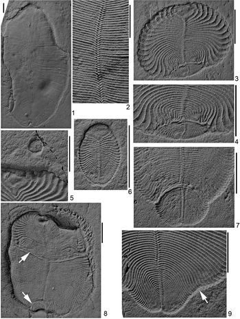
Similar to the majority of other representatives of Dickinsonia, the body of D. cf. D. menneri was flattened and ovate-elongated (Fig. 3.1, 3.6). The usual imprint of the D. cf. D. menneri body clearly shows a transverse division into two rows of right- and left-handed elements (isomers) that extended from the body axis in an alternating order (Fig. 3.2). But in our view, this structure represents only a part of the organism's body. It was a thin-walled structure that enveloped the internal parts of the body from above and below (Ivantsov et al., Reference Ivantsov, Zakrevskaya and Nagovitsyn2019b) and consisted of a substance more dense and resistant to decomposition than other materials that made up the Dickinsonia body (Bobrovskiy et al., Reference Bobrovskiy, Krasnova, Ivantsov, Luzhnaya (Serezhnikova) and Brocks2019). The ventral and dorsal sections of this structure are preserved as two layers on some imprints of Dickinsonia. This structure seems to represent a basement membrane, and was covered externally with an ectodermal tissue (Ivantsov et al., Reference Ivantsov, Zakrevskaya and Nagovitsyn2019b). An unpaired lobe was located at one end of the body. It has a subtriangular outline in small specimens of D. cf. D. menneri, and a pear-shaped outline in large specimens (Figs. 3.3, 3.6, 4.1). Judging from associated chains of feeding traces found alongside body imprints of Dickinsonia costata Sprigg, Reference Sprigg1947, the section bearing this unpaired lobe was likely the anterior end of the Dickinsonia body, judging from the direction of its movement (Gehling et al., Reference Gehling, Droser, Jensen, Runnegar and Briggs2005, Reference Gehling, Runnegar and Droser2014; Evans et al., Reference Evans, Gehling and Droser2019a). There is a shallow longitudinal depression (Figs. 3.1, 3.3, 3.7, 3.8, 4.4, 4.8), which is likely a reflection of internal axial structure of an unclear nature, possibly the digestive canal, on the surface of many imprints (Wade, Reference Wade1972; Jenkins, Reference Jenkins, Lipps and Signor1992). The growth during ontogeny of Dickinsonia involved an increase in the number of isomers, as well as their linear dimensions. This process was accompanied by a transformation of the shape of the anterior lobe, and a reduction in its relative area (Runnegar, Reference Runnegar1982; Hoekzema et al., Reference Hoekzema, Brasier, Dunn and Liu2017).
Materials
Macrofossils exhibiting evidence of damage were found in the Lyamtsa locality, located on the south coast of the Onega Peninsula, where the sediments of the Lyamtsa and Arkhangelsk Beds of the Ust-Pinega Formation of the Late Vendian are exposed (Fig. 2). The locality is characterized by the so-called Flinders- or Flinders-Belomorian-style preservation of fossil remains, in which the body imprints are located on the soles of sand layers (Narbonne, Reference Narbonne2005; Ivantsov and Zakrevskaya, Reference Ivantsov and Zakrevskaya2018). In this case, the bodies of a variety of benthic organisms were buried in life position (Seilacher, Reference Seilacher1999; Grazhdankin, Reference Grazhdankin2003, Reference Grazhdankin2004; Narbonne, Reference Narbonne2005; Droser et al., Reference Droser, Gehling and Jensen2006; Ivantsov, Reference Ivantsov, Fyodorov and Yakovlev2012). Dickinsonia menneri is the only species of this genus distributed in the lowest part of the Vendian of the White Sea section in the interval up to the upper half of the Syuzma Beds (Fig. 2) (Ivantsov, Reference Ivantsov2007). The studied material originates from the Lyamtsa Beds from a single bedding plane of the local burial event, marked as bed L2(XII).
Geographical and stratigraphic position of the Lyamtsa locality, burial L2(XII) (arrow); black vertical stripes on the right correspond to the intervals of the localities: L = Lyamtsa, SZ = Suyzma, K = Karakhta, SL = Solza, Z = Zimnie Gory.

Repository and institutional abbreviation
Type, figured, and other specimens examined in this study are deposited in the Paleontological Institute of the Russian Academy of Sciences (PIN RAS), Moscow, Russia (collection number 4716).
Results
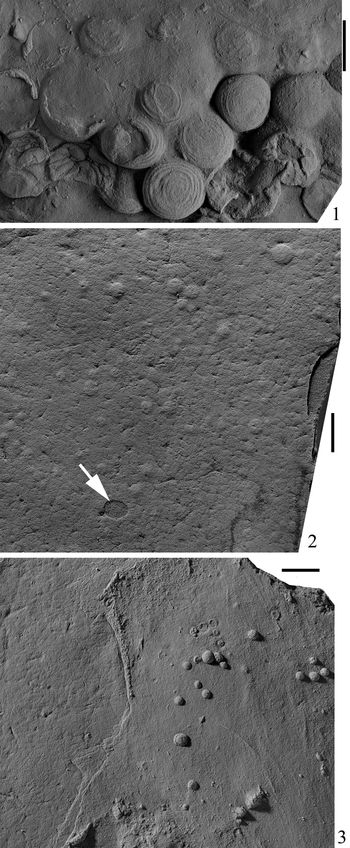
The sole of a cross-bedded sandstone of variable thickness (3–17 cm) serves as the fossiliferous surface of the L2(XII) burial event. The surface is even and has a microbially induced texture in the form of low, flat-topped tubercles separated by curving grooves (Fig. 5.2). In places, this surface bears the casts of shallow (up to 1 cm in depth) erosion channels (Fig. 5.3). The diversity of the assemblage of macrofossils on this bed is extremely poor. In addition to D. cf. D. menneri it contains Beltanelliformis brunsae Menner in Keller et al., Reference Keller, Menner, Stepanov and Chumakov1974, Parvancorina minchami Glaessner, Reference Glaessner1958, fragments of ribbon-like algae, small palaeopascichnids, rare traces of Epibaion axiferus Ivantsov in Ivantsov and Malakhovskaya, Reference Ivantsov and Malakhovskaya2002, and another two or three poorly diagnosed taxa. The deformed specimens of Dickinsonia and the specimens without visible damage are located together, without sorting by size.
In total, 146 specimens of Dickinsonia of various sizes and with different numbers of isomers were discovered in the local burial L2(XII) (Figs. 3.1, 3.5, 3.6, 8). The exact number of the isomers often cannot be calculated because their thickness is greatly reduced towards the posterior end of the body, and they become indistinguishable against the background of a granular rock. However, this indistinct zone is rather small; its area is ~0.5 mm2 and does not depend on the size of the specimen. Therefore, we consider it negligible when estimating the relative number of isomers.
Dickinsonia cf. D. menneri from the sediments of the Ust-Pinega Formation, late Vendian, southeastern White Sea area, Lyamtsa locality, burial L2(XII). (1) Specimen PIN, No. 4716/5165, one of the largest undamaged specimens of the species; the left (on the imprint) margin of the body is completely, and the right one is partially, turned up into the overlaying layer; a cross-overlapping of the dorsal and ventral parts of the dissected structure is observed at the top left of the imprint, plan view; (2) same as (1), fragment showing an alternating arrangement of isomers; (3) highly deformed specimen PIN, No. 4716/5187, plan view; (4) same as (3), posterior end; (5) specimen PIN, No. 4716/5149 (upper part of the figure), one of the smallest known specimens of Dickinsonia from Lyamtsa; its posterior end is turned up; the length of the visible fragment is 3.5 mm; (6) small, normally developed specimen without damage, specimen PIN, No. 4716/5150; (7) deformed specimen with lateral displacement of the axial structure along the break line, specimen PIN, No. 4716/5177; (8) specimen PIN, No. 4716/5282, experienced non-lethal damage twice; arrows indicate break lines; the anterior (upper in the figure) end of the body has been slightly bent and compressed during the taphonomic event, resulting in the formation of a band of scratches on the host rock surface; (9) specimen PIN, No. 4716/5181, exhibiting minor deformation, expressed as a reduction in the length of several isomers (arrow). The scale bars represent 1 cm.

Bayesian Information Criterion (BIC) analysis was used to determine the likely number of size groupings for distributions of Dickinsonia using the MCLUST package in R (Fraley and Raftery, Reference Fraley and Raftery2007; Darroch et al., Reference Darroch, Laflamme and Clapham2013; Zakrevskaya, Reference Zakrevskaya2014). As a result, the set of measured samples falls into two groups according to both univariate and multivariate analyses for logged data (Figs. 6, 7): (1) individuals of small size with body length 4.1–13.2 mm with the number of visible isomers from 15 to 38 in one row (106 specimens); and (2) medium and large individuals with length 18.3–122.0 mm with the number of visible isomers from 70 to 173 in one row in non-deformed individuals (40 specimens) (Fig. 6). Based on our observations of the communities of Ediacaran (Vendian) organisms of the White Sea region (Zakrevskaya, Reference Zakrevskaya2014), such a division indicates the existence of two or more different generations of Dickinsonia in this locality. Clear deformations were found in only 23 specimens of large and medium sizes and in one specimen of small size. A significant reduction in the relative length of the body is typical for these deformed specimens, so that the length of the body approximates to the width or becomes even smaller (Figs. 3.3, 4.1, 4.4). Meanwhile, the undamaged specimens are comparatively elongated, and the degree of their elongation increases with addition in the number of isomers (Fig. 3.1, 3.6).
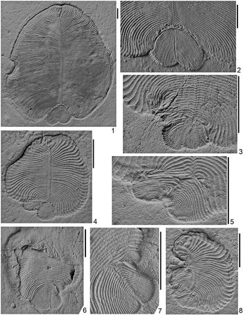
Deformed specimens of Dickinsonia cf. D. menneri from the Ust-Pinega Formation, Late Vendian, Southeastern White Sea area, Lyamtsa locality: (1) specimen PIN, No. 4716/5170, plan view, one can clearly see a gradual reduction of the curvature of the bends of the isomers’ inner ends in the anterior direction starting from the deformation zone; (2) same as (1), posterior end, latex cast from a natural mold; (3) specimen PIN, No. 4716/517, posterior end; (4) specimen PIN, No. 4716/5188, plan view; (5) same as (4), posterior end; (6) specimen PIN, No. 4716/5146, with intravital damage to the posterior end of the body, leading to splitting of the body axis and subsequent growth in two directions, plan view; (7) same as (6), fragment, the anterior part of the body was raised and located within the overlapping layer, the boundaries of the body are indicated by a sharp bend of the relief; (8) specimen PIN, No. 4716/5182, showing deep one-sided and two-sided deformations—loss of the several anterior isomers and a significant reduction in the length of the isomers of the left (in the figure) half of the body; loss of the front section of the axial structure and, possibly, the proximal ends of the adjacent isomers of the right row; lateral displacement of the posterior section of the axial structure and wave-like bending of the adjacent isomers; general curvature of the body. All scale bars represent 1 cm.

Cyanobacterial colonies Beltanelliformis brunsae Menner in Keller et al., Reference Keller, Menner, Stepanov and Chumakov1974 from the Lyamtsa locality. (1) Medium-sized specimens from talus in the lower part of the Lyamtsa outcrop; (2) small specimens from burial L2(XII), casts of depressions formed over collapsing colonies lying inside or immediately below the microbial mat and “translucent” through the structure of its surface (arrow indicates the imprint of a small Dickinsonia cf. D. menneri lying on the mat); (3) small specimens from burial L2(XII), imprint of the microbial mat's surface (left third of the figure) and cast of an erosive cutting that removed the mat and revealed a cluster of the Beltanelliformis colonies (right side of the figure). All scale bars represent 1 cm.

Statistical histograms showing size-frequency distributions, univariate BIC curves (E = equal variance, V = unequal variance, LogLik = log likelihood value), and density distribution plots for Dickinsonia from the Lyamtsa locality. (1) Length in mm; (2) logged length; (3) width in mm; (4) logged width.

Multivariate BIC plots and classifications. (1) Length and width, best BIC models are VEE, VEV, VVV with 3 clusters; (2) logged length and width, best BIC models are VEV, VVV, VEE with 2 clusters; (3) classification for length and width, the ellipses superimposed on the classification plots correspond to the covariances of the components; (4) classification for logged length and width. The BIC plot shows each BIC value for each profile in which line graphs illustrate the different types of multivariate normal distributions integrated into the model per cluster. Each model is illustrated with a different icon and a three-letter sequence. The letter sequence is a code for the geometric characteristics of volume, shape, and orientation (E = equal, V = varying, I = shape, or orientation). Models: EII = spherical, equal volume; VII = spherical, unequal volume; EEI = diagonal, equal volume and shape; VEI = diagonal, varying volume, equal shape; EVI = diagonal, equal volume, varying shape; VVI–= diagonal, varying volume and shape; EEE = ellipsoidal, equal volume, shape, and orientation; EVE = ellipsoidal, equal volume and orientation, varying shape; VEE = ellipsoidal, varying volume, equal shape and orientation; VVE = ellipsoidal, varying volume and shape, equal orientation; EEV = ellipsoidal, equal volume and shape, varying orientation; VEV = ellipsoidal, equal shape, varying volume, and orientation; EVV = ellipsoidal, equal volume, varying shape, and orientation; VVV = ellipsoidal, varying volume, shape, and orientation. For a detailed description, see Fraley and Raftery, Reference Fraley and Raftery2007; Darroch et al., Reference Darroch, Laflamme and Clapham2013.

The ratio of length to width in Dickinsonia from Lyamtsa. (1) Damaged (14 specimens) and undamaged specimens (78 specimens); (2) small undamaged specimens (71 specimens) and a posterior region of damaged specimens with two-sided deformations (12 specimens).

According to the nature of their development, the deformations observed on the imprints can be divided into two-sided and one-sided deformations. The two-sided deformations affect the isomers of both rows, with the affected isomers being more or less symmetrical about the axis of the body. The area of two-sided deformations itself is located in the posterior half of the body in all studied specimens. The one-sided deformations involve the isomers of only one row, and such changes can be found at any point along the body axis, except for the posterior end.
The area of two-sided deformations is enclosed between the anterior and posterior regions of unaltered isomers in all studied imprints (Fig. 9). Its boundary with the posterior region is here called the “break line,” which demonstrates a sharp transition from the most-deformed isomers to the isomers of normal shape. By contrast, the transition to the anterior region is continuous and accompanied by a gradual decrease in the intensity of deformation (Figs. 3.3, 4.1). We do not mean that there is an actual break in the integrity of the Dickinsonia body along this line, but we observe a break in the normal sequence of the isomers’ articulation. Only a few pairs of isomers are usually subjected to sharp deformations. The posterior region is mostly small (Fig. 9); its length varies from 3.7 to 17.1 mm, making up from 1/20 to 1/2 of the total body length of Dickinsonia. The number of isomers that can be distinguished in the posterior region varies from 10 to 35 in one row (Fig. 5.2). The peculiar specimen PIN, No. 4716/5282 has two zones of two-sided deformations (Fig. 3.8); the first one is located near the posterior end of the body, and the second one in the middle. The 20 mm long section enclosed between the two break lines contains ~40 pairs of isomers.
The tiniest observed changes (specimen PIN, No. 4716/5181) involve a small reduction in the length (the distance between the proximal and distal ends) of several isomers, causing shallow invaginations on the lateral edges of the body, along with a slight curvature of the proximal ends of these isomers (Fig. 3.9). However, often the reduction in the length of the isomers is significant and progresses towards the posterior end of the body, as in specimen PIN, No. 4716/5187 (Fig. 3.3). On the same specimen, typical fan-shaped bends of normally developed isomers are seen, which in this way cover the deformed area from the sides. Sometimes wavy and zigzag-shaped bends of isomers can be observed. They are especially distinct near the axis of the body, as in specimen PIN, No. 4716/5170 (Fig. 4.2), and increase in magnitude in the posterior isomers. In this specimen, the proximal parts of several pairs of isomers are partially destroyed and partially crumpled. These “clumps” press on the isomers of the anterior region (some harder, the others weaker), causing them to bend sharply. This bend is transmitted to the following pairs of isomers, gradually diminishing towards the anterior end of the body. Some specimens show a rupture of the axial structure and a slight displacement of the ends of the separated parts along the rupture line relative to the vertically oriented “sagittal” plane (Fig. 3.7).
We also attribute a splitting of the axis and dividing of the posterior end of the body in two (into two series of isomers) to the two-sided deformations. In specimen PIN, No. 4716/5146, the second series, consisting of small isomers, was formed on the right (in the imprint) side of the body. The symmetry axis of this secondary series is directed sideways at a large angle to the axis of the main body (Figs. 4.6, 4.7, 10). In specimen PIN, No. 4716/5188, both series of isomers have similar dimensions and are more or less symmetrical (Figs. 4.4, 4.5, 11). However, their inner rows are suppressed and partially upturned (passing into the host sandstone). Therefore, the posterior end of this specimen has the form of a two-horned tail with the tips of the “horns” facing each other.
One-sided deformations were recognized only in two specimens of medium size. In specimen PIN, No. 4716/5179 (28 mm in length), the distal ends of two dozen isomers of the left row (in the imprint) are lost in the posterior part of the body (Fig. 4.3). The proximal ends of the majority of these isomers are completely normal. The sample also displays two-sided deformation. The posterior end and the lateral margin of the organism's body were affected. Almost the entire left (in the imprint) side of the body is deformed in specimen PIN, No. 4716/5182 (23 mm in length) (Fig. 4.8). No fewer than a dozen anterior isomers are absent on the left side of the body, compared to the right one, and the lengths of the remaining ones are significantly reduced. A part of the axial structure and, possibly, the proximal ends of the isomers of the right row are also lost in this case. As a result, the body of Dickinsonia was curved to the left and the axial structure is arcuate. The collection also contains one very small specimen (specimen PIN, No. 4716/5226), the left (in the imprint) side of which is arcuate, and the entire right side is crumpled so intensely that the isomers on it are hardly visible. However, whether this may be connected with intravital damage cannot be established due to the small size of the specimen (its length is 4.8 mm).
Therefore, the following suite of deformations is observed in Dickinsonia specimens of the Lyamtsa locality: (1) a change in the body proportions with a reduction in length relative to width; (2) shortening of the isomers’ lengths with the formation of the invagination of the lateral edge of the body; (3) sharp non-typical bends of the isomers close to the midline; (4) a disappearance of the distal and proximal ends of the isomers and even entire isomers; (5) a fan-shaped bend of the distal ends of the isomers; (6) curvature of the body axis; (7) a rupture of the axial structure followed by a shifting of a part of it towards the “sagittal” plane; and (8) splitting of the body axis with the formation of an additional series of isomers.
Discussion
Many well-known imprints from Ediacara and the White Sea region show deviations from the ideal morphology of Dickinsonia, expressed as compression, stretching, curvature, wrinkling and various folding of the body, overlapping of some parts of the body with each other, intersecting of the inner layers, longitudinal wrinkling, pinch-out, and bifurcation of individual isomers. Sometimes the imprint shows the disappearance of a part of the body or its fragmentation. The occurrence of these deformations is explained by the folding, twisting, stretching, and rupture of the body during burial (Seilacher, Reference Seilacher1989; Gehling et al., Reference Gehling, Droser, Jensen, Runnegar and Briggs2005; Brasier and Antcliffe, Reference Brasier and Antcliffe2008; Evans et al., Reference Evans, Huang, Gehling, Kisailus and Droser2019b), by the lifting of some of its parts from the bottom surface (Evans et al., Reference Evans, Droser and Gehling2015), by shifting the structures of the upper and lower sides of the body relative to each other (Ivantsov et al., Reference Ivantsov, Zakrevskaya and Nagovitsyn2019b), or by penetration of the sediment into the body (Dzik, Reference Dzik2003). Concentric folds and a decrease in the body surface area relative to the feeding trace are interpreted as the result of muscle contraction preceding the death of the organism or post-mortem contraction (Glaessner and Wade, Reference Glaessner and Wade1966; Wade, Reference Wade1972; Runnegar, Reference Runnegar1982; Seilacher, Reference Seilacher1989; Gehling, Reference Gehling1991; Jenkins, Reference Jenkins, Lipps and Signor1992; Gehling et al., Reference Gehling, Droser, Jensen, Runnegar and Briggs2005; Evans et al., Reference Evans, Huang, Gehling, Kisailus and Droser2019b). The pinch-out and the bifurcation of the isomers still require explanation (Brasier and Antcliffe, Reference Brasier and Antcliffe2008). Folding, disappearance of parts, reduction in the body area during the taphonomic event, and separation into layers were also found on the fossil remains of Dickinsonia from the burial L2(XII) (Bobrovskiy et al., Reference Bobrovskiy, Krasnova, Ivantsov, Luzhnaya (Serezhnikova) and Brocks2019; Ivantsov et al., Reference Ivantsov, Zakrevskaya and Nagovitsyn2019b, Reference Ivantsov, Fedonkin, Nagovitsyn and Zakrevskayac). In the samples presented in this article, we can observe curling of the body margins and their shift along the bedding plane (Figs. 3.1, 3.5, 3.8, 4.1), lifting of a part of the body into the overlying sediment (Fig. 4.6), and overlapping of the dorsal and ventral parts of the dissected structure (Figs. 3.1, 4.4, 11).
The deformations studied here differ from those previously described in displaying a group of shortened isomers sharply “cut” from both margins. These isomers are surrounded on all sides by curved isomers of normal length. Only in cases of weak deformation do the shortened isomers have no other pronounced defects (Fig. 3.9). The attribution of this case to the studied phenomenon is determined by the symmetric arrangement of the groups of the shortened isomers on both sides of the body, and by some curvature of the corresponding section of the axial structure. Alternatively, it can be assumed that lateral compression of the Dickinsonia body in this area was the result of contraction of hypothetical muscles. However, there are no other possible signs of compression, such as the development of transverse folds in the isomers or the appearance of grooves on the surface of the host sandstone due to the displacement of the body margin (Evans et al., Reference Evans, Gehling and Droser2019a).
In some cases of deformation, it is possible to suspect folding and extrusion of the distal sections of the folds upwards into the overlapping sediment (Figs. 3.7, 4.5 [upper left quarter]). Here the difference in width and orientation of the isomers on both sides of the fold, although sharp, is nevertheless consistent with the observations of the degree of possible stretching of Dickinsonia body (Evans et al., Reference Evans, Huang, Gehling, Kisailus and Droser2019b). However, many of the described deformations (for example, Fig. 3.3, 3.4) cannot be explained by folding.
We must also consider the assumption that structures that appear to be two-sided deformations are, in fact, a consequence of the process of a vegetative reproduction, possibly present in Dickinsonia. In this case, the posterior region of normal isomers may represent a future juvenile, and the break line may be the place of its contact with the original organism. A similar mechanism of vegetative reproduction, called paratomy, occurs in some modern invertebrates, such as flatworms and polychaetes (Ruppert et al., Reference Ruppert, Fox and Barnes2004), which are groups to which Dickinsonia previously has been allied (Glaessner and Wade, Reference Glaessner and Wade1966; Termier and Termier, Reference Termier and Termier1968). This observation is supported by the similarity in the number of isomers visible in the small specimens (15–38 in one row) and in the posterior region of the deformed specimens (10–35 in one row). However, the pair of maternal and daughter individuals must exceed the non-divided organism in length or at least be equal to it. Meanwhile, as was noted above, the relative length of the deformed specimens is less than that of undeformed specimens (Fig. 8.1). The size of the young specimens present on the same bed is substantially smaller than the size of a hypothetical “not yet detached outgrowth.” While the length is quite similar (4–13 mm), the body width of free individuals reaches 11 mm, and the width of the posterior region in the non-deformed isomers is almost twice as large and reaches 18.5 mm (Fig. 8.2). In one large specimen (PIN No. 4716/5170), the length of the posterior region is 17.1 mm, and its width is 23.5 mm (Fig. 4.1, 4.2). And finally, the described structure was found only in a few Dickinsonia specimens of the same species, originating from a single local burial, whereas hundreds of specimens of different Dickinsonia species from the numerous local burials of Eastern Europe and South Australia have never shown anything like a “daughter individual.”
Elements of the structure of a deformed specimen of Dickinsonia cf. D. menneri of medium size (the number of the isomers is reduced).

It seems to us that the most probable explanation is that the considered deformations are the result of damage to some parts of Dickinsonia body, and the body's reaction to this damage, as well as, possibly, the result of some short-term depressing effect on the growth of the isomers. In the case of mild deformations (Fig. 3.9), it was probably only temporary inhibition of growth. In the cases of two-sided deformations, the damage was caused to the posterior end of the body. In addition, the organism lost its growth zone (see below), probably along with some isomers. Some of the isomers closest to the axis on both sides exhibit damage to their distal ends (Fig. 3.3). Sometimes the isomers closest to the damaged region retain their distal sections, but lose the proximal ends adjacent to the body axis (Fig. 4.2). The Dickinsonia body responded to damage by surrounding the damaged areas with normal isomers (Figs. 3.3, 3.4, 3.8, 4.2, 4.5), and through curvature of the body to compensate for the loss of part of the flesh (Fig. 4.8). This indicates the intravital, non-lethal nature of the negative impact. Traces of locomotory activity found in association with damaged specimens (Ivantsov et al., Reference Ivantsov, Nagovitsyn and Zakrevskaya2019a) show that these deformed individuals did not lose the ability to move independently.
Based on the available samples, it appears that the damaged ends of the isomers were not restored, but the isomers retained the possibility of their own growth and continued to increase in length. As a result of this growth, the damaged parts of the isomers gradually moved away from the axis to the lateral margins of the body (Fig. 3.8). The organism did not completely lose its ability to form new isomers—the growth zone was re-stored after the negative factor was removed and growth of the isomers was resumed in the usual manner. Regeneration of the growth zone was sometimes accompanied by defects. For example, a small lateral displacement of the axial structure is observed on the break line in many specimens (Fig. 3.7, 3.9). Their growth zone probably was recovered slightly away from the “sagittal” plane of the body. Two specimens show bifurcation of the body axis with the formation of an additional series of isomers (Figs. 4.4, 4.5, 4.6, 4.7, 9, 11). The process of divergence of the two newly formed growth zones could be gradual. For instance, the additional right (on the imprint) series of the isomers was formed in the specimen PIN, No. 4716/5146 (Fig. 4.6, 4.7). In this case, we can see that at first, after the resumption of the formation of the isomers, isomers of the left series appeared only on the left side, while in the case of the right series they appeared only on the right side (Fig. 10).
Schematic drawing of the deformed specimen of Dickinsonia cf. D. menneri, PIN, No. 4716/5146 from a photograph (Fig. 4.6, 4.7). (1) Plan view; (2) fragment, the dotted line shows the splitting axis (a-c and a’-c’ = successively formed isomers of the left and right rows in the initial stage of the growth zone division); the specimen demonstrates the gradual process of divergence of two newly formed growth zones; it can be seen that at first, after resumption of the formation of the isomers, the isomers of the left series appeared only on the left side (a-c), while the isomers of the right series appeared only on the right side (a’-c’).

Schematic drawing of the deformed specimen of Dickinsonia cf. D. menneri, PIN, No. 4716/5188 from a photograph (Fig. 4.4, 4.5). (1) Plan view, the arrow indicates an overlapping section of the dorsal and ventral parts of the dissected structure; (2) fragment, the dotted line shows the splitting axis.

The specimen PIN, No. 4716/5282 (Fig. 3.8) shows two episodes of restoration of the growth zone, separated by a significant period of time during which at least 40 pairs of normally developed isomers were formed. Thus, Dickinsonia from Lyamtsa was a rather resilient animal, and could survive the loss of significant areas of the body and even parts of the axial structure. They could relatively easily restore the growth zone if it was lost.
Implications for understanding anatomy and developmental processes, and the position of Dickinsonia within the Metazoa
In many deformed specimens of Dickinsonia from Lyamtsa, the distal ends of the isomers of the damaged zone are truncated by the isomers of the first pair of the posterior unchanged area, and even stand perpendicular to them (Fig. 3.3). However, the body does not break along this junction. If there was a break, then the posterior area would be practically free, and would connect with the rest of the body only in a very limited area near the axis. In this case, the posterior part is expected to be unconstrained and would easily move relative to the rest of the body. So, we would see an overlapping of the edges of the anterior and posterior areas on each other on one side of the body, and the divergence of the areas with a gap on the other. In reality, this never happens. Something prevented the areas of normal isomers from diverging. This may be a confirmation of the authors' conclusion about the presence of an ectodermal integument, which covered the body with a continuous shield from the dorsal side, in Dickinsonia and related organisms (Ivantsov et al., Reference Ivantsov, Zakrevskaya and Nagovitsyn2019b).
It is thought that an increase in the number of transverse elements of Dickinsonia was achieved by terminal addition of new ones at the posterior end of the body (Runnegar, Reference Runnegar1982; Gold et al., Reference Gold, Runnegar, Gehling and Jacobs2015). As a result, the body was extended in the posterior direction (Fig. 3.1, 3.6), while the previously formed elements moved away from the posterior end (Fig. 12.1). According to the hypothesis of the isomeric structure of the Dickinsonia body, each new isomer probably appeared in the space between its two predecessors and alternately shifted to the right and to the left side of the longitudinal axis (Ivantsov, Reference Ivantsov2008). This resembles the formation process of germ leaves in the apical meristem of plants. However, an alternative hypothesis was recently made, suggesting that growth occurred near the opposite end of the body of Dickinsonia (Hoekzema et al., Reference Hoekzema, Brasier, Dunn and Liu2017; Dunn et al., Reference Dunn, Liu and Donoghue2018). According to this hypothesis, a pair of new isomers (constituting a whole segment, rather than separated) was formed on the edge of the triangular lobe and expanded the body. In this case, the growth of Dickinsonia resembled the growth of articulate animals (Fig. 12.2). The appearance of the deformed White Sea specimens of Dickinsonia with a posterior region distinctly separated from the rest of the body along the break line and resembling, for example, the trilobite pygidium suggests the possibility of a third hypothetical variant of the formation of new isomers: insertion of new isomers somewhere within the middle of the organism, in front of some primary group of isomers (Fig. 12.3), formed behind the triangular lobe at the earliest stages of individual development. This variant can be supported by our conclusion concerning the existence of a dissected larval stage in the ontogenesis of Dickinsonia (Zakrevskaya and Ivantsov, Reference Zakrevskaya and Ivantsov2017).
Hypothetical variants of the location of the formation point of new isomers in a young specimen of Dickinsonia (marked by a dot or a bold line). (1) At the posterior end of the body (Runnegar, Reference Runnegar1982; Ivantsov, Reference Ivantsov2008; Gold et al., Reference Gold, Runnegar, Gehling and Jacobs2015); (2) on the sides of the triangular lobe (Hoekzema et al., Reference Hoekzema, Brasier, Dunn and Liu2017; Dunn et al., Reference Dunn, Liu and Donoghue2018); (3) at the front margin of some primary group of isomers; the arrow indicates the direction of apparent movement of the isomers along the axis of the growing organism.

The validity of the second and third hypotheses is contradicted by the existence of the specimens with a split axis and an additional series of isomers in the posterior region of the body of Dickinsonia. The presence of such a series is possible only with a posterior terminal location of the growth zone. Also, the last two hypotheses cannot explain the formation of the symmetry of the gliding reflection typical for D. cf. D. menneri from Lyamtsa. Thus, the most valid hypothesis is that the growth zone is located at the posterior end of the body of Dickinsonia.
The discovered ability of Dickinsonia to regenerate cannot alone be an argument in the discussion about the position of these creatures in the Metazoa system. However, the conclusion about the terminal location of the growth zone, made on the basis of the recognition of splitting of the axis of the regenerated area of the body, is very important in this regard. The posterior terminal location of the growth zone, along with the isomeric structure of D. cf. D. menneri, makes the position of Dickinsonia among the currently known branches of Bilateria unclear (see also Dunn et al., Reference Dunn, Liu and Donoghue2018).
The nature of the damaging factor
The cause of the damage to Dickinsonia from the Lyamtsa locality is mysterious. In the body of Dickinsonia, the defects of the margins and posterior end resemble healed injuries of different genesis known in fossil invertebrates. Such damage of the shields of trilobites were described by many researchers (see modern review by Bicknell and Pates, Reference Bicknell and Pates2020), including one of the authors of the present article (Ivantsov, Reference Ivantsov2003). The injuries of trilobites associated with the attacks are concentrated at the margins of the pleura of the thorax and pygidium, mainly in the posterior part of the body (Babcock, Reference Babcock1993). This localization is explained by the behavior of a predator chasing its prey from behind and the reaction of the victim to an attack, expressed as turns in one direction or another. In addition, the pleural damage was less dangerous for trilobites than damage to the head shield and rachis, where their main vital organs were located. Therefore the individuals with such injuries were more likely to survive (Babcock, Reference Babcock1993).
In Dickinsonia, as well as in trilobites, the deformations tend toward the posterior half of the body. Most of them are more likely to represent a loss of body part rather than to be a result of inhibition of the development. It is possible that Dickinsonia was attacked by an unknown predatory animal, whose remains for some reason are not preserved in the burials of the Flinders-Belomorian style. However, the damage to the Dickinsonia body does not show any consistent shape at the margins that could be associated with the morphology of offensive weapons of a predator. In addition, the predominant posterior localization of their deformations could be caused by increased vulnerability of the growth zone. It is difficult to explain the weak deformations, expressed as a small reduction in the length of the isomers, by direct physical action. Also, we cannot ignore the above-noted fact that the peculiar deformed individuals of Dickinsonia were found only in one out of many local burials known in Eastern Europe and South Australia. This information, combined with the observation that only medium-sized or large individuals were affected, inclines us to think about the formation of the damage as a result of one or two short episodes of a physicochemical, microbial, or some other similar effect, rather than occasional macrophage actions.
The following hypothesis can explain the occurrence of conditions that could have damaged Dickinsonia individuals. Large, initially spherical remains of Beltanelliformis brunsae are widely distributed in the Lyamtsa locality in general (Fig. 5.1). Their relationship to benthic colonial cyanobacteria was established by several independent methods (Steiner, Reference Steiner1996; Ivantsov et al., Reference Ivantsov, Gritsenko, Konstantinenko and Zakrevskaya2014; Bobrovskiy et al., Reference Bobrovskiy, Hope, Krasnova, Ivantsov and Brocks2018b). The average diameter of the colonies is 1–2 cm, although specimens with a diameter of up to 9 cm are known from the White Sea region (Ivantsov et al., Reference Ivantsov, Gritsenko, Konstantinenko and Zakrevskaya2014). These three-dimensional aggregates of photosynthesizing cells were enclosed in a dense matrix, and at the same time were partially submerged into the substrate. Apparently, these organisms could exist only in environments of extremely shallow waters (Ivantsov et al., Reference Ivantsov, Gritsenko, Konstantinenko and Zakrevskaya2014).
To the authors' knowledge, the local assemblage L2(XII) represents the only case of joint in situ burial of Dickinsonia and Beltanelliformis anywhere in the world. At the beginning of the taphonomic event, the bodies of the Dickinsonia were located on the upper, final surface of the microbial mat. The small (2–5 mm in diameter) collapsed colonies of Beltanelliformis were located below this surface, as evidenced by the inexpressive relief of the casts and the occurrence of microbially induced textures on them (Fig. 5.2). The erosive incisions that accompanied the taphonomic event removed the small areas of the mat and exposed the remains of these colonies (Fig. 5.3).
The absence of mineral sediment between the top of the mat and the colonies of Beltanelliformis implies the joint growth of these objects at some point, after which the colonies were inhibited and died. Perhaps here we have a record of seasonal changes in the mixed microbial community. Based on the small size of the colonies, the time favorable for their formation did not last long. It is impossible to determine the moment of the appearance of colonies, whether it was before, at the same time, or after the beginning of the microbial mat's formation and the expansion of the assemblage of macroorganisms associated with it. But, if we assume that one generation of Dickinsonia (represented by medium and large individuals in the burial) and the Beltanelliformis colonies existed together for some time, this could possibly lead us to a source of the damaging effects. The general background for the occurrence of the damage could be a temporary change of the physicochemical environmental conditions (temperature, salinity, light intensity, oxygen, or cyanotoxin concentrations, etc.), which had become less favorable for Dickinsonia, in the given part of the seabed. After death of the Beltanelliformis colonies, presumably, the conditions optimal for Dickinsonia's life were restored and their normal growth resumed. Therefore, the small individuals that likely appeared in the area after these events don't have any damage. However, at this stage it remains impossible to identify the direct factor that led to the non-lethal damage of Dickinsonia.
Summarizing our research, it should be noted that, despite the discovery of damaged individuals in the “Garden of Ediacara,” the reliable presence of macrophages in the late Precambrian has not yet been established.
Acknowledgments
The authors are grateful to P.V. Rychkov, who took an active part in the discovery and excavation of the presented material, and S.V. Bagirov (PIN RAS) who made the photographs. The authors are sincerely grateful to P. Vickers-Rich (Monash University), S. Evans (the Smithsonian Institution), A. Liu (University of Cambridge), S.A.F. Darroch (Vanderbilt University), and an anonymous reviewer for their valuable and constructive comments that improved the quality of the paper.
Field work and preparation of the fossil material were carried out with support of the Russian Foundation for Basic Research (RFBR), Grant No. 17-05-02212-a. Laboratory and theoretical studies were supported by the Russian Science Foundation (RSF), Grant No. 19-14-00346.