Introduction
A small subset of ganglion cells in the mammalian retina expresses the photopigment melanopsin. These cells are intrinsically photosensitive (reviewed by Provencio, Reference Provencio, Basbaum, Kaneko, Shepherd and Westheimer2008) and project to various brain areas including the suprachiasmatic nuclei and the olivary pretectal nuclei where they play a crucial role in nonperceptual visual processes, such as circadian photoentrainment and pupil constriction.
At least two distinct populations of melanopsin-containing ganglion cells can be distinguished anatomically. These populations have been extensively studied in rodent retina where they are referred to as M1 and M2 cells (Viney et al., Reference Viney, Balint, Hillier, Siegert, Boldogkoi, Enquist, Meister, Cepko and Roska2007; Schmidt & Kofuji, Reference Schmidt and Kofuji2009; Berson et al., Reference Berson, Castrucci and Provencio2010). The M1 cells stratify close to the inner nuclear layer (OFF sublamina); the M2 cells stratify close to the ganglion cell layer (ON sublamina). Both cell types form independent mosaics across the retina. In addition to their distinct stratification, M1 and M2 cells differ slightly in their morphology (Berson et al., Reference Berson, Castrucci and Provencio2010), show some physiological differences (Schmidt & Kofuji, Reference Schmidt and Kofuji2009), and differ in their central projections (Baver et al., Reference Baver, Pickard, Sollars and Pickard2008).
In primates, melanopsin-containing ganglion cells have been found in the retinas of humans, macaques, and marmosets (Hannibal et al., Reference Hannibal, Hindersson, Østergaard, Georg, Heegaard, Larsen and Fahrenkrug2004; Dacey et al., Reference Dacey, Liao, Peterson, Robinson, Smith, Pokorny, Yau and Gamlin2005; Jusuf et al., Reference Jusuf, Lee, Hannibal and Grünert2007) and have been distinguished into outer and inner stratifying cells. Retrograde labeling experiments in macaques showed projections to the pretectum and the dorsal lateral geniculate nucleus (Dacey et al., Reference Dacey, Liao, Peterson, Robinson, Smith, Pokorny, Yau and Gamlin2005). Electrophysiological recordings indicated that both outer and inner cells in macaque retinas have ON-type responses to light, which can be blocked by L-AP4, a group III metabotropic glutamate receptor agonist (Dacey et al., Reference Dacey, Liao, Peterson, Robinson, Smith, Pokorny, Yau and Gamlin2005), meaning that both types receive input from ON (rod and/or cone) bipolar cells.
In mouse retina, bipolar input to inner stratifying M2 cells has been found in an electron microscopic study (Belenky et al., Reference Belenky, Smeraski, Provencio, Sollars and Pickard2003), and it has been suggested that type 8 bipolar cells provide this input because type 8 cells were labeled transsynaptically through melanopsin cells (Viney et al., Reference Viney, Balint, Hillier, Siegert, Boldogkoi, Enquist, Meister, Cepko and Roska2007). In rabbit retina, M2 cells receive input from calbindin-positive bipolar axons (Hoshi et al., Reference Hoshi, Liu, Massey and Mills2009), which are equivalent to type CBb5 (MacNeil et al., Reference MacNeil, Heussy, Dacheux, Raviola and Masland2004). Neither of these studies analyzed the relationship between the two cell types quantitatively, and it is not known whether and to which extent other cone bipolar types provide input.
In primate, bipolar input to both inner and outer melanopsin cells has been demonstrated in marmoset and macaque retinas (Jusuf et al., Reference Jusuf, Lee, Hannibal and Grünert2007), but the bipolar type(s) providing this input have not been identified. Bipolar cell types stratifying in the inner third of the inner plexiform layer include rod bipolar, invaginating midget, blue cone bipolar cells, and the diffuse bipolar cell type DB6 (Boycott & Wässle, Reference Boycott and Wässle1991). Rod bipolar cells usually do not contact ganglion cells (Kolb and Famiglietti, Reference Kolb and Famiglietti1974; Strettoi et al., Reference Strettoi, Dacheux and Raviola1990). Invaginating midget bipolar cells direct their major output to ON midget ganglion cells and amacrine cells (Kolb & DeKorver, Reference Kolb and DeKorver1991; Calkins et al., Reference Calkins, Schein, Tsukamoto and Sterling1994). Blue cone bipolar axons usually form dyads onto an amacrine and a ganglion cells process (Marshak et al., Reference Marshak, Aldrich, Del Valle and Yamada1990; Kouyama & Marshak, Reference Kouyama and Marshak1992), and the small bistratified cell (blue-ON/yellow-OFF ganglion cell) is probably the major ganglion cell type contacted (Ghosh et al., Reference Ghosh, Martin and Grünert1997; Calkins et al., Reference Calkins, Tsukamoto and Sterling1998; Ghosh & Grünert, Reference Ghosh and Grünert1999). Thus, the most likely bipolar cell type providing the input to inner cells is the diffuse (ON) cone bipolar cell type DB6 (Jusuf et al., Reference Jusuf, Lee, Hannibal and Grünert2007). The main aim of the present study was to analyze the relationship between DB6 cells and inner melanopsin cells.
Recently, two studies of mouse and rabbit retinas found that outer stratifying M1 cells receive en passant synapses from descending ON cone bipolar axons (Dumitrescu et al., Reference Dumitrescu, Pucci, Wong and Berson2009; Hoshi et al., Reference Hoshi, Liu, Massey and Mills2009). This finding, that ON bipolar cells provide output in the OFF sublamina, is in contrast to the dogma, which functionally subdivides the inner plexiform layer into an outer (OFF) and inner (ON) sublamina (Famiglietti & Kolb, Reference Famiglietti and Kolb1976; Nelson et al., Reference Nelson, Famiglietti and Kolb1978). Similarly, contacts between ON bipolar cells and dopaminergic amacrine cells in the OFF sublamina have been identified (Gustincich et al., Reference Gustincich, Feigenspan, Wu, Koopman and Raviola1997; Dumitrescu et al., Reference Dumitrescu, Pucci, Wong and Berson2009; Hoshi et al., Reference Hoshi, Liu, Massey and Mills2009; Contini et al., Reference Contini, Lin, Kobayashi, Okano, Masland and Raviola2010). The question whether DB6 bipolar axons make contacts with outer stratifying melanopsin cells in primate retina was also addressed in the present study.
Material and methods
Animals
Four eyes from three adult male macaques (Macaca fascicularis) and one eye from one adult marmoset (Callithrix jacchus) were used. All procedures conformed to the provisions of the Australian National Health and Medical Research Council code of practice for the care and use of animals.
Tissue preparation
All eyes were obtained at the end of unrelated electrophysiological experiments. The animals were overdosed with sodium pentobarbitone (80–150 mg/kg, i.v.; Nembutal; Rhone Merieux ProVet, Pinkenba, Queensland, Australia).
The eyes of the macaques were perfused with physiological saline prior to perfusion with 4% paraformaldehyde in 0.1 m phosphate buffer (PB), pH 7.4, for 10 min. After perfusion, the eyes were enucleated, cut open, and the posterior eyecups were either placed in 0.1 m PB or postfixed with 4% paraformaldehyde in 0.1 m PB for 30 or 60 min. The posterior eyecup of the marmoset was immersion fixed in 4% paraformaldehyde in 0.1 m PB for 2 h. After fixation, the eyecups were rinsed in 0.1 m PB, the retinas dissected out, and immersed in 30% (w/v) sucrose in 0.1 m PB overnight. Retinas were stored at −70°C until use. Briefly fixed retinas (30 min or less) were used for triple label immunohistochemistry (melanopsin/CD15/piccolo; melanopsin/protein kinase C (PKC)/piccolo). The other retinas were used for double labeling (melanopsin/CD15). The melanopsin staining was comparable in all tissues.
Antibodies
A rabbit antiserum against 19 amino acid residues (MNPPSGPRVPPSPTQEPSC) from the N-terminus of the conceptually translated human melanopsin protein (NCBI accession number AAF24978) was provided by Dr. King-Wai Yau (Johns Hopkins University, Baltimore, MA). This antiserum was synthesized (Princeton BioMolecules, Langhome, PA) with an additional lysine residue at the amino end for cross-linking. Purified peptide was cross-linked to thyroglobulin using glutaraldehyde, then dialyzed in phosphate-buffered saline (PBS) buffer. The peptide conjugate was used to immunize rabbits (Covance, Emeryville, CA); the resulting antiserum was purified using peptide bovine serum albumin (BSA)-packed affinity column (Dacey et al., Reference Dacey, Liao, Peterson, Robinson, Smith, Pokorny, Yau and Gamlin2005). This antiserum was used to label melanopsin-containing ganglion cells in macaque retina. It did not label any cells in marmoset retina.
To label melanopsin-containing cells in marmoset retina, a rabbit antiserum against the human melanopsin protein (code number 5J68) was used at a dilution of 1:20,000 (provided by Dr. Jens Hannibal, Department of Clinical Biochemistry, Bispebjerg Hospital, Copenhagen, Denmark; Hannibal et al., Reference Hannibal, Hindersson, Østergaard, Georg, Heegaard, Larsen and Fahrenkrug2004). This antiserum was raised against a recombinant fusion protein containing the C-terminal–predicted cytoplasmic part of human melanopsin and recognizes human but not rodent melanopsin. Absorption of the antibodies with the immunization material dialyzed against PBS with 0.1% Tween abolished all staining (Hannibal et al., Reference Hannibal, Hindersson, Østergaard, Georg, Heegaard, Larsen and Fahrenkrug2004). This antiserum has previously been demonstrated to label melanopsin-containing cells in human, macaque, and marmoset retinas (Hannibal et al., Reference Hannibal, Hindersson, Østergaard, Georg, Heegaard, Larsen and Fahrenkrug2004; Jusuf et al., Reference Jusuf, Lee, Hannibal and Grünert2007).
A mouse monoclonal antibody specific for the carbohydrate epitope CD15 (provided by Prof. J. Mai, University of Düsseldorf, Düsseldorf, Germany) (Andressen & Mai, Reference Andressen and Mai1997) was used at a dilution of 1:6 to label diffuse cone bipolar (DB6) cells (Chan, Martin, Clunas, et al., Reference Chan, Martin, Clunas and Grünert2001; Chan, Martin, & Grünert, Reference Chan, Martin and Grünert2001).
A mouse monoclonal antibody against PKCα (PKCα clone MC5) was purchased from Sigma (Saint Louis, MO) (P 5704). This antibody was used at a concentration of 1:1000 to label rod bipolar cells (Greferath et al., Reference Greferath, Grünert and Wässle1990; Grünert & Martin, Reference Grünert and Martin1991; Grünert et al., Reference Grünert, Martin and Wässle1994).
A mouse monoclonal antibody against the C-terminal–binding protein 2 (CtBP2) domain of the ribbon protein RIBEYE (no. 612044; BD Biosciences, Heidelberg, Germany) was used at a dilution of 1:10,000. This antibody is raised against the C-terminal amino acids 361–445 of mouse CtBP2 (technical information; BD Biosciences) and has been characterized by Western blot analysis of mouse retina, where it labels a 110- and a 120-kDa band (representing RIBEYE B-domain) and a 50-kDa band (CtBP1; tom Dieck et al., Reference tom Dieck, Altrock, Kessels, Qualmann, Regus, Brauner, Fejtova, Bracko, Gundelfinger and Brandstätter2005). The antibody labels synaptic ribbons in the outer and inner plexiform layer of mammalian retinas, including primate (Schmitz et al., Reference Schmitz, Königstorfer and Südhof2000; tom Dieck et al., Reference tom Dieck, Altrock, Kessels, Qualmann, Regus, Brauner, Fejtova, Bracko, Gundelfinger and Brandstätter2005; Jusuf et al., Reference Jusuf, Martin and Grünert2006; Hendrickson et al., Reference Hendrickson, Troilo, Djajadi, Possin and Springer2009).
A guinea pig antiserum against the presynaptic cytomatrix protein piccolo (provided by Drs. E. Gundelfinger and W.D. Altrock, Leibniz Institute for Neurobiology, Magdeburg, Germany) was used as a presynaptic marker (dilution 1: 10,000). This antiserum was produced using bacterially expressed glutathione-S-transferase fusion protein sap44a covering amino acids 2172–2361 of piccolo protein (Cases-Langhoff et al., Reference Cases-Langhoff, Voss, Garner, Appeltauer, Takei, Kindler, Veh, De Camilli, Gundelfinger and Garner1996) and has been characterized in Western blots of rat brain extracts where it labels a protein band of 530 kDa; labeling was abolished when the antiserum was preadsorbed with the immunizing antigen (Dick et al., Reference Dick, Hack, Altrock, Garner, Gundelfinger and Brandstätter2001).
Immunohistochemistry
Immunohistochemistry was carried out on vertical (16 μm) cryostat sections and on pieces of whole retina. The sections were collected onto gelatinized slides. The pieces of unsectioned retinas were freeze cracked and subsequently processed free floating on a shaker. All steps were carried out at room temperature unless indicated otherwise.
The whole-mount piece of marmoset retina analyzed here derived from our previous study and details are given there (Jusuf et al., Reference Jusuf, Lee, Hannibal and Grünert2007). Briefly, the marmoset tissue was immersed in Target Retrieval Solution (Tris/EDTA buffer, pH 9; catalog number S 2368; Dako Australia Pty Ltd, Sydney, Australia) at 40°C for 8 h prior to immunostaining with the antiserum against the C-terminal–predicted cytoplasmic part of human melanopsin.
All retinal tissue was first rinsed in PBS pH 7.4 for 10 min and then preincubated in PBS containing 5% BSA and 0.5% Triton X-100 for 30 min (sections) or 60 min (whole mounts). For double labeling (CD15/melanopsin; piccolo/CtBP2), the tissue was incubated in a combination of two primary antibodies diluted in PBS containing 1% BSA, 0.5% Triton X-100, and 0.05% sodium azide overnight (sections) or at 4°C for up to 10 days (whole mounts). Tissue was rinsed in PBS and incubated for 60 min (sections) or at 4°C overnight (whole mounts) in a mixture of secondary antibodies. Secondary antibodies were goat anti-rabbit, goat anti-mouse, or goat anti-guinea pig IgGs coupled to Alexa 488 (1:250; Invitrogen, Melbourne, Australia) or Alexa 594 (1:500). All secondary antibodies were diluted in PBS containing 1% BSA and 0.5% Triton X-100.
For triple labeling (CD15/melanopsin/piccolo), vertical sections of macaque retina were sequentially incubated in primary and the appropriate secondary antibodies over four consecutive days. Incubation was overnight for each primary antibody and 1 h for each secondary antibody. Secondary antibodies were goat anti-mouse IgG coupled to Cy5 (1:200; JacksonImmuno Research Laboratories, West Grove, PA), goat anti-rabbit IgG coupled to Alexa 594 (1:500; Invitrogen), and goat anti-guinea pig IgG coupled to Alexa 488 (1:250; Invitrogen).
Sections and whole mounts were coverslipped in Mowiol (Hoechst, Sydney, Australia) containing 10% 1,4-diazobicyclo-[2.2.2]-octane (Harlow & Lane, Reference Harlow and Lane1988). Control sections were processed in the same way, but the primary antibodies were omitted. No specific staining was present in these control sections.
Analysis
Immunofluorescent preparations were viewed with a Zeiss (Rochester, NY) Axioplan 2 microscope. Stacks of images were taken at a step size of 0.3 μm with an Axiocam camera using a Plan Neofluar 40× oil immersion or a Plan-Apochromat 63× oil immersion objective and the appropriate filter sets (filter set 10, Ex BP450-490 wedge free; Zeiss; filter set Texas Red, 4040B, zero pixel shift, Ex562/40 BS 600 Em 624/40; Semrock; and filter set CY5 4040A, zero pixel shift, Ex628/40 BS 669 Em 692/40; Semrock, Göttingen, Germany). Images were processed using Axiovision deconvolution software (Zeiss) and the brightness and contrast of images were adjusted using Axiovision software or Adobe Photoshop (version 7 or CS4). Some stacks of images (0.7-μm intervals) were taken with a Zeiss LSM5 Pascal confocal microscope equipped with an Argon laser and a HeNe laser (filter sets BP575–615; LP 650; BP505–530). High-resolution scanning was performed with a Plan-Apochromat 63×/1.4 oil immersion objective at 1024 × 1024 pixels.
Colocalizations of dendrites and/or axons with immunoreactive puncta were quantified using three-dimensional (3D) thresholding and image segmentation functions (Object and Surface functions, Imaris V6.2.0; Bitplane, Zurich, Switzerland). Intensity maxima of synaptic marker fluorophores were detected within the 3D image stack and the distance to thresholded cell surfaces calculated. Points within 0.1 or 0.2 μm surface were considered colocalizations.
Results
Outer and inner stratifying melanopsin cells
As reported previously, two types of melanopsin-containing cells are found in macaque and marmoset retinas (Dacey et al., Reference Dacey, Liao, Peterson, Robinson, Smith, Pokorny, Yau and Gamlin2005; Jusuf et al., Reference Jusuf, Lee, Hannibal and Grünert2007). Outer stratifying (M1 in rodents) cells have somas either in the ganglion cell layer or displaced to the inner nuclear layer. The dendrites of outer cells stratify narrowly in S1 of the inner plexiform layer close to the inner nuclear layer. Inner stratifying (M2 cells in rodents) cells have their soma in the ganglion cell layer only, and their dendrites stratify in S5 of the inner plexiform layer close to the ganglion cell layer. The dendrites of inner cells are usually more weakly labeled than those of outer cells (Hattar et al., Reference Hattar, Kumar, Park, Tong, Tung, Yau and Berson2006; Jusuf et al., Reference Jusuf, Lee, Hannibal and Grünert2007; Berson et al., Reference Berson, Castrucci and Provencio2010). Fig. 1A shows a vertical section and Fig. 1C a whole mount view of inner stratifying cells in macaque retina. The cells have a sparse monostratified dendritic tree, which largely overlaps with that of outer stratifying cells.
Melanopsin ganglion cells and DB6 bipolar cells in macaque retina. (A) Vertical section labeled with the antiserum against the N-terminus of human melanopsin protein. The dendrites of an inner cell can be seen in stratum 5 of the inner plexiform layer (IPL). Collapsed stack of images. The same section was photographed with Nomarski optics to reveal the retinal layers (shown on the right). (B) Micrograph of a vertical section labeled with antibodies against CD15 showing axon terminals of DB6 cells. Collapsed stack of images. Nomarski optics shown on the right. (C) Micrograph of a whole-mounted retina labeled with the antiserum against the N-terminus of human melanopsin. The focus is on the dendrites of an inner stratifying melanopsin cell. The dendrites of outer stratifying cells are out of focus. INL, inner nuclear layer; GCL, ganglion cell layer. Scale bars = 20 μm. Scale bar in (A) valid for (A) and (B).

Spatial relationship between DB6 axons and inner melanopsin cells
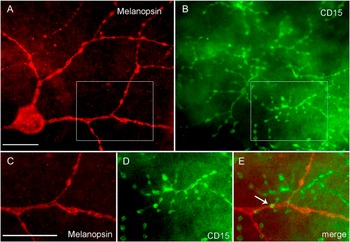
The dendrites of inner cells stratify at the same level as the axon terminals of diffuse bipolar cell type DB6, which in macaque and marmoset retinas can be identified with antibodies against the carbohydrate epitope CD15 (Fig. 1B) (Chan, Martin, Clunas, et al., Reference Chan, Martin, Clunas and Grünert2001, Chan, Martin, & Grünert, Reference Chan, Martin and Grünert2001).
The spatial relationship between the DB6 cells and inner melanopsin cells was analyzed in a double-labeled whole-mount preparation (Fig. 2). One inner stratifying cell from 4.9 mm eccentricity in marmoset retina was drawn together with the corresponding mosaic of DB6 axon terminals (Fig. 3). Because of the sparseness of the dendritic tree, only about 50% (127/276) of the DB6 axon terminals overlap with the dendrites of the melanopsin cells when the stack of images taken through the inner plexiform layer is collapsed into a single plane (Fig. 3). Nearly all (125/127) of the overlapping axon terminals have regions that are located in the same z-plane as the dendrites (presumed contact areas). In total, 365 presumed contact areas were counted and of these, about two thirds (n = 275) are located at the varicosities formed by DB6 axons; the varicosities are the presumed output regions of DB6 cells (Jusuf et al., Reference Jusuf, Lee and Grünert2004, Reference Jusuf, Lee, Hannibal and Grünert2007). This number of contact areas is in the range of bipolar input onto inner melanopsin cells as determined previously (Jusuf et al., Reference Jusuf, Lee, Hannibal and Grünert2007). However, triple label experiments including a synaptic marker (see below) and/or electron microscopy are needed to confirm the relationship between the two cell types.
Inner melanopsin ganglion cells and DB6 axons in marmoset retina. Single images of a stack of images are shown (deconvolved). (A and B) Micrographs of whole-mounted retina double labeled with the antiserum against the C-terminus of human melanopsin protein (A, red) and an antiserum against CD15 (B, green). The focus is on the dendrites of an inner melanopsin cell and the axon terminals of the CD15-labeled DB6 axon terminals. The box indicates the region shown at higher resolution in (C–E). The arrow points to a region of close contact between the two cell types. Scale bars = 25 μm. Scale bar in (A) valid for (A) and (B). Scale bar in (C) valid for (C–E).

Drawing of an inner melanopsin cell in marmoset retina together with the axon terminals of CD15-labeled DB6 bipolar cells located in the dendritic field of this cell. The same cell is shown in Fig. 2. Inset shows the region indicated by the box at higher resolution. The arrows indicate the axon of the melanopsin cell. The eccentricity of the cell is indicated in the lower left. A total of 127 (out of 276) axon terminals overlap with the dendrites in two-dimensional space. Scale bar = 200 μm.

Each of the 125 DB6 axon terminals has on average three presumed contact areas with the dendrites of the melanopsin cell (2.87 ± 1.56, mean ± s.d.). This number is lower than the number of presumed DB6 output synapses (~40 per DB6 axon terminal) estimated in central retina (Grünert et al., Reference Grünert, Lin and Martin2003), suggesting that DB6 cells also make output to other ganglion cell types and/or amacrine cells (see below and Jusuf et al., Reference Jusuf, Lee and Grünert2004; Szmajda et al., Reference Szmajda, Grünert and Martin2008).
Synaptic contacts between DB6 axons and inner melanopsin cells
In order to determine whether the areas of close contact between DB6 axons and inner melanopsin cells are indeed synaptic contacts, triple-labeling experiments were performed, including an antibody against the presynaptic cytomatrix protein piccolo.
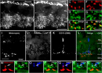
First, we established that the antibody against piccolo labels synaptic ribbons in primate retina by double labeling vertical sections with antibodies against the ribbon protein CtBP2 and antibodies against piccolo. The number of colocalized immunofluorescent puncta was determined as described previously (Grünert et al., Reference Grünert, Haverkamp, Fletcher and Wässle2002). The majority (266/337) of the CtBP2-labeled immunoreactive (IR) puncta are also labeled for piccolo (Fig. 4A–4H), indicating that ribbon synapses can be identified with antibodies against piccolo. However, only about 53% of piccolo IR puncta (266/500) are colocalized with CtBP2 IR puncta, consistent with the idea that piccolo is also expressed at many but not all amacrine synapses (Dick et al., Reference Dick, Hack, Altrock, Garner, Gundelfinger and Brandstätter2001).
Piccolo is expressed at ribbon synapses. (A–H) Micrographs of vertical sections through macaque retina double labeled with antibodies to piccolo and C-terminal–binding protein (CtBP2). Most CtBP2 IR puncta (red) are colocalized with piccolo IR puncta (green), indicating that piccolo labels synaptic ribbons. Piccolo puncta that are not colocalized with a CtBP2 IR punctum are probably located at amacrine synapses (Dick et al., Reference Dick, Hack, Altrock, Garner, Gundelfinger and Brandstätter2001). Single images of a stack of images (deconvolved) are shown. (I–L) Micrographs of a vertical section through macaque retina that was triple labeled with antibodies against melanopsin (N-terminus), CD15, and piccolo. Collapsed image of a stack of images. (M–S) High-resolution images of the boxed region indicated in (L). A piccolo IR punctum (green, arrow) is located close to a melanopsin IR processes (red, P) and is colocalized with a CD15-labeled DB6 axon terminal (blue, R). The superposition of the blue and red channel shows that the melanopsin IR process is in close contact with the CD15-labeled DB6 axon (Q) and the superposition of all three channels reveals that the synaptic punctum coincides with this region (S). Single images of a stack of images (deconvolved) are shown. Scale bar shown in (B) = 20 μm valid also for (A); scale bar shown in (H) = 2 μm valid also for (C–G); scale bar shown in (I) = 20 μm valid also for (J–L); scale bar shown in (S) = 2 μm valid also for (M–R). OPL, outer plexiform layer; INL, inner nuclear layer; IPL, inner plexiform layer; GCL, ganglion cell layer.

Despite the fact that piccolo is expressed presynaptically at both bipolar and amacrine synapses, we found that this marker is suitable for identifying synapses between DB6 cells and inner stratifying melanopsin cells for the following reasons. First, the antibody is raised in guinea pig and thus well suited for combining with the rabbit antiserum against melanopsin and the mouse monoclonal antibody against CD15 reducing the risk of cross-reactivity between antibodies. Second, bipolar synapses can be distinguished from amacrine synapses by simply considering only those puncta, which colocalize with our bipolar marker. The remaining piccolo puncta represent (most likely) presynaptic zones of other bipolar cell types and/or amacrine cells.
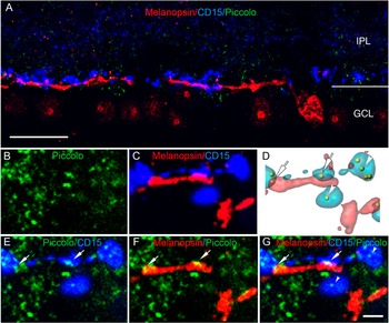
Examples of triple-labeled sections are shown in Figs. 4 and 5. Piccolo IR puncta (green) overlap with inner melanopsin processes (red) and with CD15-labeled DB6 axon terminals (blue) in the same region. A reconstruction of images in a confocal stack is shown in Fig. 5D. Piccolo IR puncta that are located at the regions of contact between DB6 axons and melanopsin dendrites can be seen (arrows) as well as additional piccolo IR puncta (arrowheads) that are colocalized with DB6 axons but are not located opposite a melanopsin-positive dendrite.
Piccolo is expressed at synapses between DB6 axons and inner melanopsin cells. Confocal micrographs of vertical sections through macaque retina triple labeled with antibodies against melanopsin (N-terminus), CD15, and piccolo. Single images of a stack of images are shown. (A) The dendrites of an inner stratifying cell (red) costratify with the axon terminals of CD15-labeled DB6 cells (blue). (B–G) High-resolution images. Piccolo IR puncta (green) are colocalized with CD15-labeled DB6 axons (E) at the region of overlap with melanopsin dendrites (F and G). (C) Superposition of the red and blue channel shows the region of overlap (melanopsin dendrite and DB6 axon) in purple. (D) A reconstruction of the images in this confocal stack. Piccolo puncta that are colocalized with DB6 axons and are located close to melanopsin dendrites are indicated by arrows. Additional piccolo IR puncta that are colocalized with DB6 axons but not close to dendrites can also be seen (arrowheads). Scale bar shown in (A) = 20 μm; scale bar shown in (G) = 2 μm valid also for (B–F).

The number of piccolo IR puncta present at contact areas between DB6 axon terminals and inner dendrites was quantified in vertical sections of macaque retina using deconvolution (Fig. 4) and confocal light microscopy (Fig. 5). It should be noted that vertical sections rarely contain inner dendrites. This is not only because of the sparseness of the dendritic tree of melanopsin cells but mainly because inner dendrites are usually labeled weakly and thus very hard to detect (Jusuf et al., Reference Jusuf, Lee, Hannibal and Grünert2007). Thus, the quantitative data deriving from vertical sections give only a rough estimate of the frequency of synaptic contacts between the two cell types. This question can only be addressed in triple-labeled whole mounts, but our attempts to obtain such whole mounts were not successful due to antibody penetration problems. Our analysis of triple-labeled vertical sections of macaque retina, however, provides some insight to the connectivity between DB6 cells and inner melanopsin cells. Six stacks of micrographs (about 12 to 30 images per stack) containing inner dendrites were analyzed. Four stacks were photographed with the deconvolution microscope (Fig. 4) and two stacks were photographed with the confocal microscope (Fig. 5). No significant differences were seen with respect to the results obtained with the different microscopes. In total, we found 44 regions of overlap between DB6 axons and melanopsin dendrites and a total of 34 piccolo IR puncta are located in these regions (77.3%). A lower percentage of puncta (50%) is found when the image is flipped in the horizontal axis. A total of 173 IR puncta are colocalized with DB6 axons, and a total of 102 IR puncta are located close to inner melanopsin dendrites. Thus, in this sample, 19.7% of the output made by DB6 cells goes to inner melanopsin cells and the synapses formed by DB6 cells make up 33.3% of the input to inner melanopsin cells.
Taken together, these results indicate that (i) when DB6 axon terminals overlap with inner dendrites of melanopsin cells, they form synapses in the majority of cases; (ii) inner stratifying melanopsin receives a low percentage (about 30%) of their input from DB6 cells; and (iii) the synapses between DB6 axons and inner melanopsin dendrites make up only a small fraction (about 20%) of the total number of outputs of DB6 axon terminal in S5. Consistently, it has been shown by electron microscopy that the large majority of the output from DB6 cells goes to amacrine cells (Jusuf et al., Reference Jusuf, Lee and Grünert2004). Furthermore, regions of close contact have also been detected between DB6 cells and large sparse ganglion cells in marmoset retina (Szmajda et al., Reference Szmajda, Grünert and Martin2008).
Synaptic contacts between rod bipolar axons and inner melanopsin cells?
It has been suggested for rat retina that melanopsin somata located in the ganglion cell layer receive input from rod bipolar axons (Østergaard et al., Reference Østergaard, Hannibal and Fahrenkrug2007). In order to determine whether rod bipolar axons make synapses with melanopsin cells in primate, we triple labeled vertical sections through macaque retina with antibodies against melanopsin, PKCα, and piccolo. In primate retina, antibodies against PKCα label rod bipolar as well as DB4 cone bipolar cells (Grünert et al., Reference Grünert, Martin and Wässle1994). The two cell types can be distinguished by the distinctive morphology of rod bipolar terminals and their stratification closer to the ganglion cell layer.
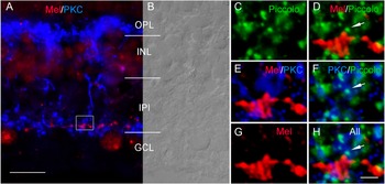
We found that the varicosities formed by rod bipolar axon terminals are usually located proximally to the dendrites of inner melanopsin cells (Fig. 6), whereas the DB6 axon terminals usually are located distally to the inner dendrites (Fig. 5). This is consistent with the finding that DB6 axons on average stratify slightly more distal than rod bipolar axons (see also Jusuf et al., Reference Jusuf, Lee and Grünert2004). As expected and shown in Fig. 6C–6H, piccolo IR puncta colocalize with PKCα-labeled axon terminals and with melanopsin-labeled ganglion cell processes. However, piccolo IR puncta do not colocalize with melanopsin and PKC at the same location (Fig. 6H), consistent with the idea that rod bipolar axons preferentially contact amacrine processes (Kolb & Famiglietti, Reference Kolb and Famiglietti1974; Strettoi et al., Reference Strettoi, Dacheux and Raviola1990; Grünert & Martin, Reference Grünert and Martin1991). We did not encounter melanopsin-labeled ganglion cell somata in our sample of sections and thus cannot exclude the possibility that some rod bipolar axon make synapses with onto melanopsin somata as has been shown for rat retina (Østergaard et al., Reference Østergaard, Hannibal and Fahrenkrug2007).
Piccolo is not expressed at synapses between rod bipolar axons and inner melanopsin dendrites. Single images of a stack of images (deconvolved) are shown. (A) Micrograph of a vertical section of macaque retina double labeled with antibodies against melanopsin (N-terminus, red) and protein kinase Cα (PKC, blue). (B) Retinal layers are shown in Nomarski optics. The varicosities formed by the rod bipolar axons are mainly located proximally to the melanopsin dendrite. (C–H) Micrographs of a vertical section of macaque retina triple labeled with antibodies against melanopsin (N-terminus, red), protein kinase Cα (PKC, blue), and piccolo (green). Piccolo IR puncta are colocalized with PKC-labeled rod bipolar axons and are found close to melanopsin-labeled dendrites but not at the same location (arrows). Scale bar shown in (A) = 20 μm; scale bar shown in (H) = 2 μm valid also for (C–G). OPL, outer plexiform layer; INL, inner nuclear layer; IPL, inner plexiform layer; GCL, ganglion cell layer.

Bipolar output to outer melanopsin cells
It has been shown in mouse and rabbit retinas that some ON cone bipolar cell types (but not rod bipolar cells) make axonal en passant synapses with M1 cells in the OFF sublamina of the inner plexiform layer (Dumitrescu et al., Reference Dumitrescu, Pucci, Wong and Berson2009; Hoshi et al., Reference Hoshi, Liu, Massey and Mills2009).
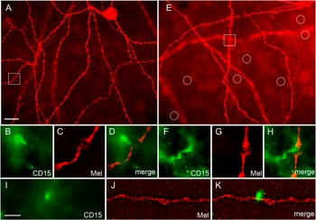
We determined potential contact areas between DB6 axons and outer melanopsin cells in double-labeled retinal whole mounts of macaque and marmoset retinas (Fig. 7). Stacks of images (n = 7 stacks in macaque and n = 3 stacks in marmoset retina) each covering an area of about 220 by 175 μm were analyzed. In these stacks, 7 out of 62 (11%) DB6 axons (macaque retina) and 2 out of 29 (7%) DB6 axons (marmoset retina) are closely associated with dendrites of outer melanopsin cells. This low percentage can be explained by the sparseness of the dendritic tree of melanopsin cells, resulting in the large majority of DB6 axons not being located in the vicinity to any outer melanopsin dendrites (see circles in Fig. 7E).
Outer stratifying melanopsin cells are in close contact with DB6 axons. (A and E) Micrographs of melanopsin cells in a whole-mounted macaque retina that was double labeled for melanopsin (N-terminus antiserum) and CD15. The focus is on the outer dendrites. The cell shown in (A) has its soma in the inner nuclear layer. Out of focus dendrites belong to inner stratifying cells. (A) Collapsed stack of 11 images; (E) single image of a stack of images. The rectangles indicate the regions shown at higher resolution in (B–H). The circles indicate the location of CD15-labeled axons as detected in the image taken with the green channel (not shown). (B–D) and (F–G) High-resolution images of the boxed regions from (A) and (E). Single images of a stack of images (deconvolved) are shown. CD15-labeled DB6 axons (green) are seen in close contact with an outer melanopsin dendrites (red). (I–K) Similar contacts between melanopsin dendrites are seen in whole-mounted marmoset retina. Scale bar in (A) = 20 μm valid also for (E). Scale bar shown in (I) = 5 μm valid for (B–K).

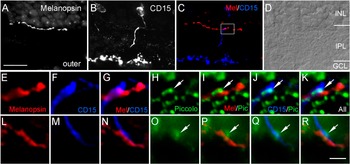
In order to see whether presumed contact areas between DB6 axons and outer melanopsin dendrites are involved with synapses, triple-labeled vertical sections of macaque retina were analyzed. We took stacks of images (each consisting of 20 to 30 micrographs) for 53 regions containing outer melanopsin dendrites. In 24 stacks, we found outer dendrites overlapping with CD15-labeled DB6 axons and in 9 of these (37.5%), a piccolo IR punctum was also present. Two examples are shown in Fig. 8. Taken together, our findings provide anatomical evidence that a subpopulation of DB6 cells provide axonal en passant synapses to outer stratifying melanopsin cells.
Piccolo is expressed at en passant synapses between DB6 axons and outer melanopsin cells. Single images of a stack of images (deconvolved) are shown. (A–C) Micrographs of a vertical section through macaque retina triple labeled for melanopsin (N-terminus), CD15, and piccolo. The box in (C) indicates the region shown in (E–K). (D) Retinal layers are shown in Nomarski optics. (E–G) and (L–N) High-resolution images showing CD15-labeled DB6 axons overlapping with outer melanopsin dendrites. (H–K) and (O–R) A piccolo-labeled IR punctum (arrows) is located at the region of overlap between the two processes. Scale bar in (A) = 20 μm valid for (A–D); scale bar in (R) = 2 μm valid for (E–R).

Discussion
This study provides anatomical evidence that DB6 ON cone bipolar cells make output synapses with inner stratifying melanopsin ganglion cells in primate retina. In addition, we provide evidence that DB6 axons make en passant synapses onto outer stratifying melanopsin cells. Our findings support and extend our previous studies of marmoset and macaque retinas, which showed that the dendrites of both outer and inner melanopsin cells are colocalized with pre- and postsynaptic markers for bipolar synapses and that both cell types receive bipolar input throughout the entire dendritic tree (Jusuf et al., Reference Jusuf, Lee, Hannibal and Grünert2007). Our findings are consistent with electrophysiological recordings obtained in macaque retina showing that both inner and outer stratifying melanopsin cells have ON-type responses mediated by synaptic inputs from rods and cones (Dacey et al., Reference Dacey, Liao, Peterson, Robinson, Smith, Pokorny, Yau and Gamlin2005).
Synaptic input to inner stratifying melanopsin cells
For one inner stratifying melanopsin cell from 4.9 mm eccentricity in marmoset retina, we found a total of 365 areas of overlap (presumed contact areas) with DB6 axons. This number is higher than the number of colocalized puncta (n = 143) determined in our previous study for an inner melanopsin cell at similar eccentricity using antibodies against the C-terminal–binding protein 2, a marker for bipolar ribbons (Jusuf et al., Reference Jusuf, Lee, Hannibal and Grünert2007). This can be explained with the possibility that not all of the overlapping areas contain a synaptic marker, as our results from vertical sections in the present study have shown. Taken together, these results imply that DB6 axons may constitute the entire bipolar input to inner stratifying melanopsin cells. Consistently, this study and our previous study (Jusuf et al., Reference Jusuf, Lee, Hannibal and Grünert2007) did not find any evidence for rod bipolar output to melanopsin cells, and rod bipolar axons have been found to contact amacrine cells almost exclusively (Kolb & Famiglietti, Reference Kolb and Famiglietti1974; Strettoi et al., Reference Strettoi, Dacheux and Raviola1990; Grünert & Martin, Reference Grünert and Martin1991) (but see Østergaard et al., Reference Østergaard, Hannibal and Fahrenkrug2007). Furthermore, as outlined in the introduction, the other cone bipolar types stratifying close to the ganglion cell layer are unlikely candidates to provide input to inner melanopsin cells.
Thus, piccolo IR puncta, which are located close to inner melanopsin dendrites but are not colocalized with DB6 axon terminals, probably derive from amacrine cells. Consistently, a result from one inner melanopsin cell in marmoset retina indicates that amacrine input to these cells occurs at a higher density than bipolar input (Jusuf et al., Reference Jusuf, Lee, Hannibal and Grünert2007).
ON bipolar input to outer stratifying melanopsin cells
Previously we showed that outer melanopsin cells in marmoset and macaque retinas receive bipolar input throughout the entire dendritic tree and discussed the possibility that this input derives from the presumed OFF bipolar cell type DB1 (Jusuf et al., Reference Jusuf, Lee, Hannibal and Grünert2007). In the present study, we analyzed double-labeled whole mounts and found that the axons of a subpopulation of DB6 cells (about 10%) make close appositions to outer melanopsin dendrites in marmoset and macaque retinas. This analysis of whole mount retina did not involve a synaptic marker, but our results from triple-labeled sections revealed that close appositions between DB6 axons and outer melanopsin cells are found in the presence of a synaptic marker in about a third of the cases. Taken together, our results from whole mounts and vertical sections suggest that en passant synapses may be formed by 3–4% of the DB6 axons. This low percentage can be explained with the sparseness of the dendritic tree and the relative low density and small diameter of bipolar axons. Likewise, in rabbit retina, axonal en passant synapses with M1 cells were found for about 6% of the calbindin-labeled bipolar cells (Hoshi et al., Reference Hoshi, Liu, Massey and Mills2009).
The synapses formed by ON bipolar axons onto dendrites of M1 cells in the OFF sublamina in mouse and rabbit retinas have been attributed to specific ON bipolar types, but the involvement of other bipolar types has not been ruled out (Dumitrescu et al., Reference Dumitrescu, Pucci, Wong and Berson2009; Hoshi et al., Reference Hoshi, Liu, Massey and Mills2009). Outer stratifying melanopsin cells in marmoset retina receive an average of two bipolar synapses per 100 μm dendritic length (Jusuf et al., Reference Jusuf, Lee, Hannibal and Grünert2007). The low percentage of DB6 axons making potential synapses with outer melanopsin cells found here cannot account for this density, suggesting that in primate, other bipolar cell types are involved in the contacts to outer stratifying melanopsin cells. In addition to DB6 cells, least four other ON cone bipolar types and one rod bipolar type are found in primate retina (Boycott & Wässle, Reference Boycott and Wässle1991; Chan, Martin, Clunas, et al., Reference Chan, Martin, Clunas and Grünert2001). Rod bipolar axons do not make ectopic synapses in mouse retina (Dumitrescu et al., Reference Dumitrescu, Pucci, Wong and Berson2009), but further studies are required to establish which other cone bipolar types may contribute to the bipolar input to outer melanopsin cells and whether this input is responsible for the ON responses found in these cells in primate retina.
Conclusion
The output synapses formed by DB6 cells onto inner as well as outer stratifying melanopsin cells may contribute to the ON response found in the two types of melanopsin cells of primate retina.
Acknowledgments
This work was supported by a Project Grant from the National Health & Medical Research Council 454460 and the Lions Vision Research Fund (U.G.). We thank Dr. Paul Martin for help with the Imaris analysis and for critical reading of the manuscript. We thank Drs. W. Altrock, E. Gundelfinger, J. Hannibal, J. Mai, and K.-W. Yau for kindly providing antibodies. We also thank Dr. Heather Young for help with confocal microscopy and Ehren Terenyi and Ana Lara for technical assistance.