


Significant outcomes
-
∙ The present study suggests amygdala alterations in adults suffering from ADHD.
-
∙ Our study also implies different role of left and right amygdala in the etiology of ADHD.
-
∙ Motor impulsivity seems to be related to smaller volumes in right amygdala.
Limitations
-
∙ The high similarity among cases provides great internal value but little external value.
-
∙ As there are no medication naïve subjects among the cases, it is we can not discard medication effects over brain structures.
Introduction
Attention-deficit hyperactivity disorder (ADHD) is the most commonly diagnosed neurodevelopmental disorder in childhood, which affects 3–7% of the population worldwide. ADHD is defined by distractibility, hyperactivity and impulsivity (DSM-IV-TR). It is a chronic neurobiological disorder with childhood onset and persistence into adolescence and adulthood. The vast majority of affected children continue to have symptoms in adulthood, resulting in an important impairment of family, work and social functioning (Reference Biederman and Faraone1). Mood and anxiety disorders, disruptive behavioural disorders, emotional dysregulation and substance abuse are common comorbidities of ADHD (Reference Faraone, Biederman and Mick2).
Although the exact mechanism has not yet been elucidated, the pathophysiology is presumed to be linked to dysfunction of the frontal–striatal–cerebellar circuits (Reference Sasayama, Hayashida and Yamasue3). An increasing number of neuroimaging studies using magnetic resonance imaging (MRI) or functional magnetic resonance imaging (FMRI) have been conducted and have demonstrated functional and structural abnormalities associated with ADHD. The early estimations showed ~4–5% overall cerebral and cerebellar volumetric reductions in children and adolescents with ADHD, compared with that of typically developing controls (TDC) (Reference Castellanos, Lee and Sharp4,Reference Carmona, Vilarroya and Bielsa5). Other structural MRI studies have reported volumetric reductions in the frontal lobe [including orbitofrontal cortex (OFC), superior frontal cortex and dorsolateral prefrontal cortex (DLPFC)], posterior and anterior cingulate gyri, precentral gyrus, caudate nuclei, corpus callosum (CC), as well as the cerebellum (Reference Seidman, Valera and Makris6–Reference Bush8).
Significantly reduced whole-brain cortical grey matter (GM) thickness has also been found in children with ADHD when compared with TDC (Reference Shaw, Lerch and Greenstein7,Reference Makris, Biederman and Valera9,Reference Shaw, Eckstrand and Sharp10). Studies also showed significantly thinner cortical thickness in regions including bilateral DLPFC and OFC, anterior and posterior cingulate cortex and the temporo–occipito–parietal junction in adults with ADHD when compared with controls (Reference Makris, Biederman and Valera9,Reference Proal, Reiss and Klein11).
The rate of cortical thinning in these regions has shown to be inversely associated with the severity of hyperactivity and impulsiveness in normal development (Reference Shaw, Gilliam and Liverpool12). The MRI findings that are most replicated in ADHD children are smaller total brain volumes, smaller cerebellum, abnormalities in prefrontal cortex and in caudate nuclei, and a smaller rostrum of the CC.
However, so far, the studies have not provided consistent findings concerning global and regional structural brain abnormalities (Reference Carmona, Vilarroya and Bielsa5). Studies using newer neuroimaging analysis techniques have identified other abnormal regions such as the temporal or parietal lobes in patients with ADHD (Reference Brieber, Neufang and Bruning13,Reference McAlonan, Cheung and Cheung14).
The amygdala and prefrontal cortex are considered to play a crucial role in the control of behaviour and impulses as well as in regulation of emotions (Reference Frodl and Bokde15–Reference Welborn, Papademetris and Reis18). A recent study evaluating voxel-based morphology has indicated significantly smaller regional GM volume in areas including the bilateral temporal polar and occipital cortices and the left amygdala in subjects with ADHD as compared with controls (Reference Sasayama, Hayashida and Yamasue3). Significantly smaller regional GM volumes were demonstrated in more extensive regions including bilateral amygdala after controlling for the confounding effect of comorbid oppositional defiant disorder and conduct disorder (Reference Sasayama, Hayashida and Yamasue3).
Our study starts from the hypothesis that smaller amygdala volumes in ADHD would be associated with less control of impulsivity and emotional instability, symptoms which are core to ADHD, oppositional defiant disorder and other conduct disorders.
Methods
A total of 18 subjects were assessed using MRI and the images were processed using FSL software. Volumetric findings were compared between patients and controls. Approval from the local ethics committee was obtained. Patients participated voluntarily in the study after written informed consent was signed. Participants were recruited from the ADHD unit of a general hospital during a period of 6 months. Each patient fulfilling the ADHD diagnostic criteria, who did not meet any exclusion criteria (see below), was offered to participate in the study. Nine ADHD patients [all male participants, average age 25.5 years (18–49)] entered the study. The control group consisted of nine healthy subjects (nine males) with no family history of schizophrenia or bipolar disorder. Their average age was 25.17 years (range from 21 to 32 years). There were no differences between the ADHD group and the control group for age (t=0.46, p=0.65) or for the educational level (16.4 vs. 17.1 years of education, respectively; t=−0.42, p=0.74).
The Barratt Impulsiveness Scale (BIS) is a questionnaire designed to assess the personality/behavioural construct of impulsiveness. The BIS was used to assess impulsivity and its punctuation was also correlated with different amygdala volumes (Reference Barratt19). The BIS questionnaire was applied to each participant before the MRI scan.
Participants were assessed for Axis I Disorders with the Structured Clinical Interview for DSM-IV (Reference First, Gibbon and Williams20) and the SCID-II for diagnosis of personality disorders. Current episodes of major depression (all patients scored below 12 in the Hamilton Depression Rating Scale) or substance dependence were considered exclusion criteria as were also a lifetime diagnosis of schizophrenic disorder, bipolar disorder or organic mental disorders. Patients were free of unstable medical conditions or neurological disorders. All patients were free of medication at least 2 weeks before the study.
The subjects were scanned with a 1.5 T magnet (GE Excite 11.0; GE Healthcare, Milwaukee, WI, USA), using the standard quadrature head coil, acquiring three-dimensional T1-weighted images using a Fast Spoiled Gradient Echo sequence with inversion pulse (TE=1.8 ms, TR=7.6 ms, TI=400 ms, BW=14.7 kHz, FOV=25×25 cm, 120 slices, slice thickness=2.0 mm, with 1.0 mm overlap, matrix size=256×256 voxels, NEX=1, scan duration=7 min 29 sec).
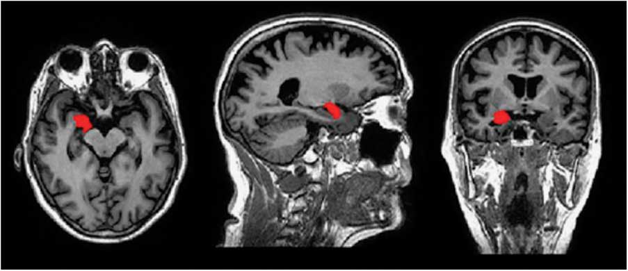
The images of each patient were later processed in a dedicated workstation. Absolute volumes of several structures and nuclei were calculated with FIRST [part of the FSL library of analysis tools for FMRI, MRI and DTI brain imaging data], a model-based segmentation/registration tool. FIRST is a fully automated segmentation method, which has been shown to accurately determine the volume of several subcortical structures, as compared with manual tracing measures (Reference Morey, Petty and Xu21). It is based on multivariate Gaussian shape/appearance models, constructed from manually segmented images (336 brains) provided by the Center for Morphometric Analysis, MGH, Boston. The manual labels are parameterised as surface meshes and modelled as a point distribution model. Deformable surfaces are used to automatically parameterise the volumetric labels in terms of meshes; the deformable surfaces are constrained to preserve vertex correspondence across the training data. Furthermore, normalised intensities along the surface normals are sampled and modelled. Shape is then expressed as a mean with modes of variation (principal components). Based on learned models, FIRST searches through linear combinations of shape modes of variation for the most probable shape instance, given the observed intensities in a T1-weighted image. The segmentation of the T1 images was performed as a two-stage affine transformation, with 12 df to the MNI152 template at 1 mm resolution, using a MNI152 amygdala mask for the second stage. The segmented images were then used to produce mesh and volumetric outputs with boundary correction. Voxels exhibiting ambiguous structural characteristics, owing to partial volume effects, which are usually located at the borders between adjacent structures, were classified as boundary voxels, to be corrected through the several iterations of a boundary correction algorithm. The vertex information was then automatically transformed back to native space using the inverse transformation matrix where the boundaries were corrected. At the end of the process, the quality of the final outputs was individually checked (Fig. 1). Values in cubic millimetres for the amygdala could be obtained for both hemispheres.
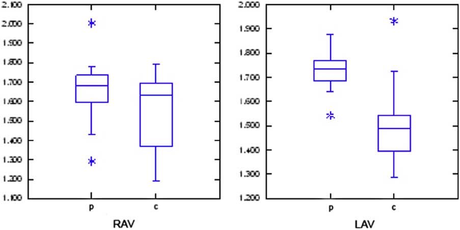
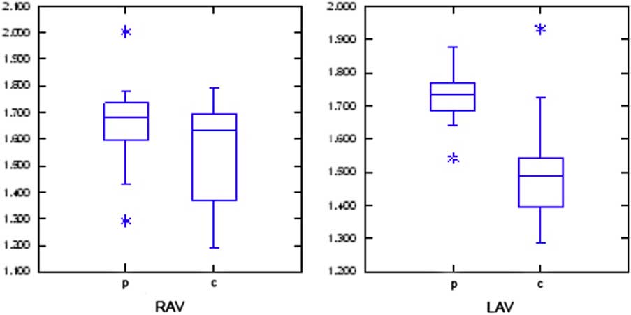
Weighted volumes for the right amygdala volume (RAV) and left amygdala volume (LAV). No significant differences were found between both groups (p and c) for the RAV, the LAV was found to be significantly smaller in the controls (c).
Moreover, brain tissue volume, normalised for subject head size, was estimated with SIENAX, which is also a part of FSL. SIENAX starts by extracting brain and skull images from the single whole-head input data. The brain image is then affine-registered to MNI152 space (using the skull image to determine the registration scaling); this is primarily in order to obtain the volumetric scaling factor, to be used as a correction for the variations of head size and to obtain a ponderated amygdala volume.
Statistical analysis was performed using SPSS 19 software (SPSS Inc., Armonk, NY, USA). Quantitative data are expressed as mean and standard deviation. Correlation between variables was done with Pearson coefficients. Null hypothesis were rejected with p<0.05.
Results
In our patients, the weighted volume for the right amygdala was 1647 mm3 (SD=203) and for the left amygdala was 1727 mm3 (SD=97). For the controls, the weighted volume for the right amygdala was 1543 mm3 (SD=205) and for the left amygdala was 1515 mm3 (SD=192). No significant differences were found between both groups for the right amygdala volumes (p=0.288, t=1.098, df=16.826); the left amygdala was found to be significantly smaller in the controls (p=0.009, t=3.069, df=13.646) (Fig. 1).
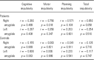
Results of the correlation analysis between weighted right and left amygdala volumes and the results of the Barratt’s impulsivity test in ADHD patients are shown in Table 1.
Correlation between right and left amygdala volumes and Barratt’s impulsivity score in attention-deficit hyperactivity disorder patients and controls

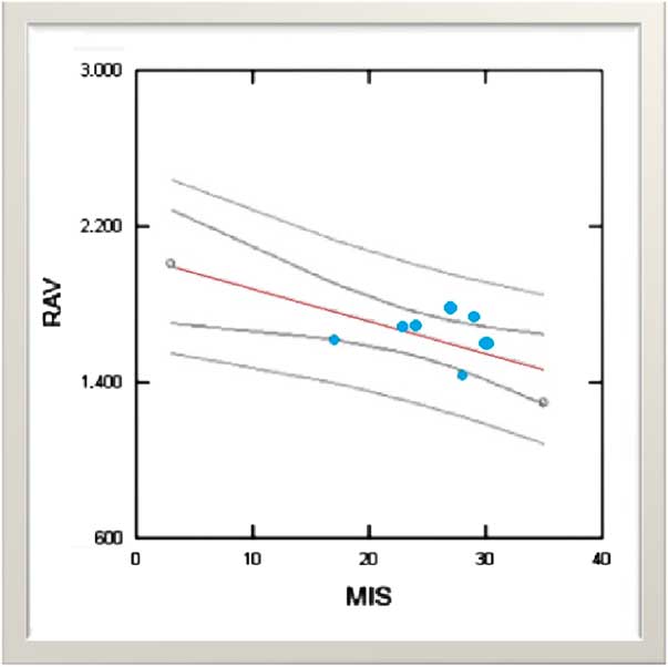
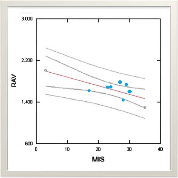
Barratt’s motor impulsivity score reached significant negative correlation with right amygdala volume (r=−0.756, p=0.018, Fig. 2). In addition, total impulsivity score showed a trend of increased scores with lower right amygdala volumes. No correlation was found between impulsivity scores and the volume of the left amygdala. Age was not found to be a contributor of the results.
Correlation between right amygdala volume (RAV) and Barratt’s motor impulsivity score (MIS) in patients (n=9). Negative correlation was observed in patients. No correlation was found in the left amygdala.
Discussion
The amygdala is one of the most critical structures in the anterior temporal region, playing a crucial role in emotional and social behaviour (Reference Morris, Frith and Perrett22). It is also considered to play a critical role in the emotional process of sensorial information as it receives projections from all the sensorial association areas (Fig. 3). These convergences of anatomic projections place the amygdala as the structure responsible for the formation of associations between the stimuli and reinforcement or punishment. In addition to the cortical projections coming from the sensorial association areas, the amygdala also receives thalamic afferences. This grouping of projections, both thalamic and cortical, to the amygdala makes it possible to give an affective meaning to the stimuli.
Application of FSL MRI in an adult ADHD case. Right amygdala volume and MRI FSL FIRST method. The right amygdala (red pot) would be involved in the recuperation of visual emotional information. MRI, magnetic resonance imaging; ADHD, attention-deficit hyperactivity disorder.

A few studies including one meta-analysis have reported significantly smaller volumes of the amygdala in subjects with ADHD (Reference Castellanos, Giedd and Marsh23–Reference Valera, Faraone and Murray25). These findings may be associated with social–cognitive impairment observed in ADHD children (Reference Nijmeijer, Minderaa and Buitelaar26,Reference Uekermann, Kraemer and Abdel-Hamid27).
Regarding to differential participation of right and left amygdala, a possible asymmetry has been suggested. The left amygdala has been related with emotional language processes and stimuli characteristics of emotion, whereas the right amygdala would imply the recuperation of emotional information, particularly the visual type (Reference Markowitsch28). However, the fact that this lateralisation has not always been found in patients with unilateral amygdala injuries suggests that this lateralisation of emotional function does not exist and that both would contribute in the same way to the emotional processes (Reference Peper, Karcher and Wohlfarth29).
The present study investigating structural brain differences between ADHD subjects and the control subjects indicates abnormal functioning of the amygdala, with a smaller volume, in ADHD participants. When right and left amygdala were assessed independently and related to the scores in the impulsivity test, smaller volume in the right amygdala was related to higher scores in the impulsiveness motor test. These results in accordance with the previous studies suggest not only the role of the amygdala in ADHD but also the lateralisation of its function, the right amygdala having a bigger role in impulsivity and behaviour control. Further studies involving larger samples of adult patients with ADHD and using multimodal designs are needed. Further studies should combine categorical definitions of patient samples based on diagnostic criteria with a dimensional assessment of the different ADHD symptoms and comorbidities to disentangle the contradictory and unreplicated findings.
Limitations
There are some limitations to our study that affect the power of this investigation and should be considered. The participants in the ADHD and control groups here matched for relevant demographic variables: they were all males, with no differences in age or education level. Although this gives the study a great internal value, the sample is little representative of the general population.
Furthermore, it is almost impossible to find adult patients with ADHD who are medication naive to participate in studies. To minimise the influence of medication, we included only patients who were medication free for at least 2 weeks before the study.
Acknowledgement
Dr. Tajima has full access to all of the data in the study and takes responsibility for the integrity of the data and the accuracy of the data analysis.
Conflicts of Interest
None of the authors acknowledge any conflicts of interest in relation to this research.