





Non-technical Summary
Thylacocephalans are a strange and little-understood group of extinct crustacean relatives. They had a large shell covering most of their body, big compound eyes, specialized grasping limbs, and multiple gills. These animals lived for a very long time—from about 430 to 66 million years ago—and have been found in many parts of the world. Despite this, scientists have not closely compared fossils from different time periods, which has made it difficult to understand how these animals are related to one another, or how they evolved. One time period that needs more attention is the Carboniferous (about 359–299 million years ago). Most known fossils from this time come from the United States, especially a genus-level group called Concavicaris, which has often been used as a catch-all category for similar fossils. In addition, several fossils from this period have been mentioned in the scientific literature but never properly described. To better understand the diversity and evolution of these animals during the Carboniferous, we carried out a detailed review of these fossils. As part of this work, we describe a new species and reorganize the group Concavicaris into several more clearly defined categories, including a new species and two new genera. This revision provides a clearer picture of how thylacocephalans evolved and diversified over time.
Introduction
Thylacocephala Pinna et al., Reference Pinna, Arduini, Pesarini and Teruzzi1982, is a puzzling group of fossil pancrustaceans characterized by a shield enveloping most of the body, large compound eyes, three pairs of raptorial appendages, eight pairs of gills, and a posterior trunk made up of 8–22 segments (Laville et al., Reference Laville, Forel, King and Charbonnier2025). With a fossil record spanning with certainty from the Silurian (Haug et al., Reference Haug, Briggs, Mikulic, Kluessendorf and Haug2014) to the Cretaceous (e.g., Charbonnier et al., Reference Charbonnier, Teruzzi, Audo, Lasseron, Haug and Haug2017) and specimens collected from all continents (Hegna et al., Reference Hegna, Vega and González-Rodríguez2014), thylacocephalans had a wide distribution, both in time and space. However, due to limited examination of specimens from different time periods, and the controversial nature of thylacocephalan systematics, more research is needed. Indeed, many questions remain regarding the phylogenetic relationships within Thylacocephala. Only one phylogenetic analysis of Thylacocephala has been presented (Schram et al., Reference Schram, Hof and Steeman1999) with a more recent systematic framework (Schram, Reference Schram2014) that is considered a working hypothesis that must be tested. This overall lack of Thylacocephala systematic knowledge has prevented a detailed study on their evolution and diversification.
Among the time periods from which thylacocephalans are known, the Carboniferous requires renewed research. Almost all known Carboniferous occurrences of thylacocephalans originate from the USA (Table 1), with two additional occurrences in Europe (Czech Republic: Rak et al., Reference Rak, Broda and Kumpan2018; France: Rolfe, Reference Rolfe1985). Across these observations, eight species have been reported in the Carboniferous, including the first ever thylacocephalan species described, Concavicaris sinuata (Meek and Worthen, Reference Meek and Worthen1868) from the Moscovian of Indiana. Seven of these eight species were reported from the USA (Table 1), including five species of Concavicaris Rolfe, Reference Rolfe1961. Because Concavicaris is considered a waste-basket group for Paleozoic thylacocephalans (Schram, Reference Schram2014) that encompasses a wide morphological variation, there is a dire need to revise this taxon. Furthermore, there are several North American Carboniferous thylacocephalans that never have been formally described (Briggs and Rolfe, Reference Briggs and Rolfe1983). To more thoroughly understand Carboniferous thylacocephalan diversity and evolution, a thorough systematic review was undertaken herein.
Occurrences of Carboniferous thylacocephalans

Table 1. Long description
The table is organized into eight columns.
1. Mississippian Series, Tournaisian Age: Líšeň Formation in Czech Republic (Moravia). Species: Paraconcavicaris viktoryni. Environment: Distal ramp. Lithology: Limestones.
2. Mississippian Series, Tournaisian Age: Haut-Béarn and Lydienne Formations in France. Species: Concavicaris aff. C. bradleyi. Environment: Outer shelf to inner slope. Lithology: Phosphatic nodules in radiolarian cherts.
3. Mississippian Series, Tournaisian to Visean Age: New Albany Shale Formation in USA (Kentucky, Indiana). Species: Concavicaris bradleyi and Paraconcavicaris elytroides. Environment: Basin. Lithology: Phosphatic nodules in grey, clay shales.
4. Mississippian Series, Visean Age: Moorefield Formation in USA (Arkansas). Species: Rotundaparma? sinuata. Environment: Shelf. Lithology: Laminated, calcareous shales.
5. Mississippian Series, Visean Age: Caney Shale Formation in USA (Oklahoma). Species: Rotundaparma indet. Environment: Deep water. Lithology: Dark shale.
6. Mississippian Series, Serpukhovian Age: Heath Formation in USA (Montana). Species: Rotundaparma cf. R. sinuata. Environment: Bay. Lithology: Lithographic limestones.
7. Pennsylvanian Series, Moscovian Age: Carbondale Formation in USA (Illinois, Indiana). Species include Convexicaris mazonensis, Eodollocaris keithflinti, Quadrangulucaris georgeorum, and Quadrangulucaris rostellata. Environments range from Bay to Lagoon. Lithology: Sideritic concretions in silty shale or dark shales.
8. Pennsylvanian Series, Moscovian Age: Linton and Staunton Formations in USA (Indiana, Illinois). Species: Rotundaparma sinuata and Triangulusinus pustulosus. Environment: Lagoon. Lithology: Dark shales.
9. Pennsylvanian Series, Kasimovian Age: Dennis Limestone and Sarpy Formations in USA (Nebraska, Iowa). Species: Quadrangulucaris cf. Q. rostellata and Concavicaris? n. sp. Environment: Marine deep-water. Lithology: Black shales.
10. Pennsylvanian Series, Gzhelian Age: Oread Formation in USA (Kansas). Species: Ostenocaris? n. sp. Environment: Marine deep-water. Lithology: Black shales.
Material and methods
Material
Twenty-seven specimens were examined herein. These consist of 18 Mississippian specimens and nine Pennsylvanian specimens.
Photography
Where possible, specimens were imaged by the authors. PRI 108750 was photographed with a Canon 6D Mark II camera using a Sigma MACRO 105mm F2.8 EX DG OS HSM lens and a Hoya UX II Polarizing filter. A Yongnuo YN24EX Macro Flash with polarizing filters was used for lighting. Focus stacks retrieved were merged using CombineZP (Alan Hadley, GPL license, no longer maintained). FMNH PE 14690 was photographed at the Faculty of Biology of Ludwig-Maximilians-Universität München, Germany, using a Keyence VHX-6000 digital microscope equipped with a VH-Z50 Keyence lens under cross-polarized light. Detailed images of FMNH PE 14690 and PRI 108750 were also made with the same set-up. Stacked images were compiled with the built-in software. Panorama images were created by recording several adjacent image details (each with a stack) and stitching them to form a unique image. Histograms of the pictures were optimized using GIMP 3.0.8 (GNU license). Plates and line drawings were made with Inkscape 1.4.3 (GNU). All other specimens were photographed by colleagues or collection staffs.
Scanning electron microscopy
FMNH PE 14690 was imaged using a ZEISS LEO-1430 VP scanning electron microscope housed at the Bavarian State Collection of Zoology (ZSM), Munich, Germany. The specimens were uncoated and were studied under back-scattered electrons and low vacuum (15Pa). The following parameters were used: beam size at 650 μm, filament current intensity at 2570 A, voltage at 20.5 kV and a 1.3-minute scan time.
Measurements
Measurements on the shield follow the scheme defined by Laville et al. (Reference Laville, Haug, Haug, Forel and Charbonnier2021c) and were made from digital photographs using Fiji (GPL v. 3; Schindelin et al., Reference Schindelin, Arganda-Carreras, Frise, Kaynig and Longair2012). Table 2 was made with R package gt (v.1.1.0.9000) (Iannone et al., Reference Iannone, Cheng, Schloerke, Hughes, Lauer, Seo, Brevoort and Roy2025; Electronic supplement S1). The following abbreviations were used for measurements: Aad = anterodorsal angle; Aav = anteroventral angle; Apd = posterodorsal angle; Apv = posteroventral angle; Ha = anterior height; Hmax = maximal height; Hp = posterior height; Lr = rostrum length; Ls = total shield length; Lw = shield length without the rostrum.
Descriptive statistics of shield measurements. Abbreviations: Aad, anterodorsal angle; Aav, anteroventral angle; Apd, posterodorsal angle; Apv, posteroventral angle; Ha, anterior height; Hmax, maximum height; Hp, posterior height; Lr, length of the rostrum; Ls, total length of the shield; Lw, length without the rostrum; NA, not applicable

Table 2. Long description
The table presents descriptive statistics for six species: Concavicaris bradleyi (n = 6), Paraconcavicaris elytroides (n = 2), Quadrangulucaris georgeorum (n = 2), Quadrangulucaris rostellata (n = 1), Rotundaparma sinuata (n = 2), and Triangulusinus pustulosus (n = 1).
Key measurement categories include:
* Ls (total length of the shield): Mean values range from 11.69 mm in Q. rostellata to 60.76 mm in Q. georgeorum.
* Lr (length of the rostrum): Mean values range from 0.52 mm in T. pustulosus to 6.44 mm in C. bradleyi.
* Lw (length without the rostrum): Mean values range from 10.91 mm in Q. rostellata to 55.46 mm in Q. georgeorum.
* Ha (anterior height): Mean values range from 3.47 mm in T. pustulosus to 17.68 mm in Q. georgeorum.
* Hmax (maximum height): Mean values range from 8.86 mm in Q. rostellata to 34.64 mm in Q. georgeorum.
* Hp (posterior height): Mean values range from 1.66 mm in T. pustulosus to 8.75 mm in Q. georgeorum.
* Angles (A ad, Aav, Apd, Apv): Aad (anterodorsal angle) means range from 88.60 to 107.08 degrees. Apd (posterodorsal angle) is highest in Q. georgeorum at 152.04 degrees.
* Ratios: Lr / Ls ratio ranges from 0.04 to 0.12. Ls / Hmax ratio ranges from 1.30 to 2.62.
Repositories and institutional abbreviations
Specimens are housed in the following institutions: Carnegie Museum of Natural History (CM), Pittsburgh, Pennsylvania, USA; Field Museum of Natural History (FMNH), Chicago, Illinois, USA; Illinois State Geological Survey (ISGS), Champaign, Illinois, USA; Northeastern Illinois State University, Mazon Creek Study Center (MCP), Chicago, Illinois, USA; Museum of Comparative Zoology (MCZ), Harvard University, Cambridge, Massachusetts, USA; the University of Oklahoma (OU) Paleontological, Norman, Oklahoma, USA; Research Institution (PRI), Ithaca, New York, USA; San Diego Society of Natural History paleontology collections (SDSNH), San Diego, California, USA; collections of the Department of Paleobiology, National Museum of Natural History, Smithsonian Institution (USNM), Washington D.C., USA.
Systematic paleontology
Euarthropoda Lankester, Reference Lankester1904
Pancrustacea Zrzavý and Štys, Reference Zrzavý and Štys1997
Thylacocephala Pinna et al., Reference Pinna, Arduini, Pesarini and Teruzzi1982
Concavicaris Rolfe, Reference Rolfe1961
Reference Meek1871 Colpocaris Meek, p. 332, 333.
Reference Meek1875 Colpocaris; Meek, p. 317, 318.
Reference Whitfield1880 Colpocaris; Whitfield, p. 36.
Reference Miller1889 Colpocaris; Miller, p. 539.
Reference Vogdes1891 Colpocaris; Vogdes, p. 3, 565.
Reference Clarke and von Zittel1900 Colpocaris; Clarke, p. 655.
Reference Gürich1929 Colpocaris; Gürich, p. 83, 87.
Reference Cooper1932 Colpocaris; Cooper, p. 347.
Reference Kobayashi1937 Colpocaris; Kobayashi, p. 339.
Reference Shimer and Shrock1944 Colpocaris; Shimer and Shrock, p. 657.
Reference Rolfe1961 Concavicaris Rolfe, p. 1243.
Reference Chlupáč1963 Concavicaris; Chlupáč, p. 110.
Reference Zangerl and Richardson1963 Concavicaris; Zangerl and Richardson, p. 129, 131, 138, 153.
Reference Rolfe and Moore1969 Concavicaris; Rolfe, p. R299, R317, fig. 122.
Reference Kent1982 Concavicaris; Kent, p. 27.
Reference Briggs and Rolfe1983 Concavicaris; Briggs and Rolfe, p. 250, 252.
Reference Arduini and Brasca1984 Concavicaris; Arduini and Brasca, p. 91.
Reference Schram1984 Concavicaris; Schram, p. 200.
Reference Rolfe1985 Concavicaris; Rolfe, p. 397.
Reference Arduini1988 Concavicaris; Arduini, p. 162
Reference Schram1990 Concavicaris; Schram, p. 2.
Reference Schram, Hof and Steeman1999 Concavicaris; Schram et al., p. 793.
Reference Lange, Schram, Steeman and Hof2001 Concavicaris; Lange et al., p. 909.
Reference Rolfe and Dzik2006 Concavicaris; Rolfe and Dzik, p. 92.
Reference Vannier, Chen, Huang, Charbonnier and Wang2006 Concavicaris; Vannier et al., p. 204, table 1., fig. 3.4, 3.5.
Reference Stigall and Hendricks2007 Concavicaris; Stigall and Hendricks, p. 104.
Reference Hegna, Vega and González-Rodríguez2014 Concavicaris; Hegna et al., p. 609
Reference Schram2014 Concavicaris; Schram, p. 350, fig. 5.
Reference Broda, Wolny and Zatoń2015 Concavicaris; Broda et al., p. 593.
Reference Ehiro, Sasaki, Kano, Nemoto and Kato2015 Concavicaris; Ehiro et al., p. 274.
Reference Ji, Tintori, Jiang and Motani2017 Concavicaris; Ji et al., p. 181.
Reference Rak, Broda and Kumpan2018 Concavicaris; Rak et al., p. 272.
Reference Braig, Haug, Schädel and Haug2019 Concavicaris; Braig et al., p. 83.
Reference Charbonnier and Brayard2019 Concavicaris; Charbonnier and Brayard, p. 41.
Reference Ehiro, Sasaki, Kano and Nagase2019 Concavicaris; Ehiro et al., p. 326.
Reference Broda, Rak and Hegna2020 Concavicaris; Broda et al., p. 914, 915.
Reference Jobbins, Haug and Klug2020 Concavicaris; Jobbins et al., p. 2.
Reference Mergl2020 Concavicaris; Mergl, p. 10.
Reference Ji, Tintori, Jiang, Motani and Confortini2021 Concavicaris; Ji et al., p. 315.
Reference Laville, Haug and Haug2021a Concavicaris; Laville et al., p. 298.
Reference Laville, Lukeneder and Lukeneder2024a Concavicaris; Laville et al., p. 717.
Reference Laville, Hitij, Forel, Gašparič, Žalohar and Charbonnier2024b Concavicaris; Laville et al., p. 236, 238.
Reference Gerbe, Hyžný and Schlögl2025 Concavicaris; Gerbe et al., p. 562, 564.
Type species
Ceratiocaris (Colpocaris) bradleyi Meek, Reference Meek1871, by subsequent designation (Miller Reference Miller1889).
Other included species
Concavicaris submarinus Jobbins, Haug, and Klug, Reference Jobbins, Haug and Klug2020; Concavicaris woodfordi (Cooper, Reference Cooper1932).
Original diagnosis (Meek, Reference Meek1871, p. 332, unmodified)
“Carapace-valves rhombic-subelliptic, more than twice as long as high, moderately convex; dorsal margins forming a very broad depressed arch from end to end, ventral margin more deeply arched, the most prominent part being near the middle, along which it is abruptly inflected, while its entire length, owing to the obliquity of the posterior end, is shorter than the dorsal margin; posterior deeply and obliquely sinuous, so as to cause the posterior extremity of the dorsal margin to terminate in an acute, downward curved projection, that extends decidedly farther back than the more obtuse termination of the lower margin; anterior end narrowed, with its margin rounded up from below, so as to connect with the dorsal nearly at right angles above. Surface smooth, but showing, under a magnifier, very minute reticulated markings. Ocular spots wanting”.
Emended diagnosis (Miller, Reference Miller1889: p. 539, unmodified)
“Carapace valves are truncated backward and upward, with a profoundly sinuous outline; posterior extremity of the dorsal margin produced, pointed and curved downward; ventral margin inflected; attached on the dorsal margin by a flexible ligament; no eye tubercle or spot”.
Emended diagnosis (Shimer and Schrock, Reference Shimer and Shrock1944: p. 657, unmodified)
“Carapace smooth, with deep anterior marginal sinus and sharp extremity; caudal plate with three spines”.
Emended diagnosis (Rolfe, Reference Rolfe and Moore1969: p. R317, unmodified)
“Posterior excavation of carapace valves semicircular, anterior margin truncate or acuminate; may bear mesolateral ridge or bluntly serrate juxtadorsal ridge; surface ?smooth, reticulate or striate; doublure may be broad; telson shorter than furca, all smooth or longitudinally striate”.
Emended diagnosis (Briggs and Rolfe, Reference Briggs and Rolfe1983: p. 252, unmodified)
“Hinge line usually fused, and produced anteriorly into rostrum; pronounced anterior excavation of carapace valves; up to three lateral ridges present”.
Emended diagnosis (Schram, Reference Schram1990: p. 2, unmodified)
“The carapace has a fused hinge line, a rostrum extended anteriorly, pronounced optic notches, and up to three lateral longitudinal ridges”.
Emended diagnosis (Schram, Reference Schram2014: p. 350, unmodified)
“Rostrum straight, pointed directly anteriad; a broadly excavated optic notch; carapace longer than it is deep”.
Emended diagnosis (Broda et al., Reference Broda, Rak and Hegna2020: p. 914, unmodified)
“Concavicarid with optic notch generally modest in size, prominently defined, dorsally delineated by a variously developed rostrum (straight or gently curved ventrally), ventrally by variously developed antero-ventral processes. Posterior aspect of carapace truncated but gently rounded. Cuticle surface ornamented by adjacent cells surrounded by distinct grooves, resembling scales or polygons”.
Emended diagnosis (this study)
Thylacocephalan with a straight anterior margin, without optic notch, a rostrum with a straight horizontal anteroventral margin, pointing forward, a convex posterior margin, a straight posteroventral margin, and a cuticle ornamented by adjacent cells surrounded by distinct grooves, resembling scales or polygons.
Occurrences
Devonian of Morocco (Jobbins et al., Reference Jobbins, Haug and Klug2020); Devonian and Carboniferous of USA (e.g., Broda et al., Reference Broda, Rak and Hegna2020; Laville et al., Reference Laville, Hegna, Forel, Darroch and Charbonnier2023b).
Remarks
Concavicaris was chosen as a substitute name for Colpocaris Meek, Reference Meek1871, which was a junior homonym of Colpocaris von Meyer, Reference von Meyer1862—a brachyuran. Colpocaris was initially assigned as a subgenus of Ceratiocaris M’Coy, Reference M’Coy1849, a taxon within Phyllocarida Packard, Reference Packard1879, to accommodate two thylacocephalan species: Concavicaris bradleyi (Meek, Reference Meek1871) and Concavicaris elytroides (Meek, Reference Meek1871). Schram (Reference Schram2014) and Broda et al. (Reference Broda, Rak and Hegna2020) unintentionally mentioned Concavicaris sinuata (Meek and Worthen, Reference Meek and Worthen1868) as the Concavicaris type species. The type species of Concavicaris is C. bradleyi, which was designated as such by Miller (Reference Miller1889).
Fourteen species are currently assigned to Concavicaris, illustrating an impressive morphological diversity. Schram (Reference Schram1990, p. 2) highlighted that this morphological variation made Concavicaris “a candidate for revision”. In the phylogenetic analysis of Thylacocephala, Concavicaris was resolved as polyphyletic (Schram et al., Reference Schram, Hof and Steeman1999, text-fig. 6). Schram (Reference Schram2014) proposed that Concavicaris should be divided into four species-groups, including Concavicaris sensu stricto; a species-group characterized by a straight rostrum pointed directly anteriad, a broadly excavated optic notch, and a carapace longer than deep. In doing so, he assigned the species C. bradleyi, C. sinuata, C. elytroides, and C. playfordi Briggs and Rolfe, Reference Briggs and Rolfe1983, to Concavicaris sensu stricto. However, there are several issues with the diagnostic character states defined by Schram (Reference Schram2014): Concavicaris bradleyi, the type species, lacks an optic notch, and instead has a straight anterior margin (Fig. 1); the length to height ratio is different between C. sinuata and the other species (see Table 2); and the C. bradleyi rostrum has a straight, horizontal, anteriorly oriented ventral margin (Fig. 1), while the rostra of C. elytroides (Fig. 2) and C. playfordi have a ventral margin pointing anteroventrally.
Concavicaris bradleyi (Meek, Reference Meek1871) from the Mississippian (New Albany Shale Formation, Carboniferous) of Kentucky, USA. (1, 2) USNM PAL 27128a (holotype); (1) part; (2) counterpart. (3) USNM PAL 27128 09 (paratype). (4–6) USNM PAL 27128 02 (paratype); (4, 5) right lateral view; (6) line drawing. Abbreviations: am, anterior margin; avm, anteroventral margin; dm, dorsal midline; pm, posterior margin; pvm, posteroventral margin; r, rostrum. Scales = 10 mm. Photographs: Nicholas Drew (USNM; CC0).

Paraconcavicaris elytroides (Meek, Reference Meek1871) n. comb. from the Mississippian (New Albany Shale Formation, Carboniferous) of Kentucky, USA. (1–4) USNM PAL27127a (holotype); (1) part; (2), counterpart; (3) line drawing; (4) close-up of ridges on counterpart. (5) USNM PAL27127d (paratype). (6, 7) USNM PAL27127b (paratype); (6) left lateral view; (7) line drawing. Abbreviations: am, anterior margin; avm, anteroventral margin; dlc, dorsolateral carina; dm, dorsal midline; ls, left side of the shield; pm, posterior margin; pvm, posteroventral margin; r, rostrum; rs, right side of the shield; vlc, ventrolateral carina. Scales = 10 mm (1–3, 5–7) or 2 mm (4). Photographs: Nicholas Drew (USNM; CC0).

Figure 2. Long description
A multi-panel display of fossil arthropods.
Panel 1: Photo of the holotype part, showing a reddish-brown elongated shield embedded in a light-colored oval matrix.
Panel 2: Photo of the counterpart with a white rectangular box labeled 4 indicating a close-up region.
Panel 3: Line drawing of the shield profile. Labels include r for rostrum at the front left, am for anterior margin, dm for dorsal midline at the top, pm for posterior margin at the right, pvm for posteroventral margin, vlc for ventrolateral carina, and avm for anteroventral margin. A scale bar represents 10 mm.
Panel 4: High-magnification photo showing fine, parallel diagonal ridges on the fossil surface. A scale bar represents 2 mm.
Panel 5: Photo of a paratype specimen showing a more weathered, yellowish-brown fossil shield.
Panel 6: Photo of another paratype in left lateral view, showing a dark greyish shield within a light matrix.
Panel 7: Line drawing of the specimen in panel 6. In addition to previous labels, it includes dlc for dorsolateral carina, ls for left side of the shield, and rs for right side of the shield. A scale bar represents 10 mm.
Based on our C. bradleyi revision and Broda et al. (Reference Broda, Rak and Hegna2020), the following character states are diagnostic of Concavicaris: straight anterior margin, without optic notch; rostrum with a straight, horizontal ventral margin, pointing anteriorly; convex posterior margin; straight posteroventral margin; cuticle ornamented by adjacent cells surrounded by distinct grooves, resembling scales or polygons. Using these character states, we determine that only C. bradleyi, C. submarinus Jobbins, Haug, and Klug, Reference Jobbins, Haug and Klug2020, from the Famennian of Morocco, and C. woodfordi (Cooper, Reference Cooper1932) (see Laville et al., Reference Laville, Hegna, Forel, Darroch and Charbonnier2023b, for a revision) from the Famennian of Oklahoma, USA, belong in Concavicaris.
Three additional species could also be placed in Concavicaris: ‘Concavicaris’ pikae Broda, Rak, and Hegna, Reference Broda, Rak and Hegna2020, ‘Concavicaris’ woodruffensis Broda, Rak, and Hegna, Reference Broda, Rak and Hegna2020, and ‘Concavicaris’ martinae Broda, Rak, and Hegna, Reference Broda, Rak and Hegna2020. ‘Concavicaris’ pikae from the Famennian of Poland shows two morphotypes: one with an optic notch (Broda et al., Reference Broda, Rak and Hegna2020, fig. 4A, B) and one without a notch (Broda et al., Reference Broda, Wolny and Zatoń2015, fig. 5B; Broda and Zatoń, Reference Broda and Zatoń2017, fig. 7A). The morphotype lacking the notch most likely belongs to Concavicaris. The presence of polygonal micro-ornamentation in C. pikae specimens supports this thesis (Broda et al., Reference Broda, Rak and Hegna2020). ‘Concavicaris’ woodruffensis from the lower Famennian of Nevada, USA, could also fit in Concavicaris due to its overall morphology and its micro-ornamentation (i.e., polygons). However, the anterior shield is not preserved in the holotype. As such, we cannot comment on the presence of an optic notch. We therefore decided to leave this species in open nomenclature. Finally, ‘Concavicaris’ martinae from the lower Famennian of Moravia, Czech Republic, also has polygonal micro-ornamentation, indicative of a Concavicaris affinity. However, the type material is poorly preserved and lacks the shield morphology indicative of the taxon. We have therefore also left this species in open nomenclature, until the discovery of better-preserved specimens.
Concavicaris bradleyi (Meek, Reference Meek1871)
Reference Meek1871 Ceratiocaris (Colpocaris) bradleyi Meek, p. 332, 333.
Reference Meek1875 Ceratiocaris (Colpocaris) bradleyi; Meek, p. 318, 319, pl. 18, fig. 6a–e.
Reference Miller1889 Colpocaris bradleyi; Miller, p. 539, fig. 986.
Reference Vogdes1891 Ceratiocaris (Colpocaris) Bradleyi; Vogdes, p. 12.
Reference Gürich1929 Colpocaris Bradleyi; Gürich, p. 83, 87, text-fig. 9.8.
Reference Shimer and Shrock1944 Colpocaris bradleyi; Shimer and Schrock, p. 657, 658, pl. 279, fig. 16.
Reference Chlupáč1963 Concavicaris bradleyi; Chlupáč, p. 113.
Reference Briggs and Rolfe1983 Concavicaris bradleyi; Briggs and Rolfe, p. 268, pl. 36, figs. 13, 15, text-fig. 6i.
Reference Schram2014 Concavicaris bradleyi; Schram, p. 350.
Reference Broda, Wolny and Zatoń2015 Concavicaris bradleyi; Broda et al., p. 593.
Reference Broda and Zatoń2017 Concavicaris bradleyi; Broda and Zatoń, p. 1046.
Reference Rak, Broda and Kumpan2018 Concavicaris bradleyi; Rak et al., p. 272, 276, 278.
Reference Broda, Rak and Hegna2020 Concavicaris bradleyi; Broda et al., p. 923, 925.
Reference Jobbins, Haug and Klug2020 Concavicaris bradleyi; Jobbins et al., p. 5.
Reference Laville, Haug, Haug, Forel and Charbonnier2021c Concavicaris bradleyi; Laville et al., p. 311.
Reference Laville, Hegna, Forel, Darroch and Charbonnier2023b Concavicaris bradleyi; Laville et al., p. 13.
Reference Gerbe, Hyžný and Schlögl2025 Concavicaris bradleyi; Gerbe et al., p. 560, table 1.
Type material
USNM PAL27128A (holotype); USNM PAL27128 02, 03, 06–10 (paratypes).
Type locality
Linietta Springs, one mile west of Junction City, Kentucky, USA.
Type deposit
New Albany Shale Formation, Mississippian, Carboniferous.
Additional examined material
USNM PAL401037.
Original diagnosis (Meek, Reference Meek1871, p. 318, unmodified)
“Carapace-valves rhombic-subelliptic, more than twice as long as high, moderately convex; dorsal margins forming a very broad depressed arch from end to end, ventral margin more deeply arched, the most prominent part being near the middle, along which it is abruptly inflected, while its entire length, owing to the obliquity of the posterior end, is shorter than the dorsal margin; posterior deeply and obliquely sinuous, so as to cause the posterior extremity of the dorsal margin to terminate in an acute, downward curved projection, that extends decidedly farther back than the more obtuse termination of the lower margin; anterior end narrowed, with its margin rounded up from below, so as to connect with the dorsal nearly at right angles above. Surface smooth, but showing, under a magnifier, very minute reticulated markings. Ocular spot wanting”.
Emended diagnosis (this study)
Same as Concavicaris.
Occurrences
Clegg Creek Member, New Albany Shale Formation, Mississippian, Carboniferous, Indiana, USA (Briggs and Rolfe, Reference Briggs and Rolfe1983); New Albany Shale Formation, Mississippian, Carboniferous, Blue Lick, Kentucky, USA (Briggs and Rolfe, Reference Briggs and Rolfe1983).
Description
Preservation
Specimens preserved in 3-dimensions in phosphatic nodules. Only shield preserved.
Shield morphology
Oval shape in lateral view (Fig. 1), about 2.3 times as long as high. Anterior margin straight, sub-vertical (89°). Anterodorsal corner expressed as a thick triangular rostrum with a straight horizontal ventral margin and a sharp tip in lateral view (Fig. 1.4–1.6). Rostrum anteriorly oriented. Dorsal midline convex. Posterodorsal corner rounded. Posterior margin straight and sub-vertical. Posteroventral corner rounded. Ventral margin divided into a posteroventral and anteroventral margins. Posteroventral margin straight, anteroventrally oriented (27°). Posteroventral and anteroventral margins merging at about two-thirds of the length of the shield posteriorly. Anteroventral margin convex, anterodorsally oriented (28°). Anteroventral corner rounded. Thin marginal fold present along posterior, anteroventral, and posteroventral margins.
Measurements
See Table 2.
Remarks
As explained in the Concavicaris discussion, C. bradleyi lacks an optic notch on its anterior margin, contrary to previously reports. The misconception on the anterior margin likely reflected the reconstruction that Meek (Reference Meek1875) illustrated. Our re-examination here demonstrates that the anterior margin is straight.
USNM PAL27128 02, USNM PAL401036, and USNM PAL402732 also have been considered to be paratype material for C. bradleyi. However, USNM PAL27128 02 represents Paraconcavicaris elytroides n. comb. and USNM PAL401036 and USNM PAL402732 are phyllocarid telsons (sensu Briggs and Rolfe, Reference Briggs and Rolfe1983).
Paraconcavicaris Broda, Rak, and Hegna, Reference Broda, Rak and Hegna2020
Reference Broda, Rak and Hegna2020 Paraconcavicaris Broda, Rak, and Hegna, p. 914.
Reference Laville, Lukeneder and Lukeneder2024a Paraconcavicaris; Laville et al., p. 717.
Reference Laville, Hitij, Forel, Gašparič, Žalohar and Charbonnier2024b Paraconcavicaris; Laville et al., p. 236, 238.
Type species
Concavicaris viktoryni Rak, Broda, and Kumpan, Reference Rak, Broda and Kumpan2018, by monotypy (Broda et al., Reference Broda, Rak and Hegna2020).
Other included species
Paraconcavicaris elytroides (Meek, Reference Meek1871) n. comb.
Original diagnosis (Broda et al., Reference Broda, Rak and Hegna2020: p. 914, unmodified)
“Concavicaridid with trapezoidal carapace with well-developed optic notch, dorsally limited by a generally straight rostrum. Ventral end of optic notch contacts with carapace ventral margin at acute angle. Posterior aspect of carapace truncated. Cuticle surface ornamented by pronounced lirae forming a complicated pattern. Micro-ornamentation of linear type consisting of densely packed ribs parallel to macro-ornamentation lirae”.
Emended diagnosis (this study)
Thylacocephalan with an anteroventrally oriented rostrum with a curved ventral margin, a well-developed optic notch, and an anteroventral margin with a shallow concave notch.
Occurrences
Devonian of USA (Cooper, Reference Cooper1932); Carboniferous of USA (Meek, Reference Meek1871) and of Czech Republic (Rak et al., Reference Rak, Broda and Kumpan2018).
Remarks
Broda et al. (Reference Broda, Rak and Hegna2020) erected Paraconcavicaris to accommodate Concavicaris viktoryni Rak, Broda, and Kumpan, Reference Rak, Broda and Kumpan2018, from the Mississippian of the Czech Republic. They justified this taxon by the presence of a linear micro-ornamentation that is distinct from the polygonal or scale-like ornamentation of Concavicaris. In a review of thylacocephalan shield ornamentation, Gerbe et al. (Reference Gerbe, Hyžný and Schlögl2025) noted that linear ornamentation comparable to P. viktoryni is present in Dollocaris ingens Van Straelen, Reference Van Straelen1923 (see Secrétan and Riou, Reference Secrétan and Riou1983), Paraostenia ambatolokobensis (Arduini, Reference Arduini1990) (see Laville et al., Reference Laville, Smith, Forel, Brayard and Charbonnier2021b), Quadrangulucaris rostellata n. gen. n. sp. (see Rolfe, Reference Rolfe and Moore1969, and below), and Paraconcavicaris elytroides n. comb. (see Briggs and Rolfe, Reference Briggs and Rolfe1983). Most of these forms have distinct morphologies from P. viktoryni and cannot be assigned to Paraconcavicaris. The validity of Paraconcavicaris may therefore be questionable. Nevertheless, there are clear morphological differences between Concavicaris and Paraconcavicaris: (1) the anterior margin is straight in Concavicaris and has an optic notch in Paraconcavicaris; (2) rostrum orientation is straight with a horizontal ventral margin in Concavicaris, while anteroventrally with an anteroventrally ventral margin in Paraconcavicaris; and (3) anteroventral margin is convex in Concavicaris, while straight with a central shallow notch in Paraconcavicaris. Therefore, we maintain Paraconcavicaris as a valid taxon, with a revised diagnosis.
Paraconcavicaris elytroides (Meek, Reference Meek1871) new combination
Reference Meek1871 Ceratiocaris (Colpocaris) elytroides Meek, p. 334.
Reference Meek1875 Ceratiocaris (Colpocaris) elytroides; Meek, p. 319, 320, pl. 18, figs. 5a–c.
Reference Miller1889 Colpocaris elytroides; Miller, p. 539.
Reference Vogdes1891 Ceratiocaris (Colpocaris) elytroides; Vogdes, p. 12.
Reference Gürich1929 Colpocaris elytroides; Gürich, p. 87.
Reference Cooper1932 Colpocaris elytroides; Cooper, p. 347, 348, pl. 52, figs. 1–4.
Reference Shimer and Shrock1944 Colpocaris elytroides; Shimer and Schrock, p. 657, 658, pl. 279, figs. 20–24.
Reference Chlupáč1963 Concavicaris elytroides; Chlupáč, p. 113.
Reference Rolfe and Moore1969 Concavicaris elytroides; Rolfe, p. R317.
Reference Briggs and Rolfe1983 Concavicaris elytroides; Briggs and Rolfe, p. 267, pl. 38, text-fig. 6m.
Reference Schram2014 Concavicaris elytroides ; Schram, p. 346.
Reference Broda and Zatoń2017 Concavicaris elytroides; Broda and Zatoń, p. 1053.
Reference Rak, Broda and Kumpan2018 Concavicaris elytroides; Rak et al., p. 272, 278.
Reference Laville, Smith, Forel, Brayard and Charbonnier2021b Concavicaris elytroides; Laville et al., p. 94, 95.
Reference Laville, Hegna, Forel, Darroch and Charbonnier2023b Concavicaris elytroides; Laville et al., p. 13, table 1.
Reference Gerbe, Hyžný and Schlögl2025 Concavicaris elytroides; Gerbe et al., p. 560, 568, table 1.
Type material
USNM PAL27127a (lectotype herein selected); USNM PAL27127b–PAL27127f (paralectotypes).
Type locality
Linietta Springs, one mile west of Junction City, Kentucky, USA.
Type deposit
New Albany Shale Formation, Mississippian, Carboniferous.
Additional examined material
USNM PAL27128 05.
Original diagnosis (Meek, Reference Meek1875: p. 319, unmodified)
“Carapace valves narrow-subelliptic, about twice and a half to three times as long as high, and rather distinctly convex; anterior end narrower than the other, and subangular or narrowly rounded above; dorsal margin gently arcuate from end to end, and terminating behind in a pointed projection that extends a little farther backward than the lower margin, and curves distinctly downward; posterior margin truncated obliquely from above forward and downward, and very deeply sinuous, the sinus being directed a little upward and forward; basal margin most prominent near the middle, and behind this straight, or sometimes very faintly sinuous, and ascending to the rather obtusely pointed, posterior basal extremity, while from near the middle forward it ascends gradually, at first, with slight convexity of outline, and farther forward with a stronger upward curve until it intersects the dorsal margin above. Surface with an obscure, linear, sometimes minutely crenulate carina, or raised line along near, but not exactly parallel, to the lower margin (being most remote from it along near the middle), and another similar but more distinctly crenulate carina, running along parallel to and about one fifth the height of the valves below the dorsal margin, otherwise appearing to the unassisted eye as if perfectly smooth, but when examined in a favorable light, by the aid of the highest power that can be conveniently used as a hand magnifier, seen to be very beautifully and minutely striated, the striae being very regular, closely arranged, and more or less divaricating from the carinae”.
Emended diagnosis (this study)
Paraconcavicaris with a crenulate dorsolateral carina.
Other occurrences
Woodford Shale, Famennian, Upper Devonian, Arbuckle Mountains, Oklahoma, USA (Cooper, Reference Cooper1932); New Albany Shale Formation, Mississippian, Carboniferous, Blue Lick, Kentucky, USA (Briggs and Rolfe, Reference Briggs and Rolfe1983).
Description
Preservation
Specimens preserved in 3-dimensions in phosphatic nodules. Only shield preserved.
Shield morphology
Oval shape in lateral view (Fig. 2), ~2.6 times longer than high. Anterior margin concave, modified as an optic notch. Anterodorsal corner expressed as a thin triangular rostrum, anteroventrally oriented, with a straight ventral margin and a rounded tip in lateral view. Dorsal midline convex. Posterodorsal corner rounded. Posterior margin vertical and convex. Posteroventral corner rounded. Ventral margin divided into a posteroventral and anteroventral margins. Posteroventral margin straight, anteroventrally oriented (25°). Posteroventral and anteroventral margins merging posteriorly about halfway along shield length. Anteroventral margin straight, anterodorsally oriented (13°), adorned with a central, shallow concavity. Anteroventral corner rounded. Thin marginal fold present along all free margins.
Shield ornamentation
Shield covered with ridges (Fig. 2.4). Ridges vertical above dorsolateral carina, becoming anteroventrally oriented. Presence of a crenulate dorsolateral carina, parallel to dorsal midline (Fig. 2.2, 2.3, 2.5). At least 23 crenulations visible. Crenulations increasing in length and diminishing in height, anteriorly to posteriorly. Presence of a ventrolateral carina (Fig. 2.6, 2.7), starting from anteroventral corner and ending near posteroventral corner. Ventrolateral carina curved, with concavity oriented dorsally.
Measurements
See Table 2.
Remarks
A series of syntypes, housed at the USNM, were used by Meek (Reference Meek1871) to erect Concavicaris elytroides. Here, we designate specimen USNM PAL27127a as the lectotype and other syntypes are paralectotypes.
The species elytroides was initially assigned to Concavicaris. However, it differs from Concavicaris in several aspects: P. elytroides n. comb. has an anterior margin expressed as an optic notch, an anteroventrally directed rostrum with an anteroventrally directed ventral margin, and a straight anteroventral margin with a central shallow notch. These characters are typical of Paraconcavicaris. We therefore assign these species to Paraconcavicaris, giving the new combination Paraconcavicaris elytroides n. comb. This material also differs from Paraconcavicaris viktoryni by the presence of a crenulate dorsolateral carina and a ventrolateral carina.
Quadrangulucaris new genus
Type species
Concavicaris georgeorum Schram, Reference Schram1990.
Other included species
Quadrangulucaris rostellata n. sp.
Diagnosis (this study)
Thylacocephala with a well-developed optic notch, a posterodorsal corner expressed as a spine, an anteroventral margin sub-horizontal posteriorly and anterodorsally oriented anteriorly. Posterior margin absent.
Occurrences
Carboniferous of USA (Rolfe, Reference Rolfe and Moore1969; Briggs and Rolfe, Reference Briggs and Rolfe1983; Schram, Reference Schram1990).
Etymology
Combination of the Latin quadrangular— quadrangulus —that reflects the rectangular shield morphology and the Latin for shrimp— caris (gender feminine).
Remarks
Quadrangulucaris n. gen. is erected to accommodate Concavicaris georgeorum Schram, Reference Schram1990, and Quadrangulucaris rostellata n. sp. Quadrangulucaris n. gen. differs from Concavicaris by the length/height ratio, an anterior margin developed as an optic notch (straight and vertical in Concavicaris), the anteroventrally oriented rostrum (anteriorly oriented in Concavicaris), a posterodorsal spine, and the absence of a posterior margin. Quadrangulucaris n. gen. also differs from Paraconcavicaris by its length/height ratio, a posterodorsal spine, the absence of a posterior margin, and the lack of a central shallow notch in the anteroventral margin.
Quadrangulucaris n. gen. displays several similarities with Ankitokazocaris Arduini, Reference Arduini1990, in particular a similar length/height ratio, an anterior margin modified into an optic notch, an anteroventrally oriented rostrum, and convex dorsal midline. However, there are distinctions: the posterior margin (present in Ankitokazocaris and absent in Quadrangulucaris), the posterodorsal corner (rounded in Ankitokazocaris and spiny in Quadrangulucaris), and the macro-ornamentation (absent in Quadrangulucaris n. gen. and transversal ridges observed in Ankitokazocaris).
Globulocaris Teruzzi and Charbonnier in Charbonnier et al., Reference Charbonnier, Teruzzi, Audo, Lasseron, Haug and Haug2017, and Kilianicaris Secrétan, Reference Secrétan1985, also have an overall circular morphology. However, Globulocaris has a slightly developed optic notch and a posterodorsal corner drawn out as two spines that are delimiting a small notch, while Kilianicaris has no optic notch, a rounded rostrum, a dorsal carina and a well-developed posterior margin. These character states are not observed in Quadrangulucaris n. gen.
Quadrangulucaris georgeorum (Schram, Reference Schram1990) new combination
Quadrangulucaris georgeorum (Schram, Reference Schram1990) n. gen. n. comb. from the Moscovian (Carbondale Formation, Pennsylvanian, Carboniferous) of Mazon Creek, Illinois, USA. (1, 2) FMNH PE 10916 (paratype); (1) right lateral view; (2) line drawing. (3, 4) FMNH PE 11020 (paratype); (3) right lateral view; (4) line drawing. (5) FMNH PE 24529. (6, 7) FMNH PE 45696; (6) right lateral view; (7) line drawing. Abbreviations: am, anterior margin; avm, anteroventral margin; ce, compound eye; dm, dorsal midline; e1–5, elements 1–5 of raptorial appendages; pts1–9, posterior trunk segments 1–9; pds, posterodorsal spine; pvm, posteroventral margin; r, rostrum; ra3, raptorial appendages 3. Scale bars = 5 mm. Photographs: Carolin Haug and Joachim T. Haug (LMU, Munich).

Figure 3. Long description
The figure consists of seven panels arranged in three rows.
Panel 1 is a photograph of specimen F MNH PE 10916 in right lateral view, showing a shield-like carapace with a cracked texture on a reddish-brown rock matrix.
Panel 2 is a line drawing of the same specimen. Labels include dm at the top center, r for rostrum at the front, and am for anterior margin. The posterior shows pds for posterodorsal spine and pvm for posteroventral margin. Below the carapace are posterior trunk segments pts 1 through 9 and a raptorial appendage ra3 with elements e2 through e 5.
Panel 3 is a photograph of specimen FMNH PE 11020 in right lateral view, showing a more circular carapace preservation.
Panel 4 is a line drawing of specimen 11020. It identifies the dm, pds, and r. Below the main body, it details fragmented p t s and a complex raptorial appendage ra3 with distinct elements e1, e2, e3, e4, and e5.
Panel 5 is a photograph of specimen FMNH PE 24529 showing a flattened, circular fossil impression.
Panel 6 is a photograph of specimen FMNH PE 45696 in right lateral view, showing a dark, well-defined carapace.
Panel 7 is a line drawing of specimen 45696. It labels the dm, r, am, avm for anteroventral margin, and pvm. A curved raptorial appendage labeled r a is positioned below the posterior section.
Each panel includes a white scale bar representing 5 millimeters in the bottom left corner.
Reference Schram1990 Concavicaris georgeorum Schram, p. 2, figs. 1–7.
Reference Rolfe1992 Concavicaris georgeorum; Rolfe, p. 301.
Reference Schram, Hof and Steeman1999 Concavicaris georgeorum; Schram et al., p. 793, table 1, text-figs. 6, 7.
Reference Lange, Schram, Steeman and Hof2001 Concavicaris georgeorum; Lange et al., p. 910.
Reference Vannier, Chen, Huang, Charbonnier and Wang2006 Concavicaris georgeorum; Vannier et al., p. 207.
Reference Schram2014 Concavicaris georgeorum ; Schram, p. 346, 351.
Reference Rak, Broda and Kumpan2018 Concavicaris georgeorum; Rak et al., p. 275.
Reference Laville, Haug and Haug2021a Concavicaris georgeorum; Laville et al., p. 298–300, figs. 3, 4.
Reference Laville, Smith, Forel, Brayard and Charbonnier2021b Concavicaris georgeorum; Laville et al., p. 79.
Reference Laville, Haug, Haug, Forel and Charbonnier2021c Concavicaris georgeorum; Laville et al., p. 311–313.
Reference Van Roy, Rak, Budil and Fatka2021 Concavicaris georgeorum; Van Roy et al., p. 1739.
Reference Gerbe, Hyžný and Schlögl2025 Concavicaris georgeorum; Gerbe et al., p. 560, table 1.
Type material
SDSNH 36777 (holotype); SDSNH 36759, 36762, 36793, 36764, 36768, 36769, 36774 (paratypes); FMNH PE 10853, 10916, 11020, 14105 15346, 15356, 23144, 23145, 23146, 24566, 24589, 24590, 24591, 29466, 29469, 30556, 30575, 31049, 31752, 32957, 40107, 45969, 51937 (paratypes); MCP 591, 595, 596 (paratypes).
Type locality
Pit 11, Mazon Creek, Illinois, USA.
Type deposit
Francis Creek Shale Member, Carbondale Formation, Moscovian (regional stage: Desmoinesian), Pennsylvanian, Carboniferous.
Examined material
FMNH PE 10916, 11020, 24529, 45696.
Original diagnosis (Schram, Reference Schram1990: p. 2, unmodified)
“The carapace is suboval in outline with a short rostrum, an optic notch prominently occupying about half of the anterior aspect, a ventral margin marked by a notch anterior of its midpoint, a pointed postero-dorsal aspect, and a single, dorsally situated, longitudinal ridge”.
Emended diagnosis (this study)
Quadrangulucaris n. gen. with a short rostrum.
Occurrences
Francis Creek Shale Member, Carbondale Formation, Moscovian (regional stage: Desmoinesian), Pennsylvanian, Carboniferous (Schram, Reference Schram1990).
Description
Preservation
Specimens preserved as impressions in siderite nodules.
Shield morphology
Parallelogram shape in lateral view (Fig. 3), about 1.7 times as long as high. Anterior margin concave, modified as a symmetric optic notch. Anterodorsal corner expressed as a short triangular rostrum with a sharp tip. Rostrum anteroventrally oriented. Ventral margin of rostrum straight. Dorsal midline convex. Posterodorsal corner expressed as a short spine. Posterior margin absent. Ventral margin divided into posteroventral and anteroventral margins. Posteroventral margin slightly convex, anteroventrally oriented (47°). Posteroventral and anteroventral margins merging posteriorly at about three-quarters of shield length. Anteroventral margin convex, sub-horizontal posteriorly, then becoming anterodorsally oriented (34°). Anteroventral corner sharp. Thin marginal fold preserved along all free margins.
Shield ornamentation
Arcuate dorsolateral carina extending from the optic notch to posteroventral margin (see Schram Reference Schram1990).
Eyes
Compound eyes protruding from optic notch. Eyes oval in lateral view.
Raptorial appendages
Raptorial appendages III preserved and protruding from horizontal part of anteroventral margin. Appendage made of two regions: a proximal consisting of two elements (elements 1, 2) and distal consisting of three elements (elements 3–5). Articulation between both parts forms an elbow. Element 1 rectangular in lateral view, ~1.7 times as long as wide. Element 2, longest, rectangular in lateral view, ~6.9 times as long as wide. Element 3 rectangular, ~2.4 times as long as wide. Element 4 rectangular, ~4.5 times as long as wide. Element 5, the shortest, ~4.6 times as long as wide, becoming thinner distally, forming a tip.
Posterior trunk
Nine posterior trunk segments protruding from posteroventral margin. Segments rectangular in lateral view.
Measurements
See Table 2.
Remarks
Quadrangulucaris georgeorum n. comb. differs from Concavicaris by its well-developed optic notch, anteroventrally oriented rostrum, posterodorsal corner expressed as a small spine, the anteroventral margin sub-horizontal posteriorly and anterodorsally oriented anteriorly, and absence of a posterior margin. These character states are diagnostic of Quadrangulucaris n. gen., of which the species georgeorum is the type species, giving the new combination Quadrangulucaris georgeorum (Schram, Reference Schram1990) n. comb. Schram (Reference Schram1990) mentioned a notch on the anteroventral margin. However, this is most likely a preservational artifact as our revision demonstrates that this region is often poorly preserved, likely appearing as a notch. Indeed, in well-preserved specimens no such notch is observed in the anteroventral margin (see Fig. 3.5).
Laville et al. (Reference Laville, Haug and Haug2021a) conducted a revision of Q. georgeorum n. comb. using previously undescribed material. They documented 6 and 10 elements within the second and third raptorial appendages, respectively. However, as illustrated here, only 5 elements are present in these appendages. The misinterpretation reflects the different preservation of the specimens because the ‘ghost’-type preservation of Mazon Creek can make differentiation more complicated, resulting in such errors.
Quadrangulucaris rostellata new species
Examples of Quadrangulucaris n. gen. (1–5) Quadrangulucaris rostellata n. gen. n. sp. from the Moscovian (Carbondale Formation, Pennsylvanian, Carboniferous) of Illinois, USA; (1, 2, 4, 5) ISGS 2955 (holotype); (1) right lateral view of part; (2) line drawing of part; (4) left lateral view of counter-part; (5) close-up of shield ornamentation (part); (3) ISGS 13242b (paratype). (6, 7) Quadrangulucaris cf. Q. rostellata from the Kasimovian (Dennis Limestone Formation, Pennsylvanian, Carboniferous) of Iowa, USA (PRI 108750); (6) left lateral view; (7) line drawing. Abbreviations: am, anterior margin; avm, anteroventral margin; dlg, dorsolateral groove; dm, dorsal midline; gi, gills; mf, marginal fold; mlc, mediolateral carina; pds, posterodorsal spine; pts?, posterior trunk segment; pvm, posteroventral margin; r, rostrum. Scales = 5 mm (1–4, 6, 7), or 0.5 mm (5). Photographs: Nichole Samson (ISGS; 1, 3–5); Thomas Laville (6).

Figure 4. Long description
A multi-panel display of fossil arthropods.
Panel 1 is a photo of specimen ISGS 2955 in right lateral view, showing a semi-circular carapace on dark rock.
Panel 2 is a line drawing of the same specimen. Labels include r for rostrum at the top right, am for anterior margin, mf for marginal fold, and avm for anteroventral margin along the bottom curve. Moving up the left side are pvm for posteroventral margin and pds for posterodorsal spine. A small notch at the top is dm for dorsal midline, and a horizontal line in the center is lc for mediolateral carina.
Panel 3 is a photo of paratype ISGS 13242b.
Panel 4 is a photo of the left lateral view of the counter-part of ISGS 2955.
Panel 5 is a high-magnification close-up of the shield ornamentation.
Panel 6 is a photo of specimen PRI 108750 in left lateral view, with a label pts? pointing to a possible posterior trunk segment at the bottom right.
Panel 7 is a line drawing of panel 6. Labels include r for rostrum and am for anterior margin on the left. Along the bottom is avm for anteroventral margin. Inside the body is gi for gills. On the right side are dlg for dorsolateral groove, mlc for mediolateral carina, and pvm for posteroventral margin.
Scale bars are 5 millimeters for panels 1 through 4, 6, and 7, and 0.5 millimeters for panel 5.
Reference Rolfe and Moore1969 Concavicaris rostellata Rolfe, p. R317, fig. 140.1b. [nomen nudum]
Reference Kent1982 Concavicaris rostellata; Kent, p. 27. [nomen nudum]
Reference Briggs and Rolfe1983 Concavicaris rostellata; Briggs and Rolfe, p. 250, 270, text-fig. 6g. [nomen nudum]
Reference Schram2014 Concavicaris rostellata; Schram, p. 350. [nomen nudum]
Reference Rak, Broda and Kumpan2018 Concavicaris rostellata; Rak et al., p. 272, 278. [nomen nudum]
Reference Broda, Rak and Hegna2020 Concavicaris rostellata; Broda et al., p. 914. [nomen nudum]
Reference Laville, Smith, Forel, Brayard and Charbonnier2021b Concavicaris rostellata; Laville et al., p. 95. [nomen nudum]
Reference Gerbe, Hyžný and Schlögl2025 Concavicaris rostellata; Gerbe et al., p. 568. [nomen nudum]
Type material
ISGS 2955A (holotype); ISGS 13242b (paratype).
Type locality
Henry County, Illinois, USA.
Type deposit
Carbondale Formation, Kewanee Group, Moscovian (regional stage: Desmoinesian), Pennsylvanian, Carboniferous.
Diagnosis (this study)
Quadrangulucaris n. gen. with a shield almost as long as high.
Occurrence
Carbondale Formation, Kewanee Group, Moscovian (regional stage: Desmoinesian), Pennsylvanian, Carboniferous.
Description
Preservation
Specimen preserved compressed. Only shield preserved.
Shield morphology
Parallelogram shape in lateral view (Fig. 4.1, 4.2), about 1.3 times as long as high. Anterior margin concave, modified as a symmetric optic notch. Anterodorsal corner expressed as a relatively thick triangular rostrum. Rostrum anteroventrally oriented. Ventral margin straight. Tip of rostrum broken. Dorsal midline convex. Posterodorsal corner expressed as a short spine. Posterior margin absent. Ventral margin divided into posteroventral and anteroventral margins. Posteroventral margin slightly convex, anteroventrally oriented (30°). Posteroventral and anteroventral margins merging at about three-quarters of the shield length posteriorly. Anteroventral margin convex, sub-horizontal posteriorly, then becoming anterodorsally oriented (34°). Anteroventral corner rounded. Thin marginal fold preserved along all free margins.
Shield ornamentation
Straight sub-horizontal mediolateral carina located in posterior half of shield. Shield entirely covered with thin ridges (Fig. 4.1, 4.2, 4.5). Ridges slightly anteroventrally oriented in posterior shield section. Ridges sub-vertical in central part of shield, except close to dorsal midline, where sub-horizontal. Close to anteroventral corner, ridges vertical then becoming parallel to anteroventral margin and follow orientation shift at margin.
Etymology
The Latin for small beak, rostellum (gender neutral).
Measurements
See Table 2.
Remarks
Concavicaris rostellata was never formally described but was first mentioned and illustrated as a line drawing by Rolfe (Reference Rolfe and Moore1969). As such, this has been considered a nomen nudum (Schram, Reference Schram2014). To redress this complication, we provide the first formal description and designate a holotype here.
Quadrangulucaris rostellata n. gen. n. sp. differs from Concavicaris by its well-developed optic notch, an anteroventrally oriented rostrum, a posterodorsal corner expressed as a small spine, an anteroventral margin sub-horizontal posteriorly and anterodorsally oriented anteriorly, and the absence of a posterior margin. These character states are diagnostic of Quadrangulucaris n. gen. We therefore assigned the new species rostellata to the new taxon Quadrangulucaris, giving the combination Quadrangulucaris rostellata n. gen. n. sp. Further, Q. rostellata is differentiated from Q. georgeorum n. comb. by the longer rostrum in Q. rostellata n. gen. n. sp.
Quadrangulucaris cf. Q. rostellata
Examined material
PRI 108750.
Occurrence
Quarry ~2.5 miles of Thayer, Union County, Iowa, USA. Stark Shale Member, Dennis Limestone Formation, Kansas City Group, Kasimovian (regional stage: Missourian), Pennsylvanian, Carboniferous.
Description
Preservation
Specimen preserved in compression. Shield and gill preserved. Cephalic, raptorial, and posterior trunk appendages not preserved.
Shield morphology
Parallelogram shape in lateral view, ~1.7 times as long as high. Shield surface generally fractured. Anterior margin concave, forming an optic notch. Anterodorsal corner expressed as a thick triangular rostrum with rounded tip in lateral view. Dorsal midline poorly preserved, appearing convex. Posterodorsal and posteroventral corners, and posterior margin not preserved. Posteroventral margin straight posteriorly, anteroventrally oriented. Posteroventral and anteroventral margins merging at about mid-length of shield. Anteroventral margin convex, sub-horizontal posteriorly then becoming anteroventrally oriented anteriorly. Thin marginal fold present along anterior and anteroventral margins.
Shield ornamentation
One sub-horizontal dorsolateral groove and one sub-horizontal dorsolateral carina located in posterior area of shield, close to posteroventral margin. Thin ridges covering posterior shield and proximal to anterior and anteroventral margins. Ridges generally vertical, slightly anteroventrally oriented close to anteroventral margin.
Posterior trunk
One rectangular structure in lateral view, possibly a posterior trunk segment, preserved posteriorly, close to posteroventral margin.
Soft anatomy
One gill preserved in anterior area of shield. Gills overall rectangular slightly leaning anteriorly.
Measurements
See Table 2.
Remarks
The specimen described here is comparable to those in Schram (Reference Schram1984) from the Papillion, Sarpy County locality of the Stark Shale. Schram (Reference Schram1984) assigned them to Concavicaris cf. sinuata. However, based on rostrum morphology and orientation (anteroventrally directed), anteroventral margin morphology (sub-horizontal posteriorly then becoming anteroventrally oriented anteriorly), and mediolateral carina, the specimens are more comparable to Quadrangulucaris n. gen., especially Q. rostellata n. gen. n. sp. We therefore assign PRI 108750 to Quadrangulucaris. However, we do not provide a formal specific designation due to the poor preservation.
Rotundaparma new genus
Type species
Ceratiocaris? sinuatus Meek and Worthen, Reference Meek and Worthen1868.
Diagnosis (this study)
Thylacocephala with a well-developed optic notch, a triangular rostrum with a straight and horizontal ventral margin, a convex posterior margin, and a V-shaped ventral margin.
Etymology
Combination of the Latin for circle— rotundus —and the Latin for small shield— parma (gender feminine)—in reference to the circular shield morphology.
Remarks
Rotundaparma n. gen. is erected to accommodate Concavicaris? sinuata Meek and Worthen, Reference Meek and Worthen1868. This material differs from Concavicaris by the length/height ratio, and the anterior margin that has an optic notch. Rotundaparma n. gen. also differs from Paraconcavicaris by its length/height ratio, the anteriorly directed rostrum with a straight horizontal ventral margin, and lack of central shallow notch in the anteroventral margin.
Rotundaparma n. gen. displays several similarities with Ankitokazocaris Arduini, Reference Arduini1990: a similar length/height ratio, an anterior margin modified as an optic notch, a convex dorsal midline, and a V-shaped ventral margin. However, they differ in the following: rostrum orientation (anteriorly oriented in Rotundaparma and anteroventrally oriented in Ankitokazocaris), posterior margin morphology (convex in Rotundaparma and straight in Ankitokazocaris), and macro-ornamentation (absent in Rotundaparma and transversal ridges in Ankitokazocaris).
Globulocaris Teruzzi and Charbonnier in Charbonnier et al., Reference Charbonnier, Teruzzi, Audo, Lasseron, Haug and Haug2017, and Kilianicaris Secrétan, Reference Secrétan1985, also have an overall circular morphology. However, Globulocaris has a slightly developed optic notch and a posterodorsal corner drawn out as two spines that are delimiting a small notch; Kilianicaris has no optic notch, a rounded rostrum, and a dorsal carina. These character states are not observed in Rotundaparma n. gen.
Rotundaparma n. gen. also displays similarities to Quadrangulucaris n. gen. These are a similar length/height ratio, an anterior margin modified into an optic notch, a convex dorsal midline, and a smooth shield. However, they differ in many other morphological features: rostrum orientation (anteriorly in Rotundaparma and anteroventrally in Quadrangulucaris), posterior margin (present in Rotundaparma and absent in Quadrangulucaris), posterodorsal corner morphology (rounded in Rotundaparma and spiny in Quadrangulucaris), and anteroventral margin morphology (sub-horizontal posterior part and anterodorsally oriented anterior part in Quadrangulucaris and entirely anterodorsally oriented anteroventral margin in Rotundaparma).
At present, we have only included Rotundaparma sinuata (Meek and Worthen, Reference Meek and Worthen1868) n. comb. within this group. “Concavicaris” incola Chlupáč, Reference Chlupáč1963, from the Famennian of Czech Republic likely also belongs to Rotundaparma n. gen. based on the drawings (Chlupáč, Reference Chlupáč1963, pl. 12, fig. 1). However, more work is needed to confirm this.
Rotundaparma sinuata (Meek and Worthen, Reference Meek and Worthen1868) new combination
Examples of Rotundaparma n. gen. (1–4) Rotundaparma sinuata (Meek and Worthen, Reference Meek and Worthen1868) n. comb. from the Moscovian (Linton Formation, Pennsylvanian, Carboniferous) of Illinois, USA. (1, 2) ISGS 13242a (holotype); (1) right lateral view; (2) drawing. (3, 4) FMNH PE 51939. (3) right lateral view; (4) drawing. (5, 6) Rotundaparma cf. R. sinuata (Meek and Worthen, Reference Meek and Worthen1868) from the Serpukhovian (Heath Formation, Mississippian, Carboniferous) of Montana, USA (CM 36001); (5) right lateral view; (6) line drawing. Abbreviations: am, anterior margin; avm, anteroventral margin; dm, dorsal midline; gi, gills; pm, posterior margin; pvm, posteroventral margin; r, rostrum. Scale bars = 5 mm. Photographs; Nichole Samson (1); Carolin Haug and Joachim T. Haug (3); Sarah N. Davis (5).

Figure 5. Long description
The figure consists of three pairs of images. Each pair contains a photograph on the left and a corresponding line drawing on the right. All specimens are shown in right lateral view. Scale bars in each panel represent 5 millimeters.
Top Row (Panels 1 and 2): Specimen ISGS 13242a. Panel 1 is a photograph of a tan and grey fossilized carapace on a dark matrix. Panel 2 is a grey-shaded line drawing. Labels include dm at the top dorsal midline, pm at the left posterior margin, pvm at the lower-left posteroventral margin, avm at the lower-right anteroventral margin, am at the right anterior margin, and r at the pointed rostrum on the top right.
Middle Row (Panels 3 and 4): Specimen FMNH PE 51939. Panel 3 is a photograph of a dark, flattened fossil. Panel 4 is a line drawing showing internal structures. In addition to the dm, pm, pvm, avm, am, and r labels, this drawing features a central pink-shaded area labeled g i for gills, consisting of several vertical, overlapping curved segments.
Bottom Row (Panels 5 and 6): Specimen CM 36001. Panel 5 is a photograph of a fossil on a light brown, fractured rock. Panel 6 is a line drawing of the specimen’s outline. It includes labels for dm, pm, pvm, avm, and am. The rostrum r is shown as a small, detached fragment to the right of the main body.
Reference Meek and Worthen1868 Ceratiocaris ? sinuatus Meek and Worthen, p. 22.
Reference Meek1871 Ceratiocaris? sinuatus; Meek, p. 332.
Reference Meek1875 Ceratiocaris (Colpocaris) sinuata; Meek, p. 319.
Reference Miller1889 Ceratiocaris sinuata; Miller, p. 537, fig. 983.
Reference Vogdes1891 Ceratiocaris? sinuata; Vogdes, p. 12.
Reference Gürich1929 Colpocaris sinuata; Gürich, p. 87, text-fig. 10.1.
Reference Zangerl and Richardson1963 Concavicaris sinuata; Zangerl and Richardson, p. 124, 128, 184, 221, pl. 22A–C.
Reference Rolfe and Moore1969 Concavicaris sinuata; Rolfe, p. R317, Fig. 140.1a.
Reference Kent1982 Concavicaris sinuata; Kent, p. 26, 27.
Reference Briggs and Rolfe1983 Concavicaris sinuata; Briggs and Rolfe, p. 263, 270, pl. 35, fig. 19, pl. 36, fig. 8, text-fig. 6d.
Reference Schram1984 Concavicaris sinuata; Schram, p. 200.
Reference Arduini1990 Concavicaris sinuata; Arduini, p. 201.
Reference Schram1990 Concavicaris sinuata; Schram, p. 13, figs. 15, 16.
Reference Schram, Hof and Steeman1999 Concavicaris sinuata; Schram et al., p. 793, table 1, text-figs. 6, 7.
Reference Haug, Briggs, Mikulic, Kluessendorf and Haug2014 Concavicaris sinuata; Haug et al., p. 2.
Reference Schram2014 Concavicaris sinuata; Schram, p. 350.
Reference Broda, Wolny and Zatoń2015 Concavicaris sinuata; Broda et al., p. 595.
Reference Ehiro, Sasaki, Kano, Nemoto and Kato2015 Concavicaris sinuata; Ehiro et al., p. 274.
Reference Rak, Broda and Kumpan2018 Concavicaris sinuata; Rak et al., p. 272, 275, 276, 278, fig. 7D.
Reference Braig, Haug, Schädel and Haug2019 Concavicaris sinuata; Braig et al., p. 76, fig. 5.28.
Reference Ehiro, Sasaki, Kano and Nagase2019 Concavicaris sinuata; Ehiro et al., p. 327.
Reference Broda, Rak and Hegna2020 Concavicaris sinuata; Broda et al., p. 911, 923, tabs 1, 2, fig. 6F.
Reference Laville, Smith, Forel, Brayard and Charbonnier2021b Concavicaris sinuata; Laville et al., p. 79, 83.
Reference Gerbe, Hyžný and Schlögl2025 Concavicaris sinuata; Gerbe et al., p. 560, table 1.
Type material
ISGS 13242a (holotype).
Type locality
Grundy County, Illinois, USA.
Type deposit
Mecca Quarry Member, Linton Formation, Moscovian (regional stage: Desmoinesian), Pennsylvanian, Carboniferous.
Diagnosis (this study)
As for Rotundaparma n. gen.
Other occurrences
Logan Quarry Member, Staunton Formation, Raccoon Creek Group, Moscovian (regional stage: Desmoinesian), Pennsylvanian, Carboniferous, Logan Quarry, Indiana, USA (Zangerl and Richardson, Reference Zangerl and Richardson1963).
Description
Preservation
Specimen preserved compressed. Only shield and gills preserved.
Shield morphology
Pentagonal shape in lateral view (Fig. 5.1–5.4), ~1.7 times as long as high. Anterior margin concave, modified as a symmetric optic notch. Anterodorsal corner expressed as a thick triangular rostrum with a straight horizontal ventral margin, and a sharp tip in lateral view (Fig. 5.1–5.4). Rostrum anteriorly oriented. Dorsal midline convex. Posterodorsal corner rounded. Posterior margin vertical and convex. Posterodorsal corner rounded. V-shaped ventral margin, divided into posteroventral and anteroventral margins. Posteroventral margin straight, anteroventrally oriented (29°). Posteroventral and anteroventral margins merging at about mid-way along shield. Anteroventral margin convex, anterodorsally oriented (29°). Anteroventral corner slightly rounded. Thin marginal fold present along all free margins.
Shield ornamentation
Shield covered with scale-like ridges (see Broda et al., Reference Broda, Rak and Hegna2020, fig. 6F).
Measurements
See Table 2.
Remarks
The species Ceratiocaris? sinuatus was erected by Meek and Worthen (Reference Meek and Worthen1868), with no diagnosis.
Rotundaparma cf. R. sinuata (Meek and Worthen, Reference Meek and Worthen1868)
Reference Briggs and Rolfe1983 Concavicaris cf. sinuata; Briggs and Rolfe, p. 270, pl. 36, fig. 14, text-fig. 6h.
Examined material
CM 36001, 63750, FMNH PE 51939.
Occurrence
SL 755, Fergus County, Montana, USA. Bear Gulch Limestone Member, Heath Formation, Big Snowy Group, Serpukhovian (regional stage: Chesterian), Mississippian, Carboniferous.
Description
Preservation
Specimen preserved compressed. Only shield preserved.
Shield morphology
Pentagonal shape in lateral view (Fig. 5.5, 5.6), ~1.7 times as long as high. Anterior margin concave, modified as symmetric optic notch. Anterodorsal corner expressed as rostrum. Rostrum anteriorly oriented. Rostrum broken and poorly preserved. Dorsal midline partly broken anteriorly, but most likely convex. Posterodorsal corner rounded. Posterior margin vertical and convex. Posterodorsal corner rounded. V-shaped ventral margin, divided into posteroventral and anteroventral margins. Posteroventral margin straight, anteroventrally oriented (29°). Posteroventral and anteroventral margins merging at about halfway along shield. Anteroventral margin convex, anterodorsally oriented (29°). Anteroventral corner broken. Thin marginal fold preserved along posterior and ventral margins. No ornamentation visible on shield.
Remarks
Briggs and Rolfe (Reference Briggs and Rolfe1983) mentioned a thylacocephalan specimen from the Bear Gulch Lagerstätte housed at Adelphi University, New York, USA, and considered this Concavicaris cf. sinuata. The specimen considered in Briggs and Rolfe (Reference Briggs and Rolfe1983) is actually deposited at the Carnegie Museum of Natural History. However, it is currently lost. Nonetheless, in searching for this material, two additional specimens were uncovered, one of which we describe herein.
The specimens have several characters similar to R. sinuata—a well-developed symmetric optic notch, convex dorsal midline and posterior margin, and a V-shaped ventral margin. However, the specimens are too poorly preserved for us to consider the rostrum morphology or shield ornamentation. Pending the documentation of better-preserved material, we leave these specimens in open nomenclature.
Triangulusinus new genus
Type species
Triangulusinus pustulosus n. sp. by monotypy.
Diagnosis (this study)
As for the type species by monotypy.
Etymology
Combination of the Latin for triangular— triangulus —and the Latin for curve— sinus —(gender masculine), referring to the triangular morphology of the optic notch.
Remarks
Triangulusinus n. gen. is erected to accommodate pustulosus n. sp. The name is introduced because the type species could not be identified as a representative of any existing group of species ranked at the genus level, nor could a direct sister relationship with another species be found. Thus, the generic name exists so that the type species is in compliance with the nomenclatorial rules governed by the ICZN.
Triangulusinus n. gen. differs from Concavicaris by its length/height ratio, the anterior margin (developed into optic notch and Triangulusinus while straight and vertical in Concavicaris), the size and orientation of the rostrum (small and ventrally oriented in Triangulusinus while anteriorly oriented in Concavicaris), and the shield ornamentation (tubercles in Triangulusinus and polygonal cells in Concavicaris). Triangulusinus n. gen. also differs from Paraconcavicaris by its length/height ratio, the size and orientation of the rostrum (short and ventrally oriented in Triangulusinus, long and anteroventrally oriented in Paraconcavicaris), the anteroventral margin morphology (smooth and convex in Triangulusinus, while a central shallow notch in Paraconcavicaris), and its shield ornamentation (tubercles in Triangulusinus and smooth in Paraconcavicaris).
Triangulusinus n. gen. displays several similarities with Ankitokazocaris Arduini, Reference Arduini1990: an anterior margin modified as an optic notch, a convex dorsal midline, and a V-shaped ventral margin. However, they differ in the morphology of their optic notch (triangular in Triangulusinus and concave in Ankitokazocaris), rostrum orientation (ventrally in Triangulusinus and anteroventrally in Ankitokazocaris), posterior margin morphology (convex in Triangulusinus and straight in Ankitokazocaris), anteroventral margin morphology (convex in Triangulusinus and straight in Ankitokazocaris), and shield ornamentation (tubercles in Triangulusinus and transversal ridges in Ankitokazocaris).
Triangulusinus n. gen. also shares similar characters with Quadrangulucaris n. gen. These are a similar length/height ratio, an anterior margin modified into an optic notch, and a convex dorsal midline. However, they differ in the optic notch morphology (triangular in Triangulusinus and concave in Quadrangulucaris), rostrum orientation (ventrally in Triangulusinus and anteroventrally in Quadrangulucaris), posterior margin morphology (present in Triangulusinus and absent in Quadrangulucaris), posterodorsal corner morphology (rounded in Triangulusinus and spiny in Quadrangulucaris), anteroventral margin morphology (sub-horizontal posterior part and anterodorsally oriented anterior part in Quadrangulucaris while entirely anterodorsally oriented anteroventral margin in Triangulusinus), and shield ornamentation (tubercles in Triangulusinus and smooth in Quadrangulucaris).
Finally, Triangulusinus n. gen. is similar to Rotundaparma n. gen. with an anterior margin modified into an optic notch, a convex dorsal midline, a convex posterior margin, and a V-shaped ventral margin. However, they differ in length/height ratio, optic notch morphology (triangular in Triangulusinus and concave in Rotundaparma), rostrum orientation (anteriorly in Rotundaparma and ventrally in Triangulusinus), anteroventral margin morphology (straight in Rotundaparma and convex in Triangulusinus), and shield ornamentation (tubercles in Triangulusinus and scale-like ridges in Rotundaparma).
Triangulusinus pustulosus new species
Triangulusinus pustulosus n. sp. from the Moscovian (Linton Formation, Pennsylvanian, Carboniferous) of Indiana, USA (FMNH PE 14690; holotype). (1, 3) Part; (1) left lateral view; (3) line drawing; (2, 4) counterpart; (2) right lateral view; (4) close-up of shield ornamentation; (5, 6) scanning electron micrographs of canal-like pores. Abbreviations: am, anterior margin; avm, anteroventral margin; dm, dorsal midline; pm, posterior margin; pvm, posteroventral margin; r, rostrum. Scales = 5 mm (1–4), 100 μm (5), 20 μm (6). Photographs: T. Laville.

Reference Briggs and Rolfe1983 Concavicaris sinuata; Briggs and Rolfe, p. 270.
Type material
FMNH PE 14690 (holotype).
Type locality
Mecca Quarry, Parke County, Indiana, USA.
Type deposit
Mecca Quarry Member, Linton Formation, Moscovian (regional stage: Desmoinesian), Pennsylvanian, Carboniferous.
Diagnosis
Thylacocephalan with a triangular-shaped optic notch, short rostrum ventrally oriented, convex dorsal midline, posterior and ventral margins, and a shield covered with tubercles.
Description
Preservation
Specimen preserved compressed. Only shield preserved.
Shield morphology
Pentagonal shape in lateral view (Fig. 6), ~1.3 times as long as high. Anterior margin modified as triangular-shaped symmetric optic notch. Anterodorsal corner expressed as a short rostrum. Rostrum ventrally oriented. Dorsal midline convex. Posterodorsal corner rounded. Posterior margin vertical and convex. Posterodorsal corner rounded. V-shaped ventral margin divided into posteroventral and anteroventral margins. Posteroventral margin straight, anteroventrally oriented (39°). Posteroventral and anteroventral margins merging at about halfway along shield. Anteroventral margin convex, anterodorsally oriented (32°). Anteroventral corner broken rounded. Thin marginal fold preserved along all free margins.
Etymology
Latin for pustules— pustulosus —referring to the small circular tubercles that cover the shield.
Measurements
See Table 2.
Remarks
The specimen was initially described as a juvenile of Concavicaris sinuata by Briggs and Rolfe (Reference Briggs and Rolfe1983). However, its morphology is distinct from C. sinuata. Further, a complete ontogenetic sequence of R. sinuata is unknown, precluding a confident interpretation of this specimen as juvenile R. sinuata. Based on these conditions, and the unique morphology, we have assigned it to the new taxon, Triangulusinus pustulosus n. gen. n. sp.
Discussion
Ornamentation of Triangulusinus pustulosus
Triangulusinus pustulosus n. gen. n. sp. has a unique form of tubercle-like micro-ornamentation that covers the entire shield. Tubercles have previously been reported in seven Thylacocephala species: Ankitokazocaris acutirostris Arduini, Reference Arduini1990 (see Laville et al., Reference Laville, Smith, Forel, Brayard and Charbonnier2021b), Atropicaris lintveri Laville et al., Reference Laville, Hitij, Forel, Gašparič, Žalohar and Charbonnier2024b, Austriocaris carinata Glaessner, Reference Glaessner1931 (see Laville et al., Reference Laville, Lukeneder and Lukeneder2024a), Eodollocaris keithflinti Laville, Haug, and Haug, 2021, Ostenocaris ribeti (Secrétan, Reference Secrétan1985) (see Laville et al., Reference Laville, Forel and Charbonnier2023a), Paraostenia ambatolokobensis (Arduini, Reference Arduini1990) (see Laville et al., Reference Laville, Smith, Forel, Brayard and Charbonnier2021b), and Paraostenia striata (Glaessner, Reference Glaessner1931) (see Laville et al., Reference Laville, Lukeneder and Lukeneder2024a). However, these tubercles are usually constrained to one region of the shield, commonly a lateral carina, margin, or dorsal midline. As such, T. pustulosus n. gen. n. sp. is distinctive here.
Ankitokazocaris parva (Ehiro et al., Reference Ehiro, Sasaki, Kano and Nagase2019) from the Early Triassic of Japan also has sub-circular ornamentation across the entire shield. The structures were originally described as pits. However, upon re-examination, we posit that they are tubercles (Ehiro et al., Reference Ehiro, Sasaki, Kano and Nagase2019, fig. 7H). These structures are slightly smaller than in T. pustulosus n. gen. n. sp. (c. 30–40 μm in diameter) but otherwise have a similar morphology with a rim and a central region. The tubercles, although they are raised, are comparable to organule-like canals found in other thylacocephalan species (Broda and Zatoń, Reference Broda and Zatoń2017; Broda et al., Reference Broda, Rak and Hegna2020). The tubercular aspect of these structures in A. parva and T. pustulosus n. gen. n. sp., in comparison to other organule-like canals, might be explained by the fact that the central portion of the canal has been filled with sediment (M. Ehiro, pers. comm., 2026). The organule-like canals in thylacocephalans are considered to be part of a sensory system characteristic of pelagic species. If these tubercles are homologous, A. parva and T. pustulosus n. gen. n. sp. may indeed represent fully pelagic taxa.
Additional occurrences of Carboniferous thylacocephalans from USA
The record of thylacocephalans from the USA is impressive, and while we aimed to address as much as possible, there are limitations in the scope of focusing on Concavicaris occurrences from the Carboniferous. In limiting our focus to this group, we have presented material outlined in Briggs and Rolfe (Reference Briggs and Rolfe1983) that was never formally described and have erected Triangulusinus pustulosus n. gen. n. sp. Additional Concavicaris specimens were also mentioned within Briggs and Rolfe (Reference Briggs and Rolfe1983) that are not considered in detail here. In particular, Concavicaris cf. C. sinuata specimens from the Mississippian of Oklahoma, USA. This material housed at the University of Oklahoma (OU 4610; Fig. 7.1) and the Museum of Comparative Zoology at Harvard University (MCZ 113322, Fig. 7.2). They most likely belong to Rotundaparma n. gen. indet., but are too poorly preserved to be assigned a species. A large Concavicaris specimen showcasing a peculiar optic notch from the Wea Shale Member (Sarpy Formation, Pennsylvanian) in Nebraska was also mentioned (Briggs and Rolfe, Reference Briggs and Rolfe1983, text-fig. 6e). Despite extensive investigation, associated with the description of new mantis shrimp from the deposit (Bicknell et al., Reference Bicknell, Smith, Klompmaker and Hegna2024), the specimen is considered likely lost (T. Hegna, pers. comm., 2025). In addition to Concavicaris, Briggs and Rolfe (Reference Briggs and Rolfe1983) reported a possible new species of Ostenocaris from the Heebner Shale Member (Oread Formation, Pennsylvanian) in Kansas, USA. This putative new species is currently under study (T. Hegna, pers. comm., 2025). Finally, four thylacocephalans species from the Mazon Creek Lagerstätte have been reported: Concavicaris remipes Schram, Reference Schram1990, Convexicaris mazonensis Schram, Reference Schram1990, Eodollocaris keithflinti Laville, Haug, and Haug, 2021, and Quadrangulucaris georgeorum n. comb., formerly assigned to Concavicaris (Schram, Reference Schram1990). We highlight that Concavicaris remipes is a myodocopan pancrustacean currently under revision by T.L.
Rotundaparma indet. from the Visean (Caney Shale Formation, Mississippian, Carboniferous) of Oklahoma, USA. (1) OU 4610. (2) MCZ 113322. Scale bars = 10 mm. Photographs: Lyndsey Farrar (1); T. Laville (2).

Figure 7. Long description
A two-panel horizontal layout of fossil photographs.
Panel 1, on the left, shows specimen OU 4610. The fossil is embedded in a dark, olive-brown shale matrix. The specimen is a flattened, roughly oval-shaped impression with faint, lighter-colored skeletal or shell fragments. A prominent vertical crack runs through the center of the rock. A black scale bar representing 10 mm is located in the bottom right corner.
Panel 2, on the right, shows specimen MCZ 113322. This specimen is more clearly defined against a lighter, tan-colored rock matrix. The fossil itself has a distinct orange-brown hue and exhibits a more complex internal structure with visible segments or plates. The rock is fractured into several pieces that fit together, with a major vertical crack and a horizontal break near the bottom. A black scale bar representing 10 mm is located in the bottom right corner.
Conclusion
We have performed a detailed review of Carboniferous Concavicaris from the USA. In doing so, we described a new taxon and species, Triangulusinus pustulosus n. gen. n. sp. We have also revised the taxon Concavicaris—a waste-basket taxon for Paleozoic thylacocephalans. Through this revision, we erected two new species-groups: Quadrangulucaris n. gen. and Rotundaparma n. gen. Despite this effort, important systematic work is needed to fully revise Concavicaris. Future directions for this research include the Devonian species from the Gogo Formation in Australia, and Daleje-Třebotov and Hády formations of the Czech Republic.
Acknowledgments
We thank S.N. Davis (Carnegie Museum, USA), P. Mayer (Field Museum, USA), N. Samson (Illinois Center for Paleontology, USA), N. Drew (Smithsonian Institution, USA), L.L. Skibinski (Paleontological Research institute, USA), L. Farrar (Oklahoma University, USA) and J.D. Cundiff (Harvard University, USA) for their curatorial assistance and help locating specimens. We also acknowledge the reviewers, M. Ehiro (Tohoku University, Japan) and S. Charbonnier (MNHN, Paris), and the editor, O. Vinn (University of Tartu, Estonia), for their detailed comments that greatly improved the manuscript. T. Laville is grateful to J. T. Haug and C. Haug (LMU, Munich) for lab space, equipment, general support and for letting us use some of their pictures.
T. Laville is supported by the Humboldt Foundation with a Postdoctoral research Fellowship. R.D.C. Bicknell is supported through an Australian Research Council grant (DE250100256) and an MAT Program Postdoctoral Fellowship.
Author contribution
TL: Conceptualization, data curation, funding acquisition, investigation, methodology, visualization, writing – original draft. RCDB: Conceptualization, resources, writing – original draft, writing – review and editing.
Competing interests
The authors declare none.
Data availability statement
The measurement data and the script to produce Table 2 are deposited in Zenodo and can be accessed at the following link: https://doi.org/10.5281/zenodo.19050852.
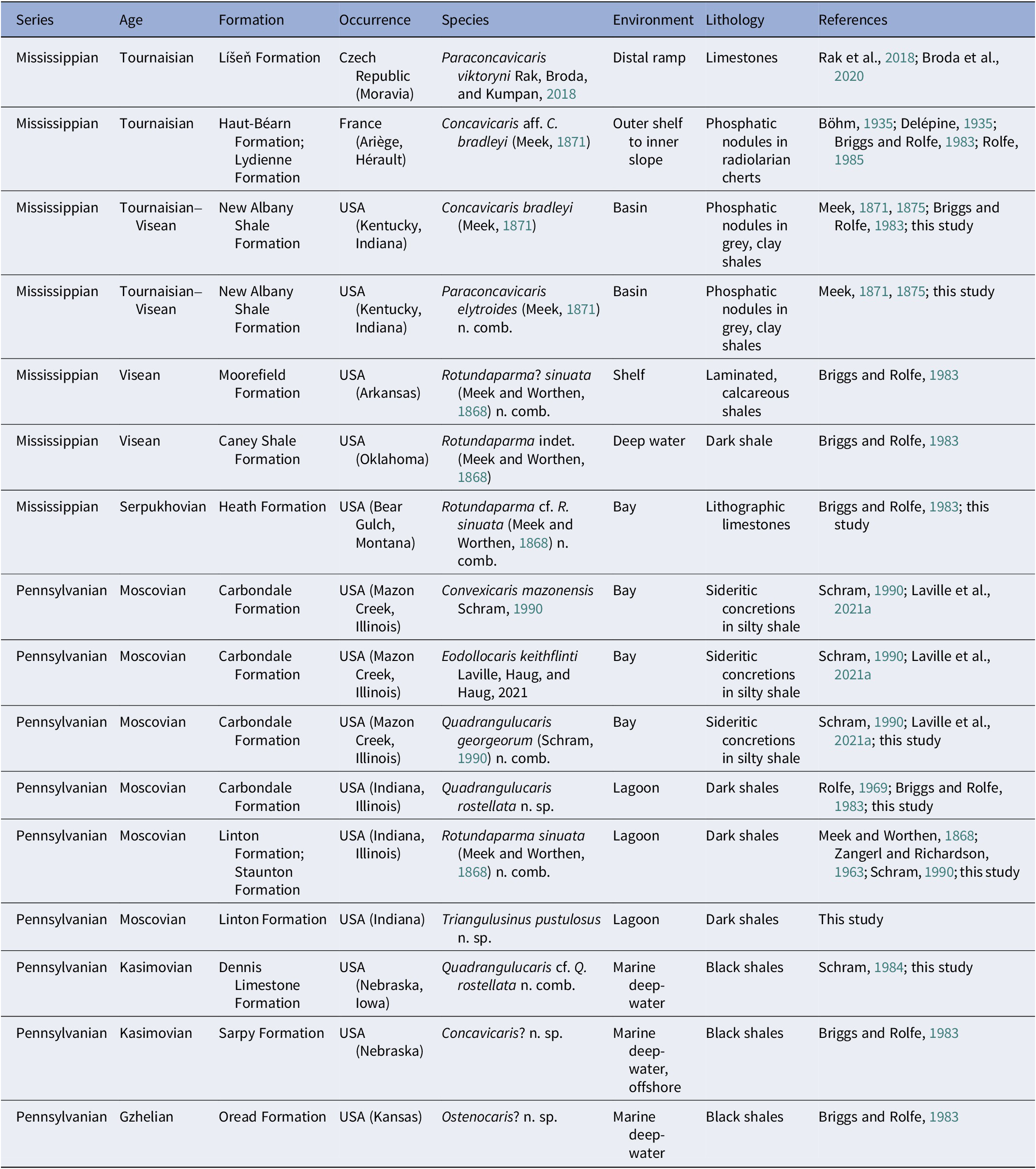
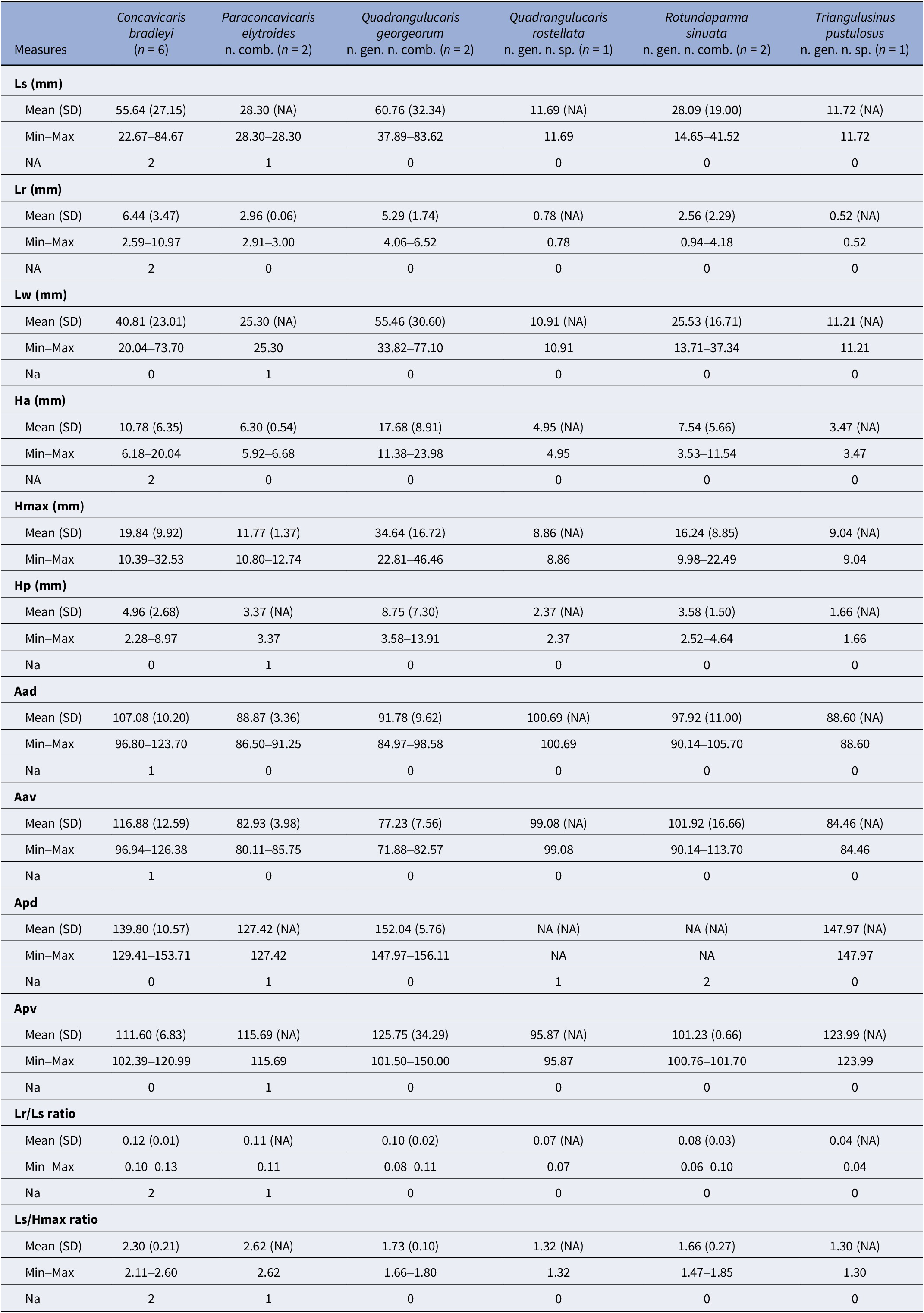
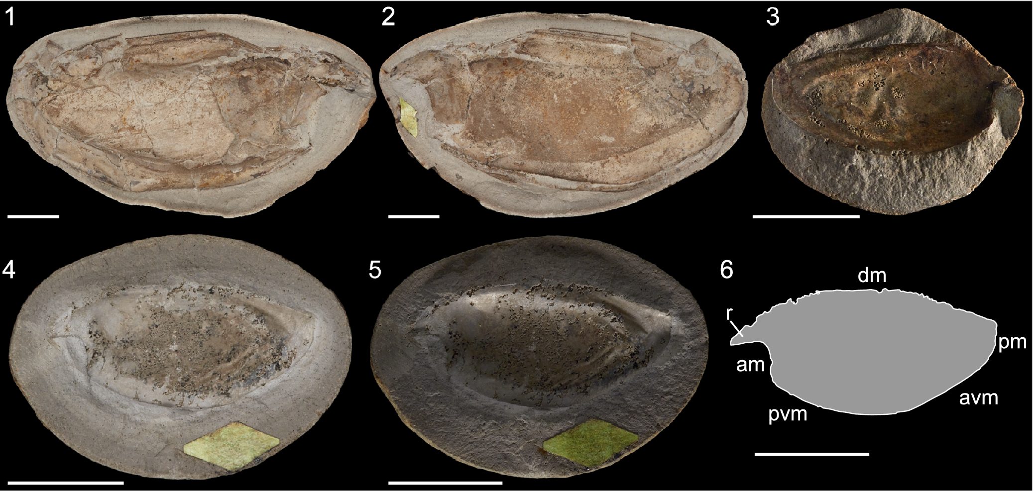
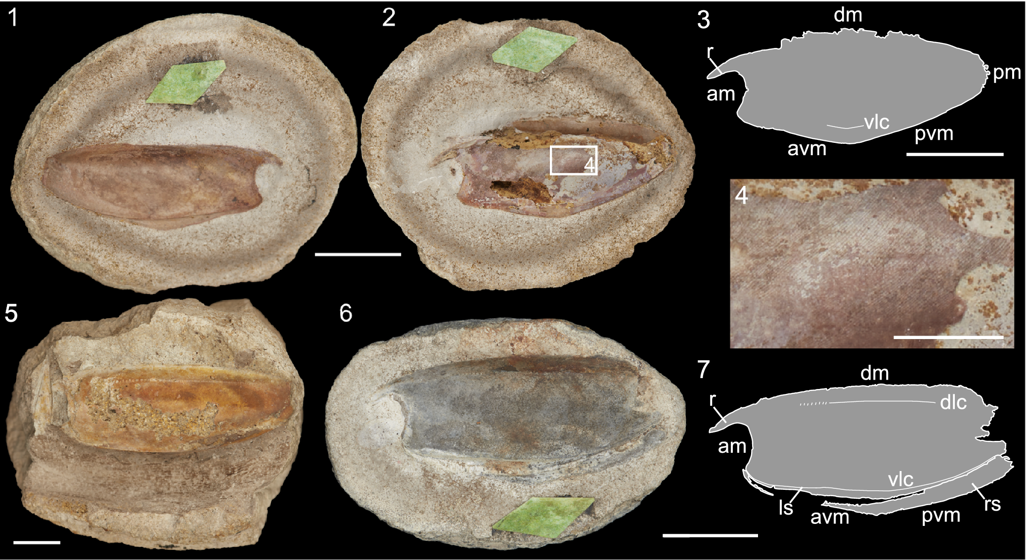
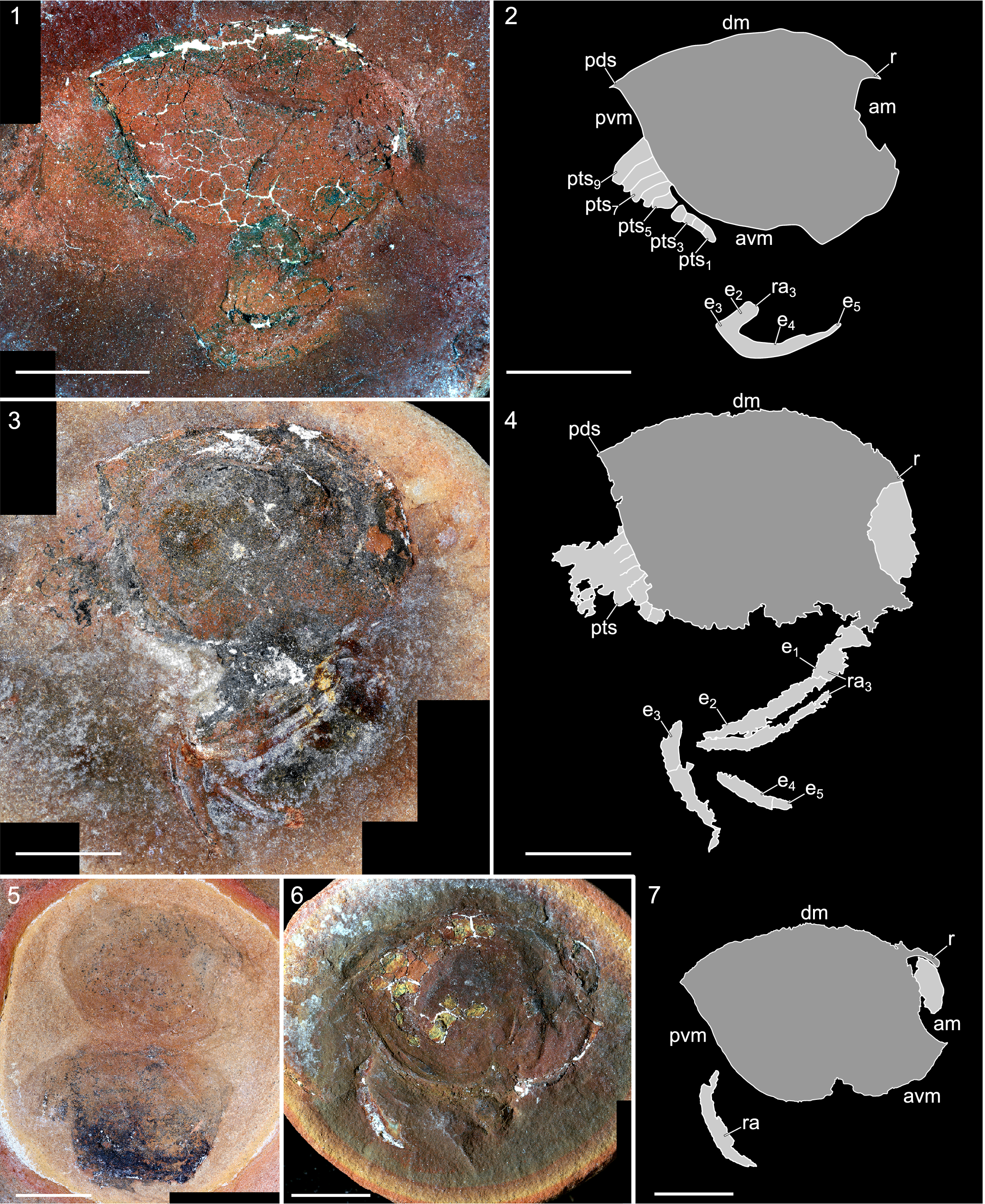
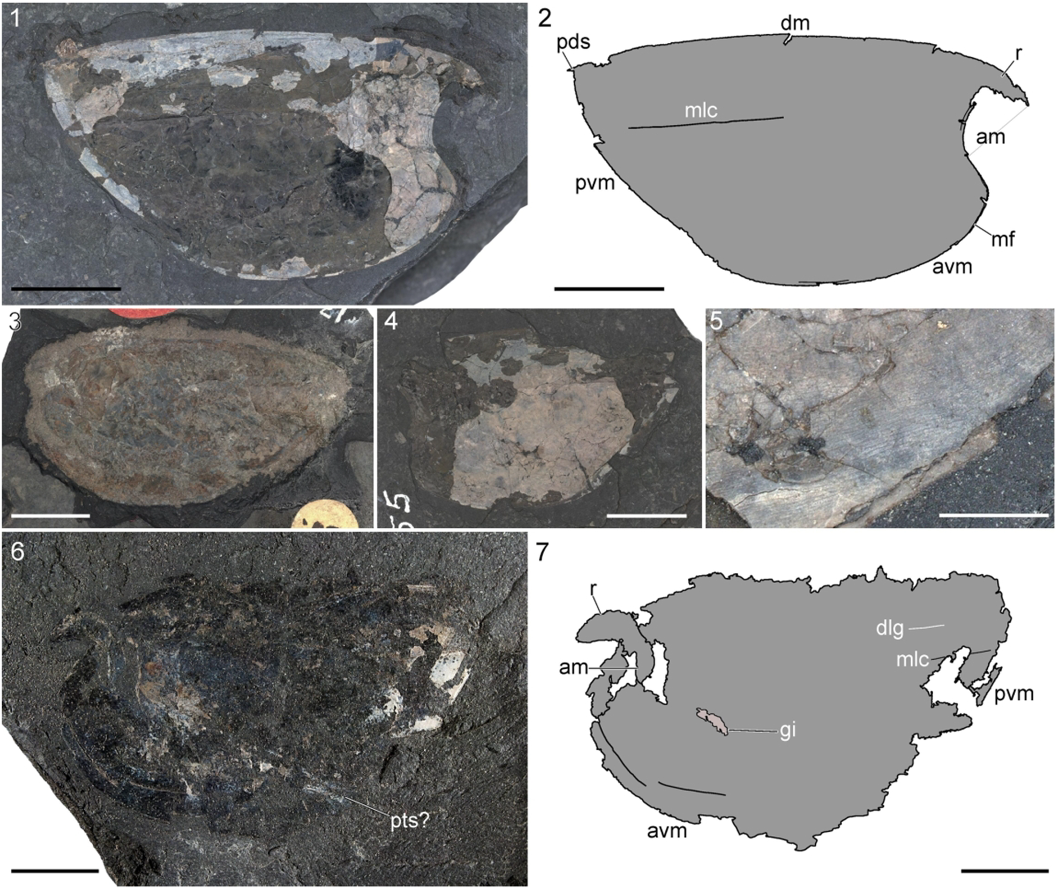
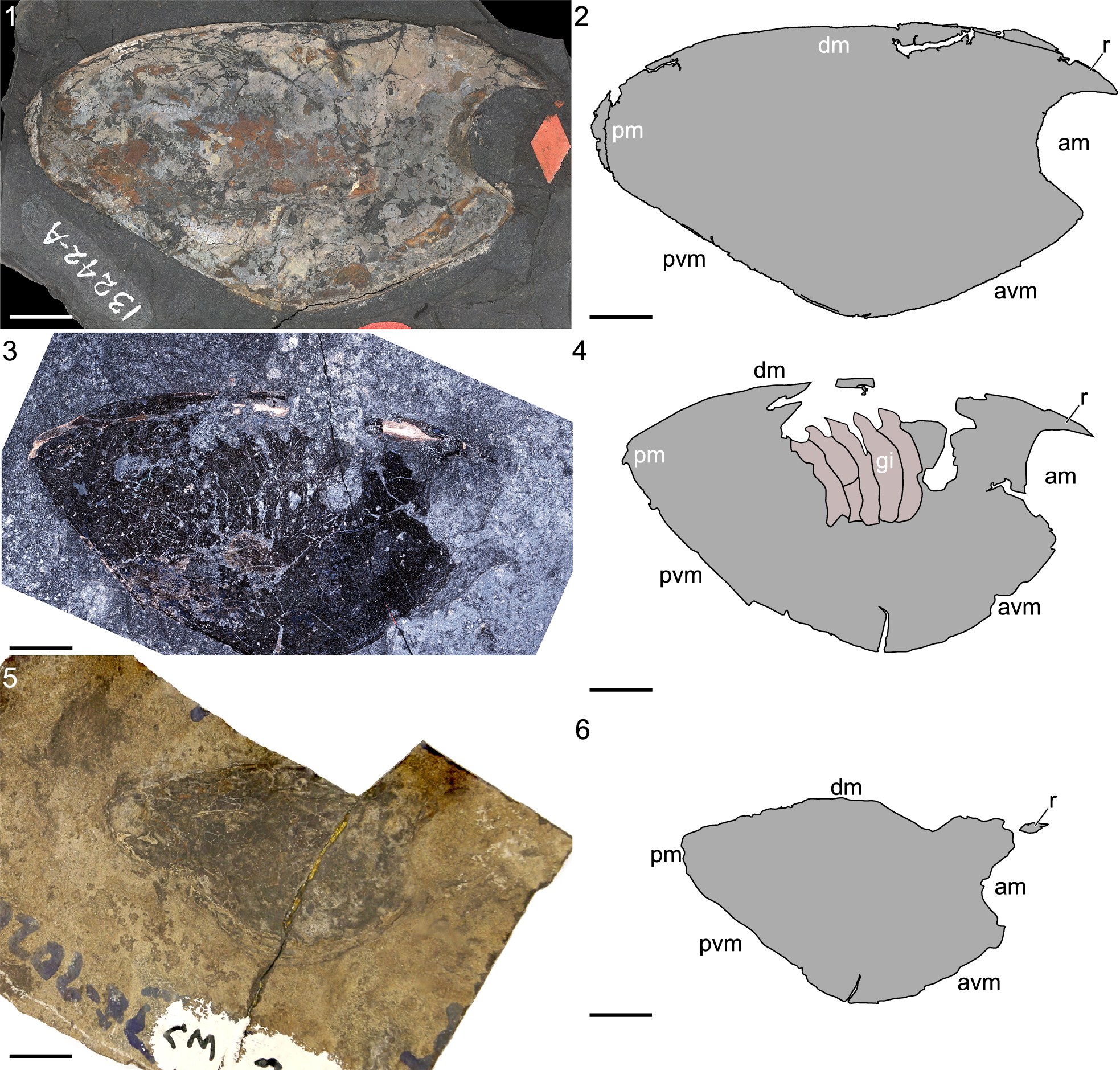
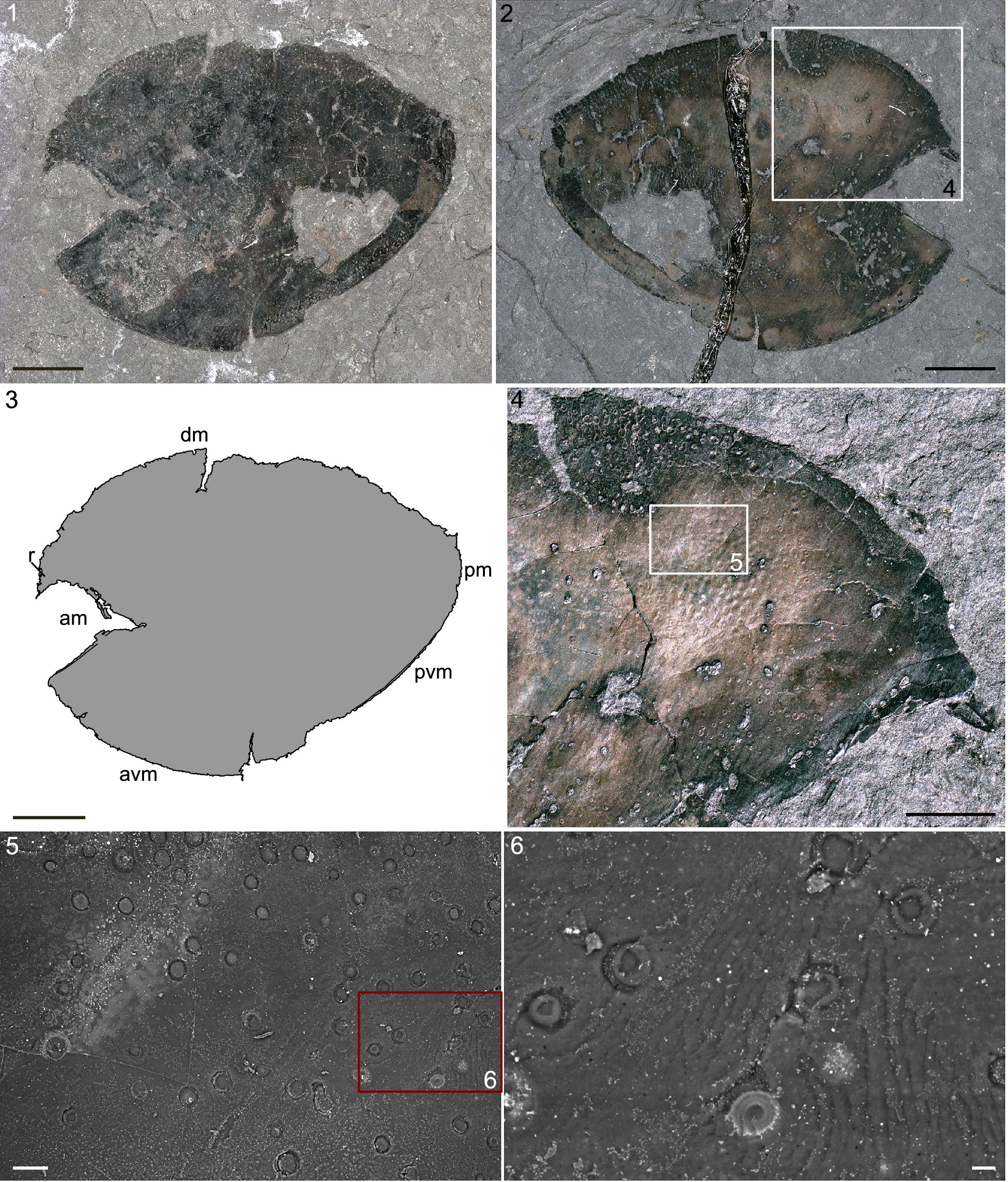
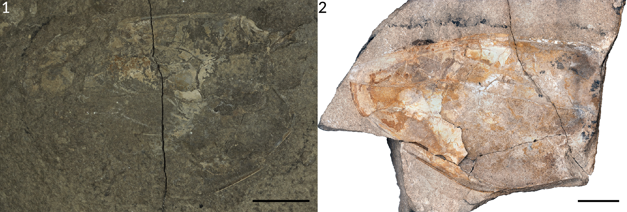

 Open access

