Introduction
Epiphora may be caused by anatomical or functional (physiological) lacrimal obstruction.Reference Paulsen1 Functional lacrimal obstruction is thought to be caused by a reduced contribution of the eyelids or puncta to the lacrimal pump mechanism.Reference Rosenstock and Hurwitz2 A diagnosis of functional obstruction may be made when syringing demonstrates no anatomical obstruction of the lacrimal system, but when a physiological test, such as dye testing or scintigraphy, demonstrates reduced passage of dye or tracer through the lacrimal system.Reference Cuthbertson and Webber3
The surgical treatment for lacrimal obstruction is dacryocystorhinostomy (DCR), which involves marsupialisation of the lacrimal sac into the nasal cavity. Dacryocystorhinostomy can be performed either externally or endoscopically; the results of both techniques are similar, with a success rate in the region of 90 per cent.Reference Sprekelsen and Barberan4, Reference Yung and Hardman-Lea5 Many authors have recommended that DCR should be performed for cases with a diagnosis of anatomical but not functional obstruction.Reference Conway6 A literature search identified five previous publications reporting the results of DCR performed for functional obstruction; an external approach was used in three reportsReference O'Donnell and Shah7–Reference Delaney and Khooshabeh9 and an endoscopic approach in two.Reference Moore, Bentley and Olver10, Reference Wormald and Tsirbas11
We performed endoscopic DCR for cases with diagnoses of anatomical and of functional obstruction. In this paper, we report results for the latter cases (we reported our results for anatomical obstruction in a previous paper).Reference Yung and Hardman-Lea12
Materials and methods
Sixty-eight primary endoscopic DCRs were performed on 44 patients for the treatment of functional obstruction, at the Ipswich Hospital National Health Service Trust between February 1998 and December 2004. The mean age of the patients was 64 years, with a range of three to 86 years. Twenty-six patients (37 per cent) were male and 42 (63 per cent) were female. Surgery was performed on the right side in 31 cases (46 per cent) and on the left side in 37 cases (54 per cent). The indication for surgery was epiphora in 66 cases (97 per cent) and recurrent dacryocystitis in two (3 per cent). Revision cases were excluded from this study.
All cases of epiphora were seen in an ‘epiphora clinic’ at the Ipswich Hospital. Cases with hyper-lacrimation or lid or punctal problems were identified and treated separately by the ophthalmology department. The remaining cases were then assessed jointly by an ophthalmologist (SHL) and an otolaryngologist (MY). All cases were investigated by lacrimal syringing. If syringing demonstrated an obstruction, a diagnosis of anatomical obstruction was made. If it demonstrated a patent lacrimal system, a scintiscan was performed. A diagnosis of functional obstruction was made if the scintiscan demonstrated reduced passage of radio-labelled tracer from the eye to the nose, in spite of a patent lacrimal system demonstrated by syringing.Reference Cuthbertson and Webber3 The present study was of cases thought to have functional obstruction of the lacrimal system.
In all cases, at operation, bone from the frontal process of the maxilla was removed to expose the lacrimal duct and sac. The medial wall of the sac was then removed with a keratome and cutting forceps and a silicone stent was inserted, as described in a previous publication.Reference Yung and Hardman-Lea5 In 32 cases (47 per cent), a septoplasty was also performed to improve access. The operation was performed under local anaesthetic (using a lacrimal fossa block)Reference Smith, Kong, Yung, Hardman-Lea and Leary13 in 59 cases (87 per cent) and under general anaesthetic in nine cases (13 per cent). Stents were removed three months post-operatively in all cases, and the minimum post-operative follow up was six months. Clinical outcome measurement was based on the percentage of cases free from epiphora, as recommended by the Royal College of Ophthalmologists.14 Results were recorded as complete cure, partial improvement or no improvement.
As part of a separate study, part of the medial wall of the lacrimal sac was sent for histological examination in 193 consecutive endoscopic DCRs performed for anatomical obstruction (178 cases) and for functional obstruction (15 cases), between January 1999 and December 2001. This study has been published previously.Reference Merkonidis, Brewis, Yung and Nussbaumer15
A literature search for previous publications reporting the results of DCRs performed for functional obstruction was performed using the Medline database. The search strategy was (dacryocystorhinostomy.tw or dacrocystorhinostomy.tw or dcr.tw) and (functional.tw or physiological.tw or pump failure.tw), limited to the English language and the years 1966 to the present. Each publication was summarised according to surgical approach (external or endoscopic), number of patients, outcome and follow up.
Results
The results of this series of 68 functional blockages, and of our previously reported series of 171 anatomical blockages, are shown in Table I. In the functional blockage series, there was complete cure in 44 cases (65 per cent), partial improvement in nine (13 per cent) and no improvement in 15 (22 per cent). Revision surgery was performed on five of the 15 cases with no improvement, after which there was an overall complete cure in 47 cases (69 per cent), partial improvement in 10 (15 per cent) and no improvement in 11 (16 per cent). In the anatomical series, there was complete cure in 152 cases (89 per cent), partial improvement in 10 (6 per cent) and no improvement in nine (5 per cent).
Results of primary endoscopic DCRs performed for functional and anatomical lacrimal obstruction

DCR = dacryocystorhinostomy
The results of histological analysis for 193 endoscopic DCRs performed for cases diagnosed with anatomical obstruction (178 cases) and functional obstruction (15 cases) are shown in Table II. In specimens from cases of anatomical obstruction, histological analysis revealed no abnormality in 44 cases (25 per cent), chronic inflammation in 132 (74 per cent) and specific pathology (sarcoidosis) in two (1 per cent). In specimens from cases of functional obstruction, histological analysis showed no abnormality in zero cases (0 per cent), chronic inflammation in 14 (93 per cent) and specific pathology (transitional cell papilloma) in one (7 per cent).
Results of histological analysis for primary endoscopic DCRs performed for functional and anatomical lacrimal obstruction

* Transitional cell papilloma; †sarcoidosis. DCR = dacryocystorhinostomy
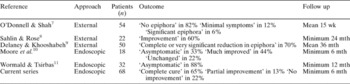
The results of previous series identified by the literature search, plus the current series, are summarised in Table III. The instruments used in the endoscopic series were: cold steel (current series), cold steel or laser,Reference Moore, Bentley and Olver10 and microdebrider.Reference Wormald and Tsirbas11
Results of six series of DCRs performed for functional lacrimal obstruction

DCR = dacryocystorhinostomy; wk = weeks; mth = months
Discussion
The definition of ‘functional obstruction’ is not robust, and many ophthalmologists relate it to pump failure of the outflow system. This could be a reflection of the imprecision of the current diagnostic tools for epiphora. Syringing is a subjective assessment and may not always separate out mild outflow obstruction and pump failure. In our institution, a diagnosis of functional blockage was made by elimination of any obvious lid or punctal problems, together with lack of any anatomical obstruction demonstrated on syringing. It is interesting that, in the present series, two cases presented with recurrent dacryocystitis suggestive of disease within the lacrimal sac rather than of pure pump failure.
Our current and previous series both show a high success rate for DCR performed in cases with a diagnosis of functional obstruction, although comparison between the series is limited by the different surgical techniques and the different reporting methods used. Such a high success rate might indicate that many cases were in fact partial anatomical obstruction.
This explanation is supported by the results of our series of biopsies taken routinely from the lacrimal sac in cases diagnosed with functional obstruction. The finding of chronic inflammation in a very high proportion of cases, and of one neoplasm of the lacrimal sac, reflects the imprecision of the diagnostic investigations used by the authors. However, the absence of a control group means that it is not possible to be sure that these histological findings differ from those of a normal population without lacrimal obstruction.
• This study reports a series of 68 endoscopic dacryocystorhinostomies (DCRs) performed for patients diagnosed with functional lacrimal obstruction
• There was complete cure in 44 cases (65 per cent), partial cure in nine (13 per cent) and no improvement in 15 (22 per cent)
• Endoscopic DCR is indicated in patients diagnosed with functional lacrimal obstruction. Currently available investigations cannot reliably distinguish partial anatomical obstruction from pump failure
There is an urgent need to improve the diagnostic investigation of epiphora, in order to differentiate genuine pump failure from outflow obstruction. Until such a method is found, these patients should not be denied surgery on the basis of syringing, dye testing and/or scintiscanning alone. One should look for other factors that may assist case selection for DCR. A history of facial palsy suggests pump failure, and a history of dacryocystitis suggests anatomical obstruction.