Introduction
Mild behavioral impairment (MBI) is increasingly recognized as a premonitory stage of dementia. Initially, conceived as a descriptor of pre-FTD behavioral changes (Taragano et al., Reference Taragano, Allegri, Krupitzki, Sarasola, Serrano and Lyketsos2009), the construct has evolved to describe a preclinical stage of all cause dementia (Ismail et al., Reference Ismail2016). MBI is characterized by later life acquired, sustained, and impactful neuropsychiatric symptoms (NPS) of any severity that cannot be better accounted for by other formal medical and psychiatric nosology. MBI is an “at risk” state for incident cognitive decline and dementia, and for some, MBI is the index manifestation of neurodegeneration, observed in advance of cognitive impairment. The International Society to Advance Alzheimer's Research and Treatment-Alzheimer's Association (ISTAART-AA) has published proposed research diagnostic criteria for MBI which operationalize the relationship between mild cognitive impairment (MCI) and MBI, in that MBI can occur in advance of MCI, or in concert with MCI, or even after MCI (but by definition, in advance of formal dementia diagnosis) (Ismail et al., Reference Ismail2016).
However, the measurement of NPS in preclinical populations remains challenging and underappreciated, where screening priorities are for cognitive symptoms and not neurological or neuropsychiatric ones (Ismail and Mortby, Reference Ismail, Mortby, Chiu and Shulman2017). NPS have been long described in dementia, and rating scales for NPS in neurodegenerative diseases are dementia focused, generally with a short 2–4 week reference range, and are silent on the natural history and onset, course, and duration of symptoms. Thus, it is not yet common practice to consider formal neurodegenerative disease NPS scales in preclinical populations, when formal psychiatric and neurological illnesses are usually the subject of concern. An MBI specific rating scale has been developed, the MBI checklist (MBI-C, www.MBItest.org) which is based on the ISTAART-AA MBI criteria. The goal of the MBI-C is to assist in the assessment of later life onset NPS, with neurodegenerative disease in mind.
Given the novelty of the MBI concept and the importance of considering NPS preceding all cause dementia (and not just FTD), we present a series of three cases from the Cognitive Neurosciences Clinics at the University of Calgary, Canada, which illustrate clinical manifestations of MBI. For all cases, MBI diagnosis was made in accordance with the ISTAART-AA MBI criteria, based on clinical history from patient and informant. The MBI-C was used to detect and describe the NPS in this clinical setting, and was completed by the informant. Case 1 presents the clinical case of a 57-year old man who met MBI criteria for affective dysregulation and impulse dyscontrol and who went on to develop Frontotemporal Dementia (FTD). Case 2 presents the clinical case of a 78-year old man who met the MBI criteria for decreased motivation and went on to develop probable Alzheimer's disease. Finally, case 3 presents the clinical case of a 64 year-old woman who presented with co-morbid MBI and MCI, meeting the MBI criteria for decreased motivation and affective dysregulation and who progressed to probable Alzheimer's disease. Informed consent letters were obtained from all patients or their formally designated substitute decision makers.
Case 1: Affective dysregulation and impulse dyscontrol
A 57 year-old male initially manifested behavioral symptoms at the age of 54. He presented with a three-year history of anxiety, irritability, and anger, followed one year later by problems with attention and planning. Ultimately, a clinical diagnosis of FTD was made, according to the International Behavioral Variant FTD Criteria Consortium clinical diagnostic criteria for behavioral variant FTD (Rascovsky et al., Reference Rascovsky2011).
Symptom presentation
The patient's wife first noticed changes in his personality about three years prior to clinic assessment, one year after they had moved from overseas. The patient became short-tempered and easily frustrated. It seemed to the patient's wife that his angered responses were out of proportion to the situation. For instance, he smashed and broke their computer after having difficulty in operating it during an online video call. There were multiple other examples of the patient causing physical damage to their home in periods of frustration, for instance by punching holes in walls. This behavior was out of keeping with his premorbid personality.
The patient's wife initially discounted the changes as situational, given their move and change in living situation, but these personality changes persisted and were followed by cognitive symptoms. Eventually, the patient also became easily distractible and less attentive, having a harder time keeping track of information and remembering details of conversations. He remained functional with respect to activities of daily living and as far as his wife was aware he remained able to perform work duties without complaints from coworkers or supervisors as to his work performance. The patient worked as a manager at a home renovation and supplies store. His wife reported the one oddity she noticed was that he had started to bring items home from work. She would find these items in the pockets of his clothes. The patient told his wife that he had forgotten to put these items back before leaving work, but it was unclear if this was due to forgetfulness or if it was new-onset shoplifting.
Clinical assessment
Cognitive testing on initial assessment included the Consortium to Establish a Registry for Alzheimer's Disease (CERAD) battery in which he scored 121. His Mini-Mental Status Exam (MMSE) score was 28/30. His Montreal Cognitive Assessment (MoCA) score was 26/30. He drew a normal clock. His Frontal Assessment Battery score was 17 out of 18. His Geriatric Depression Scale (GDS) score 3/15. All scores were within normal limits.
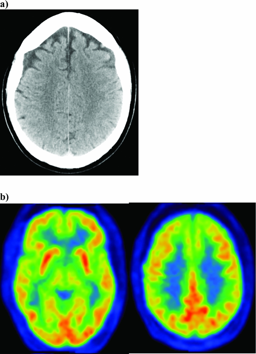
Neuroimaging ordered at initial assessment included a non-contrast CT scan of the head, which showed mild atrophy of the frontal lobes bilaterally. There were contraindications to performing MRI. An FDG PET scan of the brain, done within two months, showed hypometabolism in the frontal and temporal lobes and to a milder degree in the left insular cortex supportive of early changes related to FTD (Figure 1).
Case 1 (a) Non-contrast CT head showing mild atrophy of the frontal lobes bilaterally (b) FDG PET scan brain showed hypometabolism in the frontal and temporal lobes and to a milder degree in the left insular cortex.

Neuropsychiatric symptoms
At initial consultation, clinical assessment of NPS using the neuropsychiatric inventory questionnaire (NPI-Q), revealed moderate levels of disinhibition while levels of agitation and irritability were rated as mild. Administered at the same time, the MBI-C showed moderately severe symptoms of agitation, aggressiveness, argumentativeness, and impulsivity. Disinhibition and loss of empathy were noted to be present and mild in the severity rating. Thus, at initial consultation, the patient met ISTAART-AA criteria for MBI, exhibiting symptoms in the MBI domains of affective dysregulation and impulse dyscontrol. Neuroimaging showed changes consistent with FTD and in conjunction with further clinical decline served to support of a final diagnosis of FTD.
Treatment
The patient showed improvement in his NPS with the initiation of serotonin reuptake inhibitors but continued to experience cognitive symptoms.
Case 2: Decreased motivation
A 78 year-old man presented with a four-year history of cognitive decline, predominantly affecting short-term memory, but complicated by accompanying depressive symptoms. The patient was given a diagnosis of Alzheimer's dementia based on NIA-AA criteria (McKhann et al., Reference McKhann2011), possibly of the frontal variant type. Cholinesterase inhibitors were initiated. There was improvement of the patient's behavioral symptoms and stabilization of his cognitive performance.
Symptom presentation
The patient's wife noted a change in her husband in the spring four-years prior, when he stopped attending cancer support group meetings. He had been attending these meetings for over a decade before that. In fact, this was a support group that he had started and watched grow with success. He was proud of this achievement and his wife became worried that he no longer showed interest in remaining involved. He became socially withdrawn on many fronts, but not overtly sad, which again was unusual for him as his wife described him as a previously socially engaged person.
Clinical assessment
Around the same time, his wife noted that he was more forgetful. His cognitive screening tests at that time, including the MMSE and MOCA, were within normal range and he was treated for depression, with little effect. One year later, his cognitive symptoms became worse and began to interfere with daily function. Neuropsychological testing revealed deficits in attention, episodic memory and visuospatial function. His wife reported needing to increasingly provide him with reminders to take his medications, attend appointments, and eventually maintain hygiene. Repeat assessments showed a measurable decline in cognitive testing. Over the course of the year, his cognitive screening test scores declined to 25/30 on his MMSE, 17/30 on the MoCA, and 83 on the CERAD battery. His GDS score was 0.
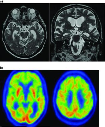
Neuroimaging included an MRI brain, which showed predominantly temporal lobe atrophy and a mild degree of chronic small vessel disease. FDG PET scan of the brain one year after he developed cognitive symptoms showed hypometabolism in the frontal lobes, including the insular cortex with a less prominent degree of hypometabolism in the temporal lobes and the inferior parietal lobes, posterior cingulate gyrus and the convexity of the temporal lobes (Figure 2).
Case 2 (a) MRI brain showed predominantly temporal lobe atrophy and a mild degree of chronic small vessel disease. (b) FDG PET scan brain showed hypometabolism in the frontal lobes, including the insular cortex with a less prominent degree of hypometabolism in the temporal lobes and the inferior parietal lobes, posterior cingulate gyrus, and the convexity of the temporal lobes.

Neuropsychiatric symptoms
Retrospectively, the patient would have met ISTAART-AA criteria for MBI at the time of initial presentation to primary care, where he demonstrated late life onset of apathy and social withdrawal, one-year prior to the emergence of cognitive symptoms. While he exhibited symptoms in the MBI domain of decreased motivation/drive, with a lack of alternative explanations, the most convenient and related psychiatric diagnosis was provided – depression. He was treated with antidepressants, but with little effect.
Case 3: Decreased motivation and MCI
A 64 year-old woman presented with a four-year history of insidious cognitive decline that was accompanied by symptoms consistent with apathy by two years, but initially diagnosed and treated as depression. A diagnosis of probable Alzheimer's dementia based on NIA-AA criteria was later made (McKhann et al., Reference McKhann2011).
Symptom presentation
Four years into the disease course, the patient began to show signs of dependency on others, and her husband took over managing the household finances. In the year following her loss of independence with respect to financial management, the rate of her decline accelerated as she displayed loss of insight, a steeper decline in daily function, and was unable to participate actively during clinic visits due to the degree of her cognitive impairment. During cognitive clinic visits, she perseverated on somatic complaints like neck and shoulder pain.
Clinical assessment
Cognitive screening tests revealed impairment in multiple domains including orientation, attention, recall, language, and construction. Cognitive screening tests included a CERAD battery on which she scored 67, MMSE on which she scored 22/30 and the Rowland Universal Dementia Assessment Scale (RUDAS) on which she scored 16/30, well below the dementia cut-off of 23. She scored 5/15 on the GDS.
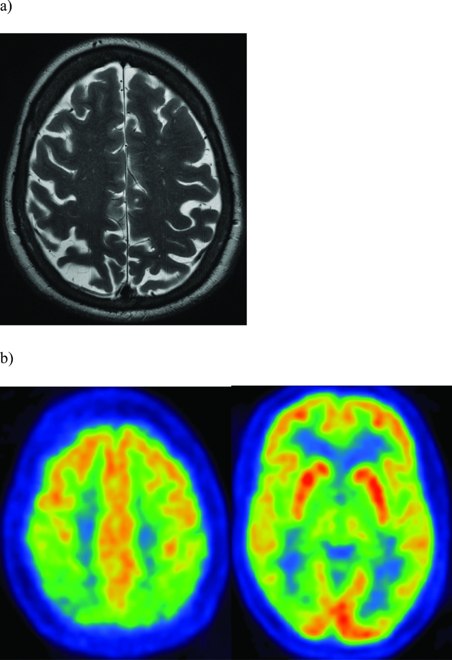
Neuroimaging included an MRI of the brain and an FDG PET scan (Figure 3). The MRI brain showed marked generalized atrophy with bilateral parietal lobe predominance. The FDG PET scan showed hypometabolism of the parietal lobes and to the lesser extent the temporal lobes, more prominently on the right then the left, a consistent with changes of neurodegenerative disorder of the Alzheimer's type.
Case 3 (a) MRI brain showed generalized atrophy with parietal lobe predominance. (b) FDG PET scan showed hypometabolism of the parietal lobes and to the lesser extent the temporal lobes, more prominently on the right then the left.

Neuropsychiatric symptoms
Retrospectively, the patient would have met ISTAART-AA criteria for MBI concurrent with MCI, exhibiting symptoms in the domain of decreased motivation/drive with initial presentation of behavioral and cognitive symptoms at age 60. Similar to case 2, in the absence of alternative nosological options, a diagnosis of depression was given, and treated unsuccessfully with antidepressant medications.
Discussion
This series of case reports aimed to illustrate that when NPS emerge in later life, and result in what is now defined as MBI, they can represent a premonitory symptom of dementia. We present narrative examples of neurodegenerative diseases affecting cognition that were preceded by NPS as outlined by the MBI criteria. These cases illustrate that early NPS occur in different forms of dementia, including but not exclusive to FTD. While FTD is more traditionally associated with early neuropsychiatric features, other dementias can also present early with NPS (Taragano et al., Reference Taragano, Allegri, Krupitzki, Sarasola, Serrano and Lyketsos2009). Often, patients with neurodegenerative disease and early NPS are given psychiatric diagnoses, which then evolve into clearer pictures of FTD, AD, or other dementias (Woolley et al., Reference Woolley, Khan, Murthy, Miller and Rankin2011). However, being given inappropriate psychiatric diagnoses can result in potential exposure to unnecessary medications or delays in the care and management of the underlying neurodegenerative disease (Jalal et al., Reference Jalal, Ganesh, Lau, Lysack and Ismail2014).
Several recent longitudinal studies have supported the consideration of later life onset psychiatric symptomatology as prodromal manifestations of dementia. A Finnish case-control study of 27,948 pairs assessed linkages between psychiatric illness and dementia risk. The findings were that the width of the time window between the exposure (psychiatric syndrome) and the outcome (AD) affected the results. The authors determined that having a mental disorder was associated with higher risk of AD with a five-year time window but not with a ten-year window, and suggested that: “some of the disorders may represent misdiagnosed prodromal symptoms of AD, which underlies the importance of proper differential diagnostics among older persons” (Tapiainen et al., Reference Tapiainen, Hartikainen, Taipale, Tiihonen and Tolppanen2017). Similarly, a 28-year follow-up of the Whitehall II cohort study in the United Kingdom assessed depression and dementia incidence in 10,308 participants. Late-phase, but not early-phase depressive symptoms, had a higher risk of dementia, suggesting that depressive symptoms are a prodromal feature of dementia or that they share common etiology (Singh-Manoux et al., Reference Singh-Manoux2017). A 14-year Australian study of 4,922 cognitively healthy men assessed baseline risk factors for development of dementia. The association between depression and dementia was only apparent during the initial five years of follow-up, suggesting that depression was more likely a marker of incipient dementia rather than a modifiable risk factor (Almeida et al., Reference Almeida, Hankey, Yeap, Golledge and Flicker2017).
The MBI construct and MBI-C fit nicely into the discussion raised by these important recent publications, and that age of onset, the degree to which symptoms are sustained, and time frames are essential to accurate diagnosis and understanding of psychiatric illness. The goal of the MBI-C is case detection, based on an explicit translation of the ISTAART-AA MBI criteria. It consists of 34 questions representing the MBI domains of apathy, emotional dysregulation, impulsivity and agitation, social cognition, and psychosis and is generally completed by an informant. The MBI-C uses non-dementia language intended for functionally independent adults, a reference range of six-months, and a stipulation that symptoms are acquired in later life (Ismail et al., Reference Ismail2017).
To illustrate the applicability of the MBI-C to the different types of dementia, our cases include patients with different dementia types, a classic behavioral variant FTD presentation, a frontal AD, and an early onset AD. The MBI-C is not meant to supplant the NPI, but rather to complement it. The NPI and NPI-Q are the gold standards for measurement of NPS in dementia, and for detecting change over time. The NPI and NPI-Q have been seminal in the evolution of our understanding of neurodegenerative disease, and are instrumental in the ongoing assessment of dementia. We hope the MBI-C will further establish the importance of recognizing NPS in dementia, and extend this awareness earlier in the disease course. The MBI-C may also serve as an educational tool to emphasize the difference between chronic symptoms and sustained new-onset psychiatric symptoms in older adults and encourage clinicians to think of neurodegenerative diseases in the differential diagnosis when symptoms emerge in late life.
Validation of the MBI-C will allow examination of the patterns of NPS among the different types of dementia. In this case report, we show how the MBI-C can be used as a tool in MBI diagnosis, and early identification of preclinical dementia. For example, in our first case, the patient presented with angry outbursts, irritability, and tendency to violent expression of anger, representing the MBI domain of impulsivity and agitation. His wife described a distinct change in personality as he progressed to developing FTD. In the latter two cases, both patients developed early symptoms consistent with apathy (representing the MBI domain of drive and motivation, with the addition of emotional dysregulation in the third), and eventually developed Alzheimer's disease.
Just as we see different patterns of cognitive impairment in the different forms of dementia, it may be that we will also see different patterns of early NPS in different pathologies. If we are able to identify these patterns with the MBI-C, we will be able to predict with higher sensitivity the etiology of the dementing illness. For instance, could MBI with predominant early apathy be the equivalent of amnestic MCI in predicting the course of illness? Future research is needed which investigates the use of the MBI-C to examine whether particular MBI domains are predominantly present among specific dementia etiologies. It will be important to link these early NPS to neurobiology. The ISTAART group of researchers have explored models that help conceptualize the neurobiology of neuropsychiatric disease in dementia, and these models will help address issues around early detection of identifying dementia and identifying NPS treatment targets (Geda et al., Reference Geda2013; Rosenberg et al., Reference Rosenberg, Nowrangi and Lyketsos2015; Lanctôt et al., Reference Lanctôt2017). Models include the frontal-subcortical circuitry, cortico–cortical networks, and the monoaminergic system; neuroimaging and biomarkers have implicated all these models in the neurobiology underlying NPS in AD (Geda et al., Reference Geda2013). The MBI-C will be advantageous in identifying the NPS that manifest at the earliest stages of AD and as such provide further insight into the neurobiology of AD. Using neuroimaging and biomarkers for lesion localization in conjunction with detecting behavioral changes earlier with the MBI-C may allow for more accurate attribution of behavioral symptomology to lesion location.
A further diagnostic strength of the MBI-C is that it can be applied as patients evolve in their dementing illness. Measuring the change that occurs in behavior will add to the information gained in measuring cognitive decline, and assist in overall illness prognostication. Understanding the evolution of NPS among the different types of dementia may lead to improved understanding and management of NPS and cognition in these patients.
Similar arguments for the utility of identifying MCI are relevant to identifying patients with MBI. Early identification of neurodegenerative illness allows an approach to studying prevention of progression to dementia and to investigating future disease modifying agents earlier in the disease course (Petersen et al., Reference Petersen, Stevens, Ganguli, Tangalos, Cummings and DeKosky2001). Furthermore, we suspect that earlier treatment of behavioral symptoms might lead to improved functional outcomes for the patient and may affect the management of caregiver stress and burden, consequently improving the patient's quality of life (Fischer et al., Reference Fischer, Ismail and Schweizer2012).
In summary, NPS can often emerge in later life, and may predict or precede the cognitive impairment and dementia. Current psychiatric nosology does not adequately frame these NPS as a dementia precursor. The construct of MBI aims to identify the importance of symptom natural history, and that later life onset of symptoms require closer consideration for alternative etiologies. The MBI-C is a tool designed for MBI case detection, and may have utility into increasing detection and awareness of later life onset NPS and their link to neurodegenerative disease.
Conflict of interest
None.
Acknowledgments
This work was supported through the Alzheimer Society of Calgary via the Hotchkiss Brain Institute.