Introduction
Kimura's disease is a chronic inflammatory disorder of unknown aetiology that is endemic in the Orient. It usually presents as painless unilateral cervical lymphadenopathy or as subcutaneous masses in the head or neck region.Reference Weiss, Goldblum, Enzinger and Weiss1–Reference Mrówka-Kata, Kata, Kyrcz-Krzemien and Helbig3 There is controversy in the literature regarding whether Kimura's disease and angiolymphoid hyperplasia with eosinophilia are the same entity. Some authors believe that Kimura's disease represents a chronic form of angiolymphoid hyperplasia with eosinophilia that involves deeper tissues; however, most recent papers distinguish between the two conditions.Reference Kuo, Shih and Chan4 This paper presents a report of eight cases of Kimura's disease and a brief review of the literature.
Materials and methods
We reviewed all cases of Kimura's disease diagnosed by the pathology department at the Government Medical College and Hospital, Chandigarh, India over a 10-year period (between January 1997 and December 2007). All the patients’ clinical records were retrieved and haematoxylin and eosin (H&E) stained slides were re-examined.
Results
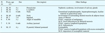
The 8 cases of Kimura's disease that were reported during the 10-year period are detailed in Table I. The mean age of patients was 22.8 years. There were six males and two females. The duration of symptoms ranged from 7 days to 5 years. Cases 2, 4 and 5 were referred from another centre and no details other than those described below were available.
Kimura's disease: summary of cases

Pt no = patient number; y = years; dur = duration of symptoms; M = male; L = left; mth = months; d = days; F = female; FNAC = fine needle aspiration cytology; M/E = microscopic examination; R = right
Case reports
Case 1
A 30-year-old male presented to the surgery out-patient department with a 4-year history of swelling in the left parotid region. The swelling was painless but had slowly increased in size. On examination, the left preauricular area showed a firm, well-defined, non-tender swelling that was 5 × 3 cm in size. The swelling was free from the underlying bone and the overlying skin was not involved. The rest of the examination was unremarkable.
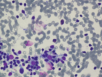
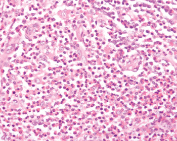
An ultrasound of the region showed a subcutaneous lesion with minimal vascularity. A previous fine needle aspiration of the lesion performed at another centre had shown adenolymphoma; however, the slides were not available for review. Repeat fine needle aspiration cytology (FNAC) performed on the preauricular swelling showed reactive lymphoid hyperplasia with eosinophils (Figure 1). The patient's medical records showed that he had been diagnosed with nephrotic syndrome. Although the swelling was painless, because it persisted and had increased in size, the surgeon suspected a tumour and excised the swelling. A pre-operative haemogram showed eosinophilia with normal total lymphocyte count and haemoglobin levels.
Photomicrograph of fine needle aspirate showing reactive lymphoid cells and some eosinophils. (May-Grünwald-Giemsa stain; ×200)

Gross examination of the excised swelling showed nodular, firm tissue pieces, which were grey-white to grey-brown in colour, and together measured 5.5 × 3.5 × 1.5 cm in size. Microscopic examination revealed salivary gland tissue infiltrated by lymphoid follicles with germinal centres, surrounded by dense, mixed inflammatory infiltrate with predominant eosinophils and eosinophilic microabscesses. Intervening dense fibrous tissue and some vascular proliferation was also reported. A diagnosis of Kimura's disease was made based on these features.
Case 2
A 30-year-old male presented to the eye out-patient department with a 5-year history of swelling over the left eyebrow. The swelling was initially painless, but then became painful. On examination, there was a swelling over the left eyebrow measuring 1.5 cm in diameter. The skin overlying the swelling showed mild erythema; it was tender to touch and not fixed to the underlying bone. Local eye examination and systemic examination showed no abnormalities. The swelling was excised and submitted for histopathological analysis.
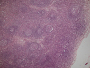
Gross examination of the excised swelling showed a single irregular grey-white to grey-brown soft tissue piece measuring 1.2 × 0.9 × 0.5 cm in size. Microscopic examination revealed that the soft tissue had numerous lymphoid follicles with prominent germinal centres (Figure 2), which are features of Kimura's disease. The intervening area was surrounded by dense, mixed inflammatory infiltrate with predominant eosinophils and fibrosis. The surrounding skeletal muscles were not infiltrated.
Photomicrograph showing numerous lymphoid follicles with reactive germinal centres. (H&E; ×40)

Case 3
A 12-year-old male presented to the paediatric out-patient department with a 6-month history of low grade fever and swelling in the neck region. On examination, there was generalised lymphadenopathy and hepatosplenomegaly. The excised cervical lymph node was sent for histopathological examination. The clinical possibilities included Hodgkin's disease, tuberculosis and crypto coccidioidomycosis.
Gross examination showed a nodular soft tissue mass measuring 1.5 cm in diameter. The cut section was greyish-white, with haemorrhagic areas. Microscopic examination showed multiple matted lymph nodes characteristic of Kimura's disease. In addition, homogeneous hyaline material was present in paracortical areas.
Case 4
A 40-year-old male presented to the ENT out-patient department with a 4-month history of swelling on the right side of the neck. On examination, the swelling was cystic and non-tender, and measured 1.8 cm in diameter. The rest of the examination was unremarkable. The excised swelling was sent to the pathology laboratory for analysis.
Gross examination revealed a single grey-brown soft tissue piece measuring 1.4 cm in diameter. The cut section was grey-white and firm. Microscopic examination showed that skeletal muscle and adipose tissue were infiltrated by numerous hyperplastic lymphoid follicles with germinal centres, and there were eosinophilic abscesses. A diagnosis of Kimura's disease was made on the basis of these observations.
Case 5
A 7-year-old male presented with fever and malaise of 7 days duration. On examination, an enlarged right inguinal lymph node was noted. Excision biopsy of the enlarged node was sent to the pathology laboratory.
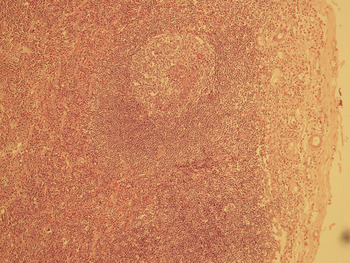
Gross examination showed a lymph node measuring 3 × 1.5 × 0.5 cm in size. Microscopic examination revealed typical features of Kimura's disease: follicular hyperplasia, eosinophilic microabscesses and vascular proliferation (Figure 3). Areas of fibrosis were also reported.
Photomicrograph showing reactive follicles, vascular proliferation and eosinophils. (H&E; ×100)

Case 6
A 40-year-old female presented with a 3-year history of swelling on the right side of her face at the angle of the mandible. The swelling was painless and did not progress in size. On examination, there was a diffuse, non-tender swelling over the lower jaw that measured 3 × 2.5 cm in size. The FNAC performed on the swelling showed lymphoid cells in various stages of maturation, along with some atypical immature cells. There was a high suspicion of malignancy, and histopathological examination was advised.
Gross examination showed a grey-brown soft tissue piece measuring 2 cm in diameter. Microscopic examination revealed skeletal muscle bundles showing the presence of numerous lymphoid follicles with prominent germinal centres. The parafollicular areas revealed eosinophilic abscesses and endothelial proliferation (Figure 4). There was focal evidence of folliculolysis indicated by eosinophils, and occasional areas of fibrinoid necrosis were present.
Photomicrograph showing endothelial proliferation and eosinophilic abscesses. (H&E; ×400)

Case 7
A 38-year-old female presented with a lump in her right breast, which had been there for 5 to 6 months. On examination, a 4 × 4 cm lump was palpated in the upper outer quadrant in the axillary tail region. The lump was firm and freely mobile. A small axillary lymph node measuring 2 cm was also noted. The mammogram indicated fibroadenoma with malignant change and axillary lymphadenopathy. The FNAC performed on the breast lump showed fibroadenosis. The FNAC conducted on the axillary lymph nodes showed reactive lymphoid hyperplasia.
During excision, the surgeon noted a 4 × 4 cm soft to firm swelling in relation to the pectoral group of lymph nodes. The excised swelling measured 4 × 3 cm in size and was encapsulated. The cut section was grey-white, firm and homogeneous. Microscopic examination revealed a lymph node showing follicular hyperplasia with eosinophilic abscesses and a few areas of folliculolysis.
Case 8
A 15-year-old male presented with a 4-year history of swelling in the right parotid area. The swelling had been excised three years previously (the records were not available), but had since recurred and slowly increased in size. On examination, a non-tender swelling measuring 4.5 × 3 cm in size was present over the parotid region. Bilateral postauricular lymph nodes measuring 1.5 to 2 cm in diameter were also reported. The clinical impression was that of lymphoma and parotid malignancy.
A haemogram showed the presence of eosinophilia. An ultrasound of the abdomen and a urine examination revealed no abnormality. The FNAC performed on the bilateral postauricular lymph nodes and the right parotid swelling showed reactive lymphoid hyperplasia with a large number of eosinophils. The right parotid swelling and left postaural lymph nodes were excised and sent for histopathological examination, which revealed features of Kimura's disease. The deposition of eosinophilic material was reported. No parotid tissue was identified.
Discussion
Kimura's disease was first reported in China by Kimm and Szeto in 1937. They described seven cases of a condition they termed ‘eosinophilic hyperplastic lymphogranuloma’.Reference Kimm and Szeto5 The disorder received its current name in 1948, when Kimura et al. noted the vascular component and referred to it as an ‘unusual granulation combined with hyperplastic changes in lymphoid tissue’.Reference Kimura, Yoshimura and Ishikawa6
Kimura's disease is a rare, localised, chronic inflammatory disease. This benign disease involves subcutaneous tissues, the major salivary gland, and lymph nodes primarily in the head and neck area.Reference Weiss, Goldblum, Enzinger and Weiss1, Reference Abuel-Haija and Hurford2 Renal involvement is its only systemic manifestation. There are reports of patients with both Kimura's disease and nephrotic syndrome, but the basis of this association is unclear.Reference Matsuda, Makiguchi, Ishibashi, Chida, Ida and Matsuda7 The typical presentation is that of a slowly enlarging painless mass with occasional pruritus of the overlying skin. Adherences of mass to surrounding structures can mimic tumours. Most of the cases are reported in Asians, and adult males are more commonly affected. Peripheral blood eosinophilia is seen in 98 per cent of the cases and serum immunoglobulin E levels are often raised.Reference Weiss, Goldblum, Enzinger and Weiss1
The cause of Kimura's disease is unknown; however, an allergic reaction or an alteration of immune regulation is suspected. Proposed theories include persistent antigenic stimulation following arthropod bites, and parasitic or candidal infection. To date, none of these theories has been substantiated.Reference Chen, Thompson, Aguilera and Abbondanzo8–Reference Mitsui, Ogino, Ochi and Ohashi10
Our eight cases illustrate some of the variations in the mode of presentation and in the histological features of Kimura's disease. Case 1 is notable as it is a sporadic case in a non-oriental. The patient had presented with a well-defined swelling which led to suspicion of a tumour. There was associated nephrotic syndrome and peripheral blood eosinophilia. Cases 2, 5 and 8 are unusual as Kimura's disease is uncommon in children. These cases showed the typical histopathological features of the disease. However, in case 5, presentation of the disease was in the inguinal region, which is an uncommon site. A study of Kimura's disease in children showed that the postauricular region is the commonest site of involvement. Incidence of the disease is more common in the second decade of life. In this study, recurrence was seen in 16.6 per cent of cases.Reference Viswanatha11 Presentation as bilateral soft tissue masses and recurrence after surgery (as seen in case 8) are also uncommon.Reference Tseng, Lin, Huang and Su12
Kimura's disease was initially regarded as a type of angiolymphoid hyperplasia with eosinophilia that involves deeper tissues. However, most recent publications differentiate between the two conditions on the basis of clinical and histopathological features. Males are more commonly affected by Kimura's disease than females, whereas angiolymphoid hyperplasia with eosinophilia shows a female predominance. Kimura's disease is usually seen in young adults with a median age of 28 years. Patients with angiolymphoid hyperplasia with eosinophilia tend to be in their 30s or 40s. On microscopic examination, Kimura's disease shows lymphoid nodules with discrete germinal centres which can occupy an area extending from the reticular dermis to the fascia and muscle. There is marked eosinophilic infiltrate and eosinophilic abscesses are present. Capillary proliferation is not characteristic, but when present, it manifests as masses of canalised vessels with flat endothelial cells. Fibrosis usually surrounds and may extend into lesions, as seen in our cases 1 and 2. Dense hyaline fibrosis can be seen in the later stages of the disease, as was seen in case 3. In contrast, lesions of angiolymphoid hyperplasia with eosinophilia are more superficial, with lymphoid follicles that lack germinal centres. Although eosinophils are present, eosinophilic abscesses are not observed. Masses of uncanalised vessels are lined with plump, atypical endothelial cells, which are often described as epithelioid in appearance. Fibrosis is less evident in angiolymphoid hyperplasia with eosinophilia.Reference Abuel-Haija and Hurford2, Reference Kung, Gibson and Bannatyne13
• Kimura's disease should be considered in patients with head and neck swellings and lymphadenopathy
• It predominantly affects adult males but should not be ruled out in females and children
• Pathologists need to be able to differentiate Kimura's disease from disorders requiring aggressive therapy
• Kimura's disease has an indolent course and good prognosis
Conservative surgical excision is considered the treatment of choice for Kimura's disease; however, lesions often recur after excision.Reference Yuen, Goh, Low and Lim-Tan14, Reference Day, Abreo, Hoajsoe, Aarstad and Stucker15 There is no potential for malignant transformation and the main concern is disfigurement due to large lesions. Intralesional or oral steroids can shrink the nodules but seldom result in cure. Cyclosporine was recently reported to induce remission in a patient with Kimura's disease of the earlobe.Reference Kaneko, Aoki, Hattori, Sato and Kawana16 In addition, radiotherapy has been used to treat recurrent or persistent lesions.Reference Day, Abreo, Hoajsoe, Aarstad and Stucker15, Reference Hareyama, Oouchi, Nagakura, Asakura, Saito and Satoh17