Introduction
Diplostomum von Nordmann, 1832 is a large genus of trematodes widely distributed around the world. Their complex life cycles involve freshwater snails as the first intermediate hosts (sporocysts and cercarial stage), various fish species and frogs as the second intermediate hosts (metacercarial stage), and fish-eating birds and mammals as definitive hosts (adults) (Achatz et al. Reference Achatz, Martens, Kostadinova, Pulis, Orlofske, Bell, Fecchio, Oyarzún-Ruiz, Syrota and Tkach2022; Shigin Reference Shigin1986, Reference Shigin1993). The members of the genus Diplostomum are important fish pathogens, the most pathogenic stage being metacercariae infecting fish eyes or brain, which leads to cataract formation and impaired vision in wild and farmed fish (Karvonen and Marcogliese Reference Karvonen, Marcogliese, Woo PTK Leong and Buchmann2020; Ondračková et al. Reference Ondračková, Valigurová, Hodová, Bartáková, Tkachenko, Janáč and Vetešník2025; Shigin Reference Shigin1986).
The validity of various species of Diplostomum and their distribution have been extensively studied in recent years. Their morphological identification is notoriously difficult, particularly at the larval stage, and therefore, they are often identified using molecular methods. The molecular data made it clear that the complex molecular and morphological description is indispensable for elucidating the diversity of Diplostomum spp. It is desirable to obtain as much information as possible about the characteristics of parasites from different locations and different hosts, especially specific ones, taking into account the data accumulated in earlier studies (Niewiadomska Reference Niewiadomska2010; Shigin Reference Shigin1986, Reference Shigin1993). The use of molecular data together with the material from the original descriptions of different developmental stages and the information on distribution and localisation has made it possible to restore the validity and to elucidate parts of the life cycles and distribution of different Diplostomum species (Achatz et al. Reference Achatz, Martens, Kostadinova, Pulis, Orlofske, Bell, Fecchio, Oyarzún-Ruiz, Syrota and Tkach2022; Faltynkova et al. Reference Faltýnková, Kudlai, Pantoja, Yakovleva and Lebedeva2022; Lebedeva et al. Reference Lebedeva, Chrisanfova, Ieshko, Guliaev, Yakovleva, Mendsaikhan and Semyenova2021, Reference Lebedeva, Popov, Yakovleva, Zaicev, Bugmyrin and Makhrov2022; Locke et al. Reference Locke, Drago, Núñez, Souza and Takemoto2020; Pérez-del-Olmo et al. Reference Pérez-del-Olmo, Georgieva, Pula and Kostadinova2014; Schwelm et al. Reference Schwelm, Georgieva, Grabner, Kostadinova and Sures2021; Selbach et al. Reference Selbach, Soldánová, Georgieva, Kostadinova and Sures2015; Sokolov et al. Reference Sokolov, Ieshko and Lebedeva2023).
About 20 species and 30 species-level lineages of the genus Diplostomum have been characterised so far (Achatz et al. Reference Achatz, Martens, Kostadinova, Pulis, Orlofske, Bell, Fecchio, Oyarzún-Ruiz, Syrota and Tkach2022; Behrmann-Godel Reference Behrmann-Godel2013; Blasco-Costa et al. Reference Blasco-Costa, Faltýnková, Georgieva, Skírnisson, Scholz and Kostadinova2014; Faltýnková et al. Reference Faltýnková, Georgieva, Kostadinova, Blasco-Costa, Scholz and Skírnisson2014; Galazzo et al. Reference Galazzo, Dayanandan, Marcogliese and McLaughlin2002; Georgieva et al. Reference Georgieva, Soldánová, Pérez-Del-Olmo, Dangel, Sitko, Sures and Kostadinova2013; Gordy and Hanington Reference Gordy and Hanington2019; Haarder et al. Reference Haarder, Jørgensen, Kania, Skovgaard and Buchmann2013; Hoogendoorn et al. Reference Hoogendoorn, Smit and Kudlai2020; Kudlai et al. Reference Kudlai, Oros, Kostadinova and Georgieva2017; Locke et al. Reference Locke, McLaughlin and Marcogliese2010a, Reference Locke, McLaughlin, Dayanandan and Marcogliese2010b, Reference Locke, Al-Nasiri, Caffara, Drago, Kalbe, Lapierre, McLaughlin, Nie, Overstreet, Souza, Takemoto and Marcogliese2015, Reference Locke, Drago, Núñez, Souza and Takemoto2020; Pérez-del-Olmo et al. Reference Pérez-del-Olmo, Georgieva, Pula and Kostadinova2014; Selbach et al. Reference Selbach, Soldánová, Georgieva, Kostadinova and Sures2015; Soldánová et al. Reference Soldánová, Georgieva, Rohacova, Knudsen, Kuhn, Henriksen, Siwertsson, Shaw, Kuris, Amundsen, Scholz, Lafferty and Kostadinova2017; etc.). However, despite considerable advances in research on Diplostomum spp., many species, including metacercariae of Cottus spp. fishes in European part of Palearctic, remain unstudied.
The European Bullhead Cottus gobio sensu lato is a species complex (about 12 species/lineages) widely distributed in the rivers and lakes of Europe (Freyhof et al. Reference Freyhof, Kottelat and Nolte2005; Sideleva et al. Reference Sideleva, Kesminas and Zhidkov2022). In the northern Baltic Sea basin including southern Finland, Cottus gobio Linnaeus, 1758, Cottus koshewnikowi Gratzianov, 1907, and Cottus sp. are present, and can be recognised by their genetic and some morphological characteristics (Kontula and Väinölä Reference Kontula and Väinölä2001, Reference Kontula and Väinölä2004; Sideleva et al. Reference Sideleva, Naseka, Nowak and Palandačić2019). Bullhead populations in central parts of Finland and neighbouring Karelia (part of North-western Russia; Figure 1) represent the component species Cottus koshewnikowi (Sideleva et al. Reference Sideleva, Kesminas and Zhidkov2022; Risto Väinölä, personal communication; https://laji.fi/en). A disjunct northern population in the River Teno system of the Barents Sea basin has also been recognised as Cottus koshewnikowi (Freyhof et al. Reference Freyhof, Kottelat and Nolte2005; Kontula and Väinölä Reference Kontula and Väinölä2001). The bullheads arrived in this river system in the late 1970s and are considered as an invasive species there (Pihjala et al. Reference Pihlaja, Julkunen, Niemela and Erkinaro2003).
Sampling places. Russia, Karelia (light blue): 1. River Syuskyuyanyoki; 2. River Vuokinyoki; 3. River Nemina; 4. River Lososinka; 5. River Keret. Finland: 6. River Utsjoki – invasive population boxed. The map was designed using SimpleMappr (Shorthouse Reference Shorthouse2010).

The diversity of Diplostomum metacercariae of Cottus bullheads, mostly considered as Cottus gobio, in different parts of northern Europe has been studied for a long time (Dorovskih Reference Dorovskih1997; Ellendt Reference Ellendt2016; Evseeva Reference Evseeva2001; Freyhof et al. Reference Freyhof, Kottelat and Nolte2005; Ieshko et al. Reference Ieshko, Lebedeva, Barskaya, Shul’man and Niemela2013; Kirjušina and Vismanis Reference Kirjušina and Vismanis2007; Petrushevsky et al. Reference Petrushevsky1940; Rumyantsev et al. Reference Rumyantsev, Ieshko and Shulman2003; Shulman Reference Shulman1962; Sulman et al. 1974; Valtonen et al. Reference Valtonen, Hakalahti-Sirén, Karvonen and Pulkkinen2012; Yushkov and Ivashevsky Reference Yushkov and Ivashevsky1999). In these studies, metacercariae Diplostomum spp. have often been reported in various parts of its eyes, mostly in the lens and the vitreous humour, and described under various names. Most parasites from the lens were identified as D. gobiorum (Rumyantsev 2008; Rumyantsev et al. Reference Rumyantsev, Ieshko and Shulman2003). Some metacercariae were identified as the species D. spathaceum sensu lato or as Diplostomum sp. (Dorovskih Reference Dorovskih1997; Evseeva Reference Evseeva2001; Kirjušina and Vismanis Reference Kirjušina and Vismanis2007; Petrushevsky et al. Reference Petrushevsky1940; Rumyantsev et al. Reference Rumyantsev, Ieshko and Shulman2003; Shulman et al. Reference Shulman, Malakhova and Rybak1974). In some water bodies, parasites from bullhead eyes were identified as Diplostomum volvens (Dorovskih Reference Dorovskih1997; Ieshko et al. Reference Ieshko, Lebedeva, Barskaya, Shul’man and Niemela2013; Rumyantsev Reference Rumyantsev2007). In the eyes of C. gobio from the rivers Dvina and Mesen, D. commutatum, D. mergi, and D. pungiti were noted (Dorovskih Reference Dorovskih1997). This information about the species diversity of Diplostomum metacercariae is based on morphological data according to the classification suggested by Shigin (Reference Shigin1986), but without any morphological evidence confirming species identification. This means that the actual species diversity of the metacercariae of Diplostomum from bullheads in the study area remains largely unknown.
In this paper, we present new data on the infection of bullhead Cottus koshewnikowi with metacercariae of Diplostomum spp. from five rivers of Karelia (north-western Russia) and one river of northern Finland. We provide morphological descriptions and molecular characteristics (ITS1-5.8S-ITS2 and cox1) of Diplostomum metacercariae found in the lens and the retina of eyes of this fish. Considering that the bullhead population in the River Utsjoki (a tributary of the River Teno, Finland) is considered invasive for the river system, we tried to understand how the introduction of the new fish host influenced the parasite fauna of aboriginal fish.
Materials and methods
The parasites for this study were collected from 55 individuals of Cottus koshewnikowi in five rivers of Karelia (Russia) and one river in Finland (Table 1, Figure 1).
Sampling places

Diplostomum metacercariae found in different parts of the eyes of fish were removed under a preparation stereomicroscope, washed in distilled water, and counted. Immediately after that, they were fixed in 96% ethanol. Metacercaria paragenophores (morphological vouchers) stained with acetocarmine were mounted in Canadian balsam for the morphological investigation (Sudarikov and Shigin Reference Sudarikov and Shigin1965). Twenty molecular vouchers fixed in ethanol were used for molecular studies and measurements.
The ecological metrics of fish infection, prevalence (P), and mean abundance (A) were calculated according to Bush et al. (Reference Bush, Lafferty, Lotz and Shostak1997). The taxonomic nomenclature of Achatz et al. (Reference Achatz, Martens, Kostadinova, Pulis, Orlofske, Bell, Fecchio, Oyarzún-Ruiz, Syrota and Tkach2022), Schwelm et al. (Reference Schwelm, Georgieva, Grabner, Kostadinova and Sures2021), Faltýnkova et al. (Reference Faltýnková, Kudlai, Pantoja, Yakovleva and Lebedeva2022), and Sokolov et al. (Reference Sokolov, Ieshko and Lebedeva2023) for the species and lineages of Diplostomum spp. was applied.
Morphological investigation
Photomicrographs of paragenophores of metacercariae were made with a Levenhuk C1400 NG digital camera attached to Olympus BX-53 microscope using LevenhukToupView image analysis software (V 3.5) at the Core Facility of the Karelian Research Centre of the Russian Academy of Sciences, Petrozavodsk, Russia. The morphology of 66 metacercariae from the eyes of C. koshewnikowi stained with acetocarmine (33 from lens and 33 from retina) was investigated. Additionally, 42 larvae fixed with 96% ethanol were measured. Thirteen morphological characteristics according to Shigin (Reference Shigin1986) were scored (in μm): body length (BL), body width (BW), oral sucker length (OSL), oral sucker width (OSW), ventral sucker length (VSL), ventral sucker width (VSW), holdfast organ length (HL), holdfast organ width (HW), pseudosucker length (PSL), pharynx length (PHL), pharynx width (PHW), distance from centre of ventral sucker to anterior end of body (O), and number of excretory concretions. Six indices of the relative values of these parameters were used: BW × BL/HW × HL, BW × BL/VSW × VSL, OSW × OSL/VSW × VSL, HW × HL/ VSW × VSL, BW/BL (%), O/BL (%).
Morphological characteristics of the parasites were assessed with the help of discriminant analysis in PAST v. 4.05 (Hammer et al. Reference Hammer, Harper and Ryan2001). Values of ten morphometric parameters of metacercariae of different Diplostomum spp. were chosen for the canonical discriminant analysis (BL, BW, OSL, OSW, VSL, VSW, HL, HW). The choice of species was based on the availability of published data and their relevance for morphological study (Faltýnková et al. Reference Faltýnková, Georgieva, Kostadinova, Blasco-Costa, Scholz and Skírnisson2014; Kudlai et al. Reference Kudlai, Oros, Kostadinova and Georgieva2017; Shigin Reference Shigin1969, Reference Shigin1986).
Slides of paragenophore specimens (DiplCg1-DiplCg8) are deposited in the Helminthological Collections of the Karelian Research Centre, Russian Academy of Sciences (Petrozavodsk, Karelia, Russia).
Molecular and phylogenetic analysis
Genomic DNA was isolated from individual ethanol-fixed specimens using the DNA-Extran kits (Synthol, Moscow, Russia). The ITS1-5.8S-ITS2 cluster of the rRNA gene was amplified using primers designed by Galazzo et al. (2001): D1F (5′-AGGAA TTCCT GGTAA GTGCA AG-3′) and D2R (5′-CGTTA CTGAG GGAAT CCTGG-3′). Fragments of cytochrome c oxidase I gene were generated by the primers of Moszczynska et al. (Reference Moszczynska, Locke, McLaughlin, Marcogliese and Crease2009): Plat-diploCOX1F (5′-CGTTT RAATT ATACG GATCC-3′) and Plat-diploCOX1R (5′-GCATA GTAAT MGCAG CAGC-3′). Cycling parameters of PCR amplification followed those of Moszczynska et al. (Reference Moszczynska, Locke, McLaughlin, Marcogliese and Crease2009) for cox1 and those of Gallazo et al. (2001) for ITS1-5.8S-ITS2. The amplified products were purified with ColGen Purification Kits (Eurogen, Moscow, Russia) following the manufacturer’s instructions and sequenced using the same primers of PCR reactions with MegaBACE 1000 DNA Analysis System (‘Beagle’, St. Petersburg, Russia). Consensus sequences were assembled within MEGA v. 12 (Kumar et al. Reference Kumar, Stecher, Suleski, Sanderford, Sharma and Tamura2024) and deposited in GenBank with accession numbers PV138821–PV138840 for cox1 and PV137969–PV137988 for ITS1-5.8S-ITS2 fragment. The identity of the newly generated sequences was checked with the Basic Local Alignment Search Tool (BLASTn) (www.ncbi.nih.gov/BLAST).
To assess the phylogenetic relationships of the newly found Diplostomum metacercariae based on partial cox1 and ITS1-5.8S-ITS2 regions, we aligned the novel sequences with those of 176 members of the genus Diplostomum (Supplement S1). Alignments were trimmed to the shortest lengths (985 bp for ITS1-5.8S-ITS2 and 407 bp for cox1 in MEGA v. 12 (Kumar et al. Reference Kumar, Stecher, Suleski, Sanderford, Sharma and Tamura2024). Then the alignments were transformed for the analyses using Fa-Box (V 1.61) online toolbox (Villesen Reference Villesen2007).
Bayesian Inference (BI) and Maximum Likelihood (ML) analyses were applied. Maximum Likelihood analyses of HKY + G parameter distances for ITS1-5.8S-ITS2 region and HKY + G + I for cox1 were carried out using MEGA v12; nodal support was estimated using 1,000 bootstrap resampling. The best-fitting model for BI was identified as GTR + G + I for both regions with jModelTest v2.1.2 (Darriba et al. Reference Darriba, Taboada, Doallo and Posada2012). Bayesian Inference analyses were conducted using MrBayes (v3.2.3) (Ronquist et al. Reference Ronquist, Teslenko, Van Der Mark, Ayres, Darling, Höhna and Huelsenbeck2012). Markov chain Monte Carlo (MCMC) simulations were run for 10,000,000 generations, log-likelihood scores were plotted, and only the final 75% of trees were used to produce the consensus tree. Posterior probability was calculated to estimate nodal support. The sequences of Tylodelphys clavata (Nordmann, 1832) (JQ665459 for the ITS1-5.8S-ITS2; JX986908 for cox1) were used as an outgroup (Supplementary Table S1). FigTree v1.4 (Rambaut Reference Rambaut2012) was used to visualise the trees. Genetic distances (uncorrected p-distance) were calculated with MEGA v. 12 (Kumar et al. Reference Kumar, Stecher, Suleski, Sanderford, Sharma and Tamura2024). The cox1 haplotypes of Diplostomum spp. were identified with Fa-Box (V 1.61) online toolbox (Villesen Reference Villesen2007). The haplotype network was reconstructed and visualized using the Median-Joining method in PopART software v 1.7 (Leigh et al. Reference Leigh and Bryant2015).
Results
Overview of infection with Diplostomum spp
Metacercariae of Diplostomum spp. were found in the eyes of bullheads in all the rivers examined in our study except for the River Syuskyuyanjoki, which flows into Lake Ladoga (Table 2). Bullheads in the other rivers were infected with Diplostomum larvae, but infection rates varied between locations. In the River Keret (White Sea basin) and the River Vuokinjoki (River Kem catchment area, White Sea basin), infections in fish were sporadic. Bullheads from the rivers Nemina and Lososinka, which flow into Lake Onego, had a high infection intensity of metacercariae (Table 2). The highest prevalence and infection intensity were recorded in fish of the River Utsjoki (Teno River catchment area, Barents Sea basin).
Infection indices of bullheads C. koshewnikowi with metacercariae of Diplostomum spp

In fish from the rivers Keret and Vuokinjoki, metacercariae were localised only in the retina, whereas in fish from the rivers Nemina and Lososinka, they were found both in the retina and in the lens. In fish from the River Utsjoki, the parasites were localised only in the lens.
Molecular identification and phylogenetic analysis
We generated partial mitochondrial (cox1) and ribosomal (ITS1-5.8S-ITS2) sequences (410 and 1229 nt) for Diplostomum isolates recovered from bullheads of five northern rivers. These sequences were analysed together with other representatives of Diplostomum spp. retrieved from GenBank (Supplementary Table S1) for phylogenetic analysis with
Bayesian Inference (BI) and Maximum Likelihood (ML) methods for two molecular markers (Figures 2, 3).
Maximum likelihood (ML) and Bayesian inference (BI) tree for Diplostomum spp. based on the partial cox1 sequences. Nodal supports from both analyses are indicated as ML/BI. The scale bar indicates the expected number of substitutions per site. Newly generated sequences are highlighted with different colours.

Maximum likelihood (ML) and Bayesian inference (BI) tree for Diplostomum spp. based on the partial ITS1-5.8S-ITS2 sequences. Nodal supports from both analyses are indicated as ML/BI. The scale bar indicates the expected number of substitutions per site. Newly generated sequences are highlighted with different colours.

Both ML and BI analyses of the cox1 datasets recovered several reciprocally monophyletic lineages where the newly obtained sequences were recognised as three species: Diplostomum spathaceum, D. mergi Lineage 3, and Diplostomum sp. Lineage 6 (Figure 2). This is the first molecular evidence of the occurrence of these three parasite species in C. koshewnikowi.
The ten lens isolates ex C. koshewnikowi of the rivers Lososinka and Utsjoki were clustered with the isolates comprising D. mergi Lineage 3 of D. mergi species complex sensu Georgieva et al. (Reference Georgieva, Soldánová, Pérez-Del-Olmo, Dangel, Sitko, Sures and Kostadinova2013). This clade also included the isolates ex R. auricularia, Salmo trutta fario L., Gobio gobio L., and Gobio niger L. (Georgieva et al. Reference Georgieva, Soldánová, Pérez-Del-Olmo, Dangel, Sitko, Sures and Kostadinova2013; Martinek and Hernández-Orts 2024). This clade was sister for two other lineages of D. mergi complex: D. mergi Lineage 2 sensu Georgieva et al. (Reference Georgieva, Soldánová, Pérez-Del-Olmo, Dangel, Sitko, Sures and Kostadinova2013) and D. mergi Lineage 4 (Figure 2).
The single isolate from the lens of the bullhead in the River Nemina joined the sequence (JX986887) of adult D. spathaceum (Georgieva et al. Reference Georgieva, Soldánová, Pérez-Del-Olmo, Dangel, Sitko, Sures and Kostadinova2013). The nine larvae from the retina of the bullhead in the rivers Lososinka, Nemina, Keret, and Utsjoki clustered with metacercarial and cercarial isolates of the Diplostomum sp. Lineage 6 sensu Blasco-Costa et al. (Reference Blasco-Costa, Faltýnková, Georgieva, Skírnisson, Scholz and Kostadinova2014) ex Gasterosteus aculeatus, Pungitius pungitius, Radix balthica, and R. peregra (Blasco-Costa et al. Reference Blasco-Costa, Faltýnková, Georgieva, Skírnisson, Scholz and Kostadinova2014; Kuhn et al. Reference Kuhn, Kristoffersen, Knudsen, Jakobsen, Marcogliese, Locke, Primicerio and Amundsen2015; Rahn et al. Reference Rahn, Krassmann, Tsobanidis, MacColl and Bakker2016; Soldanova et al. 2017).
The intraspecific divergence within the cox1 dataset of D. mergi Lineage 3 ranged between 0% and 2.2 %. The p-distance of the samples was 0–0.5%. The p-distances between isolates from Karelia and Germany were 0.78 %. The lowest value (0.4%) was obtained for the parasites of C. koshewnikowi and G. niger from Finland. Selbach et al. (Reference Selbach, Soldánová, Georgieva, Kostadinova and Sures2015) noted p-distance 0.65 % for isolates of D. mergi Lineage 3 from snails and fishes of the Ruhr river system in Germany.
The isolates of Diplostomum sp. Lineage 6 exhibited 0–1.3% divergence value with average 0.61% between Karelian samples. The p-distances of the cox1 marker between the Karelian isolates and worms from Norway, Iceland, and Scotland were 1.3–1.5 %, 1.6–1.9%, and 1.1–2.4%, respectively.
The analysis of the ITS1-5.8S-ITS2 data supported the molecular identification of these isolates from cox1 gene trees (Figure 3). In general, the topology of both obtained trees, especially the clade of the D. mergi complex, was consistent with that found previously by Georgieva et al. (Reference Georgieva, Soldánová, Pérez-Del-Olmo, Dangel, Sitko, Sures and Kostadinova2013), Selbach et al. (Reference Selbach, Soldánová, Georgieva, Kostadinova and Sures2015), Kudlai et al. (Reference Kudlai, Oros, Kostadinova and Georgieva2017).
Haplotype diversity
The parasites of the lens of C. koshewnikowi from the rivers Lososinka and Utsjoki were grouped into the clade with representatives of Diplostomum mergi Lineage 3 (Figures 2, 3). There are relatively few data on the distribution of the D. mergi Lineage 3. Together with the previously published cox1 sequences of metacercariae, 11 haplotypes have been identified in D. mergi Lineage 3 (Figure 4; Supplementary Table S2). No haplotype specificity by the host species was noted, but some haplotypes from fish and molluscs in Germany, Norway, Russia, and Finland were shared. The greatest number of haplotypes (4) not found in other regions was noted in Gobius niger L. from the Finnish Archipelago (Martinek and Hernández-Orts 2024).
Haplotype network for Diplostomum mergi Lineage 3 sensu Georgieva et al. (Reference Georgieva, Soldánová, Pérez-Del-Olmo, Dangel, Sitko, Sures and Kostadinova2013) based on previously published and newly obtained cox1 sequences (Supplementary material S2). Haplotypes found in this study are marked with an asterisk. Unsampled intermediate haplotype is represented by a short intersecting line; each branch corresponds to a single mutational difference and connective lines represent one mutational step. Circle size is proportional to the number of isolates sharing a haplotype; black circles indicate transitive haplotypes that have not been found.

The newly examined helminths represented by five haplotypes, none of which was unique. Metacercariae from bullheads in the Lososinka were represented by four haplotypes, and those from bullheads in the Utsjoki were represented by three haplotypes (Figure 4; Supplementary Table S2).
The most common haplotype (represented by PV138823, PV138823, PV138826, PV138828, PV138830) found in both rivers was shared with numerous isolates from the lens of Black Goby Gobius niger L. from the Finnish Archipelago (Martinek and Hernández-Orts 2024) and from the lens of Gobio gobio, Salmo trutta fario, and the pancreas of Radix auricularia from several water bodies of Germany (Georgieva et al. Reference Georgieva, Soldánová, Pérez-Del-Olmo, Dangel, Sitko, Sures and Kostadinova2013; Sellbach et al. 2015). One more haplotype (PV138829) was shared with that from Radix auricularia from Germany. Another haplotype (PV138827) was shared with two specimen of Salmo trutta fario from Germany. Sequences PV138821 from Lososinka and PV138825 from Utsjoki coincided with those of two parasites from Germany (Figure 4; Supplementary Table S2).
The parasites of the eye retina of C. koshewnikowi from the Karelian rivers (Lososinka, Nemina, Vuokinjoki, and Keret) were grouped into the clade with representatives of Diplostomum sp. Lineage 6 (Figures 2, 3). This is the first record of this parasite in C. koshewnikowi. Its metacercariae have been mostly reported from three-spined Stickleback Gasterosteus aculeatus in Iceland, Norway, and Scotland and in molluscs Radix spp. and occasionally from nine-spined stickleback Pungitius pungitius (Blasco-Costa et al. Reference Blasco-Costa, Faltýnková, Georgieva, Skírnisson, Scholz and Kostadinova2014; Rahn et al. Reference Rahn, Krassmann, Tsobanidis, MacColl and Bakker2016; Soldánová et al. Reference Soldánová, Georgieva, Rohacova, Knudsen, Kuhn, Henriksen, Siwertsson, Shaw, Kuris, Amundsen, Scholz, Lafferty and Kostadinova2017). Together with the previously published cox1 sequences of metacercariae from fish and molluscs from the areas listed above, 49 haplotypes were found in Diplostomum sp. Lineage 6 (Figure 5; Supplementary Table S3).
Haplotype network for Diplostomum sp. Lineage 6 sensu Blasco-Costa et al. (Reference Blasco-Costa, Faltýnková, Georgieva, Skírnisson, Scholz and Kostadinova2014) based on previously published and newly obtained cox1 sequences (Supplementary material S3). Haplotypes found in this study are marked with an asterisk. Unsampled intermediate haplotype is represented by a short intersecting line; each branch corresponds to a single mutational difference and connective lines represent one mutational step. Circle size is proportional to the number of isolates sharing a haplotype; black circles indicate transitive haplotypes that have not been found.

Newly studied parasites were represented by eight haplotypes. Metacercariae from bullheads in the Lososinka River were represented by one haplotype which was shared with isolates from three-spined stickleback and Radix balthica in Norway and Iceland (Blasco-Costa et al. Reference Blasco-Costa, Faltýnková, Georgieva, Skírnisson, Scholz and Kostadinova2014; Kuhn et al. Reference Kuhn, Kristoffersen, Knudsen, Jakobsen, Marcogliese, Locke, Primicerio and Amundsen2015; Rahn et al. Reference Rahn, Krassmann, Tsobanidis, MacColl and Bakker2016; Soldanova et al. 2017). Four metacercariae (PV138836–PV138839) from the retina of the only bullhead examined in the River Nemina had the single unique haplotype (Figure 5). The only metacercaria from bullhead of the Keret River also had a unique haplotype (PV138833). The parasite from fish in the Vuokinjoki (PV138840) had the same haplotype as the several isolates from G. aculeatus and Radix balthica in Norway, Scotland, and Iceland (Kuhn et al. Reference Kuhn, Kristoffersen, Knudsen, Jakobsen, Marcogliese, Locke, Primicerio and Amundsen2015; Rahn et al. Reference Rahn, Krassmann, Tsobanidis, MacColl and Bakker2016; Soldánová et al. Reference Soldánová, Georgieva, Rohacova, Knudsen, Kuhn, Henriksen, Siwertsson, Shaw, Kuris, Amundsen, Scholz, Lafferty and Kostadinova2017).
Morphological descriptions and discriminant analysis of Diplostomum spp. morphometry
Metacercariae of two species, Diplostomum sp. Lineage 6 sensu Blasco-Costa et al. (Reference Blasco-Costa, Faltýnková, Georgieva, Skírnisson, Scholz and Kostadinova2014) and D. mergi Lineage 3 sensu Georgieva et al. (Reference Georgieva, Soldánová, Pérez-Del-Olmo, Dangel, Sitko, Sures and Kostadinova2013), were recorded in Cottus koshewnikowi for the first time (Figures 6, 7). The measurements of specimens from different locations are given in Supplementary Tables S5 and S6. Their main morphological characteristics are given in Table 3 together with the previous data.
Metacercaria of Diplostomum mergi Lineage 3 sensu Georgieva et al. (Reference Georgieva, Soldánová, Pérez-Del-Olmo, Dangel, Sitko, Sures and Kostadinova2013) from lens of C. koshewnikowi. Scale bar 100 μm. A – specimen stained with acetocarmine; B – ethanol-fixed specimen.

Metacercaria of Diplostomum sp. Lineage 6 sensu Blasco-Costa et al. (Reference Blasco-Costa, Faltýnková, Georgieva, Skírnisson, Scholz and Kostadinova2014) from retina of C. koshewnikowi. Scale bar 100 μm. A – specimen stained with acetocarmine; B – ethanol-fixed specimen.

Metrical data for metacercariae of Diplostomum spp. from Cottus koshewnikowi in comparison with some previously published ones (in μm).

Abbreviations: BL – body length, BW – body width, OSL – oral sucker length, OSW – oral sucker width, PHL – pharynx length, PHW – pharynx width, VSL – ventral sucker length, VSW – ventral sucker width, HL – holdfast organ length, HW – holdfast organ width.
*Abramis brama (L.), Alburnus alburnus (L.), Ballerus sapa (Pallas), Blicca bjoerkna (L.), Chondrostoma nasus (L.), Vimba vimba (L.)
Diplostomum sp. Lineage 6 sensu Blasco-Costa et al. (Reference Blasco-Costa, Faltýnková, Georgieva, Skírnisson, Scholz and Kostadinova2014) has large, elongate-oval body, with maximum width at anterior third of body. Oral sucker large, elongate-oval. Pseudosuckers strongly muscular, ear-like, 74–88 (82) long; some specimens (presumably old ones) could have retracted pseudosuckers. Pharynx elongate-oval and prepharynx absent. Ventral sucker massive, transversely oval, slightly larger than oral sucker. Distance from anterior extremity of body to ventral sucker middle 190–470 (310). Holdfast organ massive, transversely oval, bipartite, contiguous with ventral sucker, in third quarter of body. Excretory vesicle small, V-shaped; reserve excretory system of diplostomid type; excretory concretions medium-sized, 800–900 (770) in number, located throughout body. Ratios are BW × BL/HW × HL 9–36 (14); BW × BL/VSW ×VSL 16–60 (33); OSW × OSL/VSW ×VSL 0.4–1.8 (1); BW/BL (%) 52–81 (68); О/BL (%) 44–65 (58). Hindbody not developed.
The only available morphometric data on metacercariae of Diplostomum sp. Lineage 6 are from the retina of G. aculeatus in Iceland (Faltýnková et al. Reference Faltýnková, Georgieva, Kostadinova, Blasco-Costa, Scholz and Skírnisson2014). Based on measurements of fixed metacercariae, those from bullhead appear slightly larger in most morphometric characters than those from three-spined stickleback.
Diplostomum mergi Lineage 3 sensu Georgieva et al. (Reference Georgieva, Soldánová, Pérez-Del-Olmo, Dangel, Sitko, Sures and Kostadinova2013) has elongate body with maximum width just anterior to ventral sucker. Oral sucker elongate-oval. Pseudosuckers muscular, mostly prominent, ear-like, 50–65 (56) long. Pharynx elongate-oval, preferring absent. Ventral sucker transversely oval, nearly equal to oral sucker, slightly posterior to mid-body length. Distance from anterior extremity of body to ventral sucker middle 162–353 (278). Holdfast organ transversely oval, bipartite, contiguous with ventral sucker. Excretory vesicle small, V-shaped; reserve excretory system of diplostomid type; excretory concretions large, 180–387 (263) in number, located in the anterior two-thirds of body. Hindbody well developed, 25–50 (35) long. Ratios are BW × BL/HW × HL 6–18 (11); BW × BL/VSW ×VSL 25–75 (45); OSW × OSL/VSW ×VSL 0.6–2.2 (1); BW/BL (%) 44–65 (52); О/BL (%) 42–63 (58).
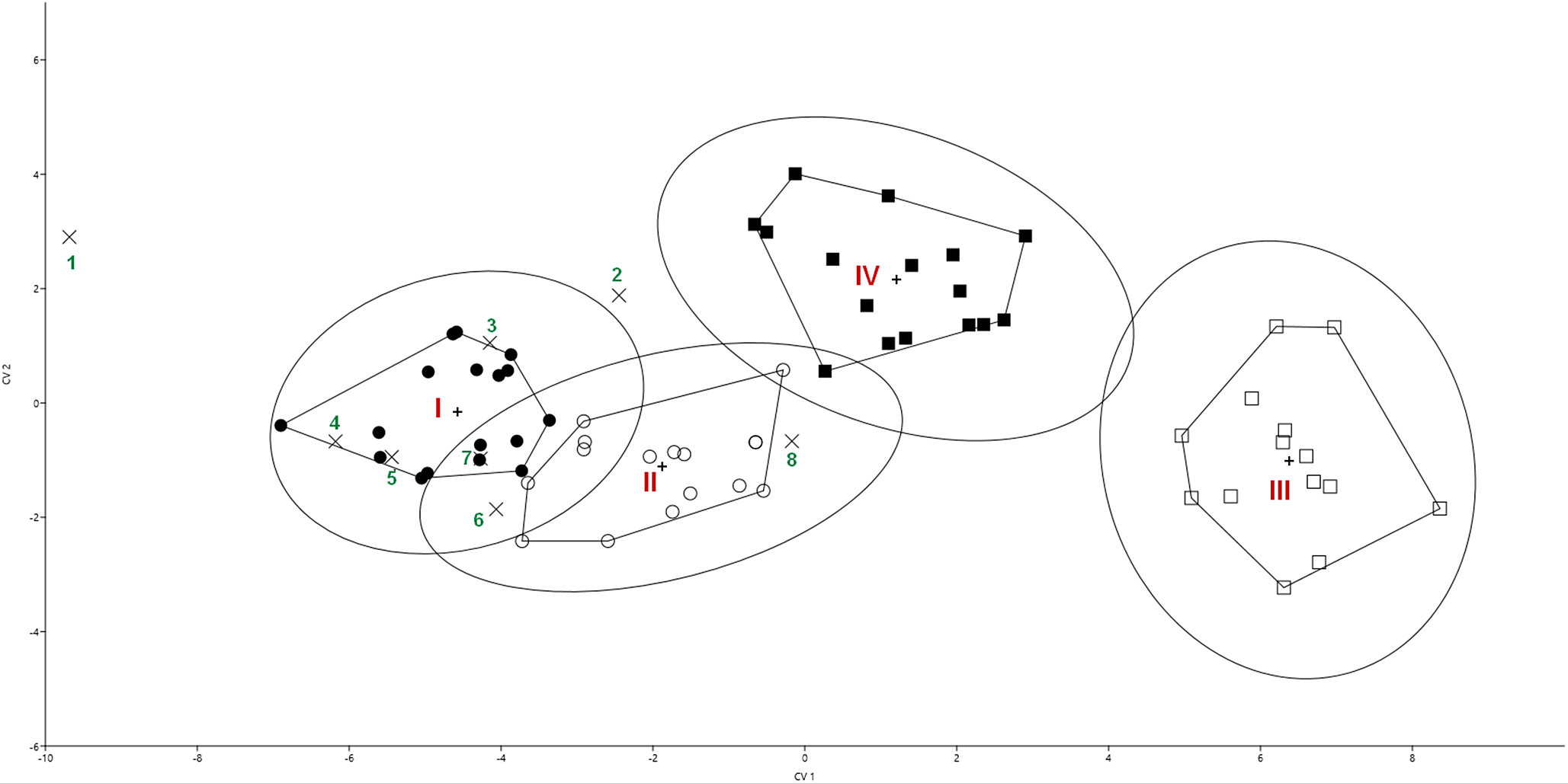
No comparative data for the metacercariae of D. mergi Lineage 3 are available. We can only compare their morphological characteristics with those of molecularly close metacercariae of D. mergi Lineage 2 described from various fishes in the River Danube (Kudlai et al. Reference Kudlai, Oros, Kostadinova and Georgieva2017) and with the different species found earlier in eyes of C. gobio s.l.: D. gobiorum, D. mergi, D. nordmanni, D. parviventosum, D. pungiti, D. volvens (Table 3). The comparison of the morphometric dataset by ten dimensions of different Diplostomum larvae was conducted with the use of discriminant analysis (Figure 8).
Ordination of traits of Diplostomum spp. metacercariae from eyes of Cottus koshewnikowi and related species in two canonical variates (CV) by discriminant analysis. The means of 10 traits, including morphometry, from the present study and the literature were included (Table 3). Data set for discriminant analysis: Groups I – Diplostomum mergi Lineage 3, River Utsjoki; II – D. mergi Lineage 3, River Lososinka; III – Diplostomum sp. Lineage 6, River Lososinka; IV – Diplostomum sp. Lineage 6, River Nemina. Species from the literature: 1 – Diplostomum gobiorum (Shigin Reference Shigin1969); 2 – Diplostomum sp. Lineage 6 (Faltýnková et al. Reference Faltýnková, Georgieva, Kostadinova, Blasco-Costa, Scholz and Skírnisson2014); 3 – Diplostomum mergi Lineage 2 (Kudlai et al. Reference Kudlai, Oros, Kostadinova and Georgieva2017); 4 – Diplostomum mergi (Shigin Reference Shigin1986); 5 – Diplostomum parviventosum (Shigin Reference Shigin1986); 6 – Diplostomum nordmanni (Shigin Reference Shigin1986); 7 – Diplostomum volvens (Shigin Reference Shigin1986); 8 – Diplostomum pungiti (Shigin Reference Shigin1986).

All metacercariae from the present study were arranged into four groups based on species and geographic location (Figure 8): I – Diplostomum mergi Lineage 3 from lens of bullheads (River Utsjoki), II – D. mergi Lineage 3 from lens of bullheads (River Lososinka), III – Diplostomum sp. Lineage 6 from retina of bullheads (River Lososinka), IV – Diplostomum sp. Lineage 6 from retina of bullheads (River Nemina). A group was not given for the parasite species whose measurements were taken from the literature (Table 3).
Discriminant analysis based on 10 dimensions showed that the four groups of metacercariae diverged along the first and the second canonical axis (Figure 8). Total success of the classification of the discriminant function was assessed as 95%. The first canonical axis carried the greatest functional load (86%), mostly reflecting the linear size of the parasites. Body length (BL) and body width (BW) of metacercariae made the greatest contribution into the divergence of the groups. The second canonical axis determined about 9% of the variability, being associated with the body shape: ratio of BL to BW and ratio of holdfast organ length (HL) to holdfast organ width (HW). Morphological differences between populations of metacercariae of D. mergi Lineage 3 from the lens were smaller than those of metacercariae of Diplostomum sp. Lineage 6 from the retina (Figure 8). Out of the eight Diplostomum species for which the average measurements of metacercariae were taken from the literature, six (D. mergi Lineage 2, D. mergi, D. parviventosum, D. nordmanni, D. volvens, D. pungiti) were placed into the confidence cloud of Diplostomum mergi Lineage 3. Diplostomum sp. Lineage 6 sensu Blasco-Costa et al. (Reference Blasco-Costa, Faltýnková, Georgieva, Skírnisson, Scholz and Kostadinova2014) and D. gobiorum (Shigin, Reference Shigin1969) were placed outside the groups considered in our study (Figure 8).
Discussion
Metacercariae from bullheads in the rivers examined in our study belonged to three species. The only parasite individual from the lens of a single bullhead examined in the River Nemina was identified as D. spathaceum. The most common (and possibly host-specific) species of metacercariae from bullheads were D. mergi Lineage 3 from the lens and Diplostomum sp. Lineage 6 from the retina. These two species had fairly high infection indices in geographically distant parts of the distribution (Figure 1, Table 2) in rivers belonging to different sea basins of the northern latitudes (Scotland, Norway, Iceland, Finland, north-western Russia). The second intermediate hosts of Diplostomum sp. Lineage 6 are various fishes from the families Gasterosteidae and Cottidae occurring under similar ecological conditions – that is, in cold lotic waters. In Scotland, Iceland, and Norway, where European bullhead is absent, the role of the second intermediate host is probably played by three-spined stickleback. Among the hosts of D. mergi Lineage 3, there also are some cold-loving fish species such as Salmo trutta, Gobio gobio, Cottus gobio, and C. koshewnikowi, whose distribution is confined to the northern latitudes. Therefore, we may suppose that these two Diplostomum species are also distributed in the northern and the middle European part of the Palaearctic.
Our morphometric data (Figure 8) show the importance of detailed information about the morphology of these parasites in addition to their molecular characteristics. The size of the larvae of Diplostomum sp. Lineage 6 examined in our study varies considerably between populations, which could lead to misidentification without the use of molecular methods. The fact that Diplostomum sp. Lineage 6 from three-spined stickleback in Iceland (Faltýnková et al. Reference Faltýnková, Georgieva, Kostadinova, Blasco-Costa, Scholz and Skírnisson2014) was different from the newly studied group in metrical features may be explained by two considerations. First, the morphometry of the metacercariae in Faltýnková et al. (Reference Faltýnková, Georgieva, Kostadinova, Blasco-Costa, Scholz and Skírnisson2014) was based on unstained ethanol-fixed material, whereas the metacercariae in our study were stained with acetocarmine. Secondly, the larvae in Faltýnková et al. (Reference Faltýnková, Georgieva, Kostadinova, Blasco-Costa, Scholz and Skírnisson2014) were collected from three-spined stickleback, while those in our study were taken from bullheads, and it is known that the host influences the morphology of metacercaria (Field and Irwin 1995; Kudlai et al. Reference Kudlai, Oros, Kostadinova and Georgieva2017; Niewiadomska Reference Niewiadomska1987; Niewiadomska and Szymański Reference Niewiadomska and Szymański1991, Reference Niewiadomska and Szymański1992; Shigin Reference Shigin1986; Sokolov et al. Reference Sokolov, Ieshko and Lebedeva2023).
In an earlier study of bullheads in the River Utsjoki (Ieshko et al. Reference Ieshko, Lebedeva, Barskaya, Shul’man and Niemela2013), we misidentified D. mergi Lineage 3 as D. volvens. This misidentification, though partly due to the erroneously recorded localisation, mostly arose from the fact that we used only morphological data, and the average dimensions of D. volvens, as can be seen in Figure 8, fall within the confidence interval of the size characteristics of D. mergi Lineage 3.
In addition, our data suggest that D. mergi Lineage 3 may actually be D. parviventosum. This conjecture is supported by the fact that the measurements of metacercariae of D. parviventosum fell within the confidence limit of D. mergi Lineage 3 individuals found in our study (Figure 8). Besides, some of these metacercariae were found in the eyes of European Bullhead G. gobio by Georgieva et al. (Reference Georgieva, Soldánová, Pérez-Del-Olmo, Dangel, Sitko, Sures and Kostadinova2013), and it is from this fish that D. parviventosum was described (Shigin et al. Reference Shigin, Chupilko and Klochkova1985). This instance highlights the importance of the integrated approach for studies of Diplostomum metacercariae.
It cannot be ruled out that many of the parasites identified in our study as D. mergi Lineage 3 actually belong to D. gobiorum, which has been noted in C. gobio before (see above). According to Shigin (Reference Shigin1969, Reference Shigin1986), D. gobiorum is a specific parasite of gobiid fishes, and its distribution is associated with that of its fish hosts such as Ponticola, Neogobius, Proterorhinus etc. living in fresh and freshened water bodies. In the former USSR, these were the coasts of the Caspian, the Azov, the Black and the Baltic Sea, and the lower reaches of rivers flowing into them. However, in Figure 7, the data on average measurements of D. gobiorum obtained by Shigin (Reference Shigin1969, Reference Shigin1986) are quite far from the group of D. mergi Lineage 3, which indicates significant morphological differences. In addition, Shigin (Reference Shigin1969) described D. gobiorum from the gobiid fish Ponticola gorlap (Iljin, 1949) (syn. Neogobius kessleri), which is distributed in the Caspian Sea basin (Kottelat Reference Kottelat1997). Therefore, it is likely that D. gobiorum and D. mergi Lineage 3 are two valid species with a different distribution and a different range of hosts.
However, the morphological differences between D. gobiorum and D. mergi Lineage 3 may also be associated with the different host species and different infection indices in the study by Shigin (Reference Shigin1969) and in our study. Detailed comprehensive studies of Diplostomum metacercariae from fishes of the families Cottidae and Gobiidae are needed to solve this problem. According to Kvach et al. (Reference Kvach, Boldyrev, Lohner and Stepien2015), gobiid fishes in the basins of the Black and the Azov seas have high infection rates with Diplostomum spp., which means they can be easily identified using integrative approach. Such studies would provide new data on the diversity of Diplostomum spp. in the region as well as insights into the Diplostomum taxonomy. They would also be useful for controlling invasive species of both fish and helminths. A case study of Cottus koshewnikowi invasion in the River Utsjoki showed a marked increase in the abundance of its dominant parasite species, such as Schistocephalus solidus (Müller, 1776) Steenstrup, 1857, Apatemon gracilis (Rudolphi, 1819) Szidat, 1928, and Diplostomum spp. (Ieshko et al. Reference Ieshko, Lebedeva, Barskaya, Shul’man and Niemela2013; Pihlaja et al. Reference Pihlaja, Julkunen, Niemela and Erkinaro2003; Seppala et al. Reference Seppala, Chubb, Niemela and Valtonen2007).
The River Utsjoki is a tributary of the Tena, one of the most important rivers in Europe for the conservation of Atlantic salmon Salmo salar L. Bullhead C. koshewnikowi was introduced into the Utsjoki in the late 1970s (Jørgensen et al. Reference Jørgensen, Avundsen, Gabler, Halvorsen, Erkinaro and Niemela1999; Pihlaja et al. Reference Pihlaja, Julkunen, Niemela and Erkinaro2003), and segregation of trophic niches between naturalised bullheads and native salmon juveniles has been shown (Jørgensen et al. Reference Jørgensen, Avundsen, Gabler, Halvorsen, Erkinaro and Niemela1999). However, the naturalisation of the bullhead in the Utsjoki River, an increase in its numbers, and the attraction of fish-eating birds may contribute to the formation of a focus of infection with trematode larvae, which might be dangerous for the native Atlantic salmon juveniles.
The composition and diversity of parasites in bullheads and native Atlantic salmon juveniles in the Utsjoki River was shown to be different (Ieshko et al. Reference Ieshko, Lebedeva, Barskaya, Shul’man and Niemela2013). At the same time, over the period of ten years (1997–2007), the numbers of bullheads increased considerably, and so did the infection levels: the prevalence of Diplostomum metacercariae increased 10-fold, and that of Apatemon gracilis metacercariae increased 40-fold. The infection rates of Diplostomum spp. and A. gracilis metacercariae in young salmon examined in the 1990s and 2000s increased, too. The prevalence of diplostomids in young salmon was 7% according to Ieshko and Shulman (Reference Ieshko and Shulman2001) and already as high as 20–93% according to Barskaya et al. (Reference Barskaya, Ieshko and Lebedeva2008). Similarly, the prevalence of A. gracilis in salmon in the Teno River system ranged from 27 to 87% with an abundance of 1–8 specimens per fish (Barskaya et al. Reference Barskaya, Ieshko and Lebedeva2008). In rivers with no invasive bullheads, the prevalence was much lower or even zero. For instance, it was 1% with an abundance of 0.9 in the River Namsen (Norway), while in the Vefsna River, this parasite was not found in salmon at all (Shulman et al. Reference Shulman, Ieshko, Shchurov, Yonsen and Yensen2004, Reference Shulman, Ieshko, Shchurov, Yonsen and Yensen2005). Bullheads are closely associated with benthic invertebrates such as gastropod molluscs, and there are good reasons to believe that they play a leading role in maintaining the foci of fish infection with Diplostomum spp. A similar study has been conducted on round goby (Neogobius melanostomus), which invaded Canadian water bodies in the 1990s and can assist in maintaining the infection of Diplostomum spp. in native fish (Marcogliese and Locke Reference Marcogliese and Locke2021). Martinek and Hernández-Orts (Reference Martinek and Hernández-Orts2023) note that metacercariae D. mergi Linage 3 are the most prevalent and abundant parasite (nearly 40%) of black goby Gobius niger of Finnish Archipelago (Baltic Sea), even though black gobies exist in marine or brackish environments. These data indicate that the infection of bullheads with metacercariae of Diplostomum spp. in any natural ecosystem may lead to increased infection levels in resident fish and needs to be controlled.
Supplementary material
The supplementary material for this article can be found at http://doi.org/10.1017/S0022149X25100515.
Data availability statement
The data supporting the findings of this study are available in the supplementary material. Any additional data supporting the findings of this study are available from the corresponding author upon reasonable request.
Acknowledgements
The authors are grateful to the Drs. Igor Schurov, Vyacheslav Shirokov, Alexey Veselov † (IB KRC RAS), and Era Niemela (Finnish Game and Fisheries Research Institute, Helsinki, Finland) for help with the fish sampling.
Special thanks are due to Drs. Olga Novokhatskaya (Unicaf University, Cyprus) and Yulia Barskaya (IB KRC RAS) for assistance with parasitological investigation of fish from the River Utsjoki.
We thank Natalia Lentsman for linguistic assistance.
The authors are grateful to two anonymous reviewers for helpful comments and suggestions.
Author contribution
Concept and design of the study: D.L. Writing: D.L., S.B., E.I. Review and editing: D.L., E.I., S.B., D.Z. Fieldwork: E.I. and D.L. Morphological analysis of parasites: D.L. Molecular analyses of parasites: D.L. and D.Z. Statistical analysis: S.B. All the authors have read and approved the final version of the manuscript.
Financial support
The study was carried out under the state order of the Ministry of Science and Higher Education of the Russian Federation (122032100130-3).
Competing interest
The authors declare no conflict of interest.
Supporting information
Additional supporting information may be found in the online version of the article at the publisher’s website.