




No CrossRef data available.
Article contents
Effect of parasitism on plasma sex-specific proteins in Cyphocarax gilbert (Teleost, Curimatidae)
Published online by Cambridge University Press: 03 February 2005
Abstract
Cyphocarax gilbert (Szidat, L., 1948) is a fish commonly found in coastal drainage of eastern Brazil. This fish is sometimes caught with signs of infection by the crustacean Riggia paranensis, a haematophagous parasite. A remarkable feature of infected fish is that they lack gonads. In this paper we have analysed the frequency of parasitism, the gonadal development of non-infected fish and the profile of plasma proteins in both infected and non-infected specimens. Two reproductive periods/year were observed, beginning in February and August. On average, 40% of fish were infected, in the Itabapoana River (Brazil). Sex-specific proteins were identified by electrophoresis. SDS-PAGE analysis demonstrated that a 143 kDa female-specific glycolipoprotein (FSP) is a calcium-binding phosphoprotein. FSP was isolated through ultracentrifugation and SDS-PAGE analysis showed that the native protein is composed of three polypeptides of 143, 100 and 70 kDa. Both FSP and a 33 kDa male-specific protein (MSP) are absent from infected fish plasma. FSP levels in female plasma changes with the developmental stage of gonads. Altogether these data suggest that the FSP corresponds to fish vitellogenin. Furthermore, the absence of the above-mentioned proteins in infected fish suggests that R. paranensis might interfere with the regular hormonal process of fish vitellogenesis.
Keywords
Information
- Type
- Research Article
- Information
- Copyright
- © 2005 Cambridge University Press
INTRODUCTION
The diversity of freshwater isopods of the Cymothoidae in South America is greater than in other continents but the knowledge of this taxonomic group is very limited (Huizinga, 1972; Thatcher, 1991; Schaefer, 1993). Several species of Cymothoidae have been described as blood-sucking fish parasites (Radujkovic, 1991; Trilles, 1991). Some of these parasites have a considerable impact on the growth and physiological state of their hosts (Romestand, Janicot & Trilles, 1977; Bragoni, Romestand & Trilles, 1983; Radujkovic, 1991). Cymothoidae species cause metabolic damage to their host, such as a significant reduction in the number of erythrocytes (Adlard & Lester, 1995), body weight loss (Marks et al. 1996), reduction of female host fecundity and increase of juvenile mortality (Adlard & Lester, 1994). The main physiological effects of R. paranensis on the fish Cyphocarax gilbert, is the inhibition of the host gonadal development, as has been described in northern Rio de Janeiro state drainage basins, at the Itabapoana River, and lakes of Cima and Feia (Thomé, 1997; Azevedo et al. 2002). The C. gilbert population have a spawning period with 2 annual reproductive cycles. The reproductive periods occur with the increase in the temperature and the rains (Thomé, 1997; Azevedo et al. 2002). Only R. paranensis females are haematophagous parasites that obtain blood from the gills of the host. Like other species of the family, females of R. paranensis maintain an opening in the host body to obtain oxygen from water and to deliver their brood (Szidat, 1948; Huizinga, 1972; Thacther, 1991). This parasite causes significant damage to C. gilbert gonadal development. This damage is so extensive that it is difficult to distinguish the sex of captured infected specimens. Nevertheless, the mechanism of inhibition of gonadal development is unknown.
Gonadal development in oviparous animals requires some plasma components such as proteins and hormones (Schneider, 1996). The role of vitellogenin (Vg) and other lipoproteins in yolk deposition is now widely recognized (Bujo et al. 1997). In general, Vgs are female-specific phospholipoglycoproteins of high molecular mass, synthesized in the liver, released in the blood and takenup by the developing oocytes (Wallace & Selman, 1985; Tyler & Lubberink, 1996; Utarabhand & Bunlipatanon, 1996). Inside the oocytes of non-mammalian vertebrates Vg is the target of proteases which then gives rise to a set of smaller yolk polypeptides: lipovitellins and phosvitins (Wallace & Selman, 1985). Despite the fact that the molecular details of Vg synthesis and storage in the oocytes are available in the literature, the influence of parasitism on fish plasma protein biology has not been investigated.
The objective of this study was to describe the frequency of parasitism in the Itabapoana River, the reproductive cycles of this species of fish and to analyse the changes in the plasma proteins profile induced by R. paranensis. The main effects of the parasite on growth and development of host ovaries are also described.
MATERIALS AND METHODS
Animals
Cyphocarax gilbert specimens were collected monthly from 1999 to 2000 in the middle Itabapoana River (Rio de Janeiro/Espírito Santo states, Brazil; 21°15′S and 42°30′W), using gillnets of 20 mm mesh. Live fish were placed in plastic sacks under constant oxygenation and immediately transported to the laboratory to sample blood, quantify the frequency of infected fish, and analyse gonadal development considering ovarian and testicular morphological characteristics.
Gonadal development
Gonads of males and females were morphologically classified in 4 development stages: initial development, maturing, mature, and post-spawning phase (Andrade & Godinho, 1983; Nagahama, 1983; MacKenzie, Thomas & Farrar, 1989).
Collection of plasma
The fish were maintained for 1 min in water at 0 °C for anaesthesia. Blood samples were then taken by caudal vessel puncture with disposable syringes containing Alserver solution (114 mM glucose, 27 mM sodium citrate, 71 mM NaCl, pH 6·1). The blood was then centrifuged at 5000 g for 10 min, at room temperature. The supernatant was aliquoted and stored at −22 °C.
Polyacrylamide gel electrophoresis (PAGE)
Non-denaturing gel electrophoresis (7·5%) was carried out as described by Davis (1964). The gels were stained either with Coomassie blue to develop total proteins or with Sudan black to develop only lipoproteins.
PAGE was also performed in the presence of SDS in 5–15% gels as described by Laemmli et al. (1970). The gels were stained with either Coomassie blue or the Schiff – PAS reagent (Sigma) to develop total proteins or glycoproteins respectively. Whenever indicated, gels were also stained with methyl green in order to develop phosphoproteins Cutting & Roth (1973). The molecular mass of the proteins was estimated from a calibration curve obtained with the standard proteins provided in the MW-SDS-200 kit (Sigma). Protein content of samples was determined by the method of Lowry et al. (1951) using BSA as a standard.
Calcium-binding assay
Qualitative calcium binding was assayed as described (Maruyama, Mikawa & Ebashi, 1984), as modified by Silva-Neto et al. (1996). Briefly, after electrophoresis, proteins were electroblotted onto nitrocellulose membranes. After transfer, membranes were washed for 1 h in 3 changes of a buffer containing 60 mM KCl, 10 mM imidazole (pH 6·8), and 5 mM MgCl2, followed by a 5 min incubation at room temperature in the same buffer containing 45Ca obtained from DuPont NEN and adjusted to 0·25 μM (1 mCi/l). Finally, membranes were washed with distilled water for 5 min, dried, and exposed to Kodak X-Omat AR-5 film for 48 h at 70 °C. The EF-hand calcium-binding protein calmodulin and cytochrome c were used as positive and negative controls respectively in calcium-binding assays (Silva-Neto et al. 1996).
FSP isolation
FSP was isolated by density-gradient ultracentrifugation. Aliquots of plasma were overlayered on KBr solution (density 1·25 g/ml) and centrifuged at 100000 g for 20 h in a Hitachi ultracentrifuge (model CP85β). The total volume of the tubes (5 ml) was fractionated from top to bottom into 0·5 ml aliquots. The protein content of each fraction was monitored by spectrophotometry at 280 nm. Three zones of the gradient were isolated: top, medium and bottom. The fractions were analysed by non-denaturing or SDS-PAGE, after extensive dialysis against PBS in order to remove KBr. Whenever indicated, after electrophoresis, FSP was extracted from the gel according to the method of Retamal, Thiebaut & Alves (1999). The extracted protein was resubmitted to SDS-PAGE for polypeptide composition analysis.
RESULTS
Parasite frequency and reproductive cycle of Cyphocarax gilbert
A total of 737 C. gilbert specimens was analysed. The mean prevalence of infected fish was 40%, highest in the autumn and winter (54·1%) and lowest in the spring and summer (32·7%). All infected fish had small, undeveloped gonads.
The gonadal development of C. gilbert was verified in 449 uninfected specimens (Table 1). Two annual reproductive cycles were observed in the middle Itabapoana river C. gilbert population. Reproductive periods began in February and August. Mature male and female specimens were observed in April and November. Spawning females were observed from May to June, at the middle of autumn, before the dry season and from December to January during the summer at the rain season. Post spawning males were not identified in the study area (Table 1).
Table 1. Frequencies of Cyphocarax gilbert in different gonadal development stages at middle Itabapoana River (Specimens were collected in the Itabapuana River (Rio de Janeiro/Espirito Santo, Brazil) during 12 months. The gonads of males and females were observed and classified into 4 gonadal development stages: initial, maturing, mature, post-spawning.)

PAGE analysis of the fish plasma
The plasma proteins of the male, female and infected fishes were analysed by non-denaturing PAGE and differences on protein profile are depicted in Fig. 1. A male-specific protein (MSP), with high mobility in the gel, was observed (Fig. 1, lanes 1, 2 and 3). A female-specific protein (FSP), with low mobility in the gel, was also observed (lanes 4, 5 and 6). FSP and MSP are both absent from the plasma of infected fish (lanes 7, 8 and 9). The same plasma protein pattern was observed when male or female infected specimens were identified (data not shown).

Fig. 1. Plasma protein profiles ([male ], [female] and infected fish) by non-denaturing PAGE. (5–15%). Lanes 1, 2, 3=non-infected male plasma of 3 different fish showing testes in stage 2; Lanes 4, 5, 6=non-infected female plasma of 3 different fish showing ovaries in stage 2; Lanes 7, 8, 9=infected fish plasma of 3 different fish showing atrophic gonads. The gel was stained with Coomassie Blue. Arrows indicated the sex-specific proteins: MSP, male-specific protein; FSP, female-specific protein.
Analysis by SDS-PAGE of the total plasma of the male, female and infected specimens was carried out next (Fig. 2). Male plasma showed a 33 kDa specific polypeptide (Fig. 2A, lane M). In the females, a 143 kDa specific polypeptide was also observed (lane F). Both of them were absent in the plasma of infected fishes (lane I). The 143 kDa female-specific polypeptide is a glycoprotein as shown by PAS-Schiff staining (Fig. 2B, lane 2). The 33 kDa male-specific polypeptide was not stained by this reagent (data not shown).

Fig. 2. Plasma protein profiles ([male ], [female] and infected fish) by SDS-PAGE (5–15%). (A) Coomassie Blue-stained gel: S, molecular weight markers; M, non-infected male plasma; I, infected fish; F, non-infected female plasma; B, non-infected female plasma; 1, Coomassie Blue-stained gel; 2, PAS-Schiff reagent-stained gel.
143 kDa polypeptide and gonadal development
Plasma was collected from females in different phases of their reproductive cycle. Changes in the profile of plasma proteins were verified by SDS-PAGE (Fig. 3). The results show that the 143 kDa FSP concentration increased with the gonadal development index, whereas other proteins did not change at that time, indicating a positive association between FSP concentration and gonadal development of C. gilbert females. These data suggest that FSP could be involved in oogenesis. The plasma proteins of the male, or of the infected fishes, were not modified during the same period (data not shown).

Fig. 3. Plasma protein profiles at 3 gonadal development stages. SDS-PAGE (5–15%). 0, Females presenting post-spawing gonads; 1, females presenting gonads in initial development; 2, females presenting gonads in maturation or mature. Arrow indicates FSP.
Isolation of the FSP
Lipoproteins from the plasma of the male and female normal and infected fish were isolated by ultracentrifugation under KBr gradient. Fig. 4 shows a non-denaturing PAGE analysis of the obtained fractions. FSP was found in the medium of the gradient (Fig. 4A, lane 2). The same fractions obtained from the plasma of male (Fig. 4A, lane [male ]) and infected fish (Fig. 4A, lane I; Fig. 4C, lane 3) did not contain this protein. The Sudan black stained gel shows the lipoprotein features of proteins found in the fractions (Fig. 4B). The 143 kDa FSP was strongly stained under these conditions (Fig. 4B, lane 2).

Fig. 4. Non-denaturing PAGE analysis of the KBr gradient fractions (A) Coomassie Blue-stained gel; (B) Sudan Black-stained gel. [male ], total male plasma; [female], total female plasma; I, total infected fish plasma. 1, Top of the gradient – plasma female; 2, medium of the gradient-plasma female; 3, bottom of the gradient – plasma female. (C) Coomassie Blue- stained gel of the infected fishes fractions: 1 and 2, top of the gradient; 3, medium of the gradient; 4, 5 and 6, bottom of the gradient.
The subunit composition of the isolated FSP was analysed by SDS-PAGE (Fig. 5). Results showed that the protein is composed by 143, 100 and 70 kDa subunits (Fig. 5B, lane 3). On lane B, the 143 kDa polypeptide, extracted from the another SDS-PAGE, was re-submitted to SDS-PAGE in order to reject the hypothesis of proteolysis.

Fig. 5. SDS-PAGE analysis of the FSP isolated by KBr gradient ultracentrifugation. The band of the non-denaturing PAGE corresponding to FSP was detached and submitted to SDS-PAGE. (A) The 143 kDa subunit of FSP was detached from another SDS-PAGE gel and re-submitted to SDS-PAGE. (B) FSP was detached from a non-denaturing PAGE and analysed by SDS-PAGE. Arrows indicate the molecular weight of FSP subunits.
FSP is a calcium-binding phosphoprotein
One recurrent feature of non-mammalian vertebrate Vgs is the presence of anionic groups such as acidic amino acids or phosphorylated residues (Silva-Neto et al. 1996). Due to this property, Vgs are able to transport cations such as iron and calcium to growing oocytes. Later on during embryo development these ions will be required for haemeprotein, bone and tooth formation. Analysis of fish plasma by SDS-PAGE following methyl green staining (Fig. 6, panel A) showed only 1 phosphopeptide present exclusively in the plasma female. This band is absent from male plasma and corresponds to FSP. Furthermore, calcium-binding assays demonstrated that this phosphorylated female-specific protein also binds calcium as shown in Fig. 7. Altogether these data demonstrate that FSP displays the main biochemical features of non-mammalian vertebrates Vgs.

Fig. 6. Identification of phosphoproteins from Cyphocharax gilbert plasma. Non-denaturing PAGE of the fish plasma was stained with methyl green. (A) Non-denaturing PAGE stained with methyl green. (B) Non-denaturing PAGE stained with Coommassie Blue. M, plasma males; F, plasma females. Arrow indicates FSP.

Fig. 7. Binding of 45Ca to FSP and calmodulin. Fish plasma samples were subjected to SDS-PAGE (5–15%) and then transferred to nitrocellulose membranes. In (A) the gel was stained with Coomassie Blue. (B) Autoradiogram of the nitrocellulose membrane incubated for 5 min with 45Ca, as described in the Materials and Methods section. M, Male plasma; F, female plasma; Ca, calmodulin as a positive control; Cc, cytochrome C as a negative control.
DISCUSSION
The effect of R. paranensis on the host, C. gilbert, is remarkable due to the inhibition of the gonadal development and variation of parasitism frequency. The parasite infection rate in the Itabapoana River is higher than in other areas in southeast of Brazil. In lakes (de Cima, Feia) and Paraíba do Sul River the frequency of parasitism is lower, varying from zero to 12% over 1 year (Thomé, 1997). A similar low frequency of isopod prevalence was observed for the catfish Microlepidogaster maculipinnis collected in Paraguay. This species was infected by 2 cymothoids (Artystone minima and Nerocila armata) causing body tissue damage and atrophy (Schaefer, 1993). The high prevalence of parasitism in the Itabapoana River might be due to environment quality, the host immune response, the host and parasite genetic characteristics, and the time of coexistence between them (Toft, 1991; Lafferty & Kuris, 1999).
The present study identified 2 biochemical markers in the plasma of C. gilbert, comparing proteins of normal male and female, and infected specimens. The protein profile is very similar among different fish of the same sex, because they are in the same reproductive stage. However, this profile, which was consistently reproducible, is different between male and female. In the parasitized male or female fish plasma the presence of sex-specific proteins was not observed. We identified a male 33 kDa protein, the function of which remains unknown, and a female-specific lipoglycoprotein composed of 143, 100 and 70 kDa subunits. The level of this protein increases in the plasma, probably its synthesis, is synchronized with the female reproductive cycle. In Zoarces elongatus, the level of vitellogenin changes from 0·2 mg/ml in the immature females to 6·4 mg/ml during the vitellogenesis process (Koya et al. 1997). Methyl Green, PAS-Schiff, Sudan-Black positive staining and calcium-binding assay showed that FSP is a calcium-binding phosphoglycolipoprotein. Thus, our data suggest that FSP might be a fish vitellogenin. The absence of this protein in the plasma of the infected fishes can indicate an indirect effect induced by the parasite, on the hormones controlling vitellogenin synthesis (Tata & Smith, 1978; Callard, Riley & Peres, 1990).
Effects of cymothoide parasitism have been described as reversible and temporary. Fish parasitized by Neurocila orbignyi showed a decrease in protein and lipid concentrations in the blood and experienced a high mortality rate (Bragoni et al. 1983). In our study less than 1% of the specimens of C. gilbert presented the body-opening lesion without Riggia paranensis within the opening. Presumably, the parasite died because R. paranensis females are usually bigger than the host body opening and the parasite cannot escape through it. The specimens that lost the parasite were scarred and developed male or female gonads, showing that the effects of the parasite were reversible. However, FSP was not observed in the post-infected female specimens (data not shown). This suggested that the effect of the parasite under the FSP expression can be irreversible and that probably the oocytes grow with the utilization of the other plasma proteins. This redevelopment of gonads thus requires further study.
The absence of sex-specific proteins in the infected fish, and the inhibition of gonadal development in both male and female specimens, suggested the R. paranensis might affect hormonal metabolism of C. gilbert. Reproductive steroidal hormones, such as oestradiol, regulate vitellogenesis in fish (MacKenzie et al. 1989). These are currently being analysed in this C. gilbert population.
The reproductive cycle of uninfected C. gilbert in the Itabapoana river was similar to the other populations in northern Rio de Janeiro state such as the population in Cima Lake, Feia Lake, and Paraíba do Sul River (Menezes, 1994; Thomé, 1997). A low frequency of post-spawning males and females was observed in the study area, suggesting that this phase might be very short.
In conclusion, the reproductive cycle of uninfected C. gilbert seems to maintain the population at Itabapoana River despite high prevalence of a parasitic castrator cymothoide isopod. The two sex-specific proteins absent in infected specimens represent biochemical markers that could be useful in further experimental procedures to understand this unusual parasite-host relationship.
This work was supported by FAPERJ, FENORTE and CNPq. Dr Marilvia Dansa-Petretski (F/2693-2) and Dr Mario A. C. Silva-Neto (F/2887-2) are also supported by the International Foundation for Science (IFS, Stockholm, Sweden). We acknowledge the technical support of Cristovão Barros Pinheiro, Lacy de Souza Pita (Laboratorio de Quimica e Função de Proteinas e Peptideos) and Antonio Carlos de Oliveira Pessanha (Laboratório de Ciências Ambientais).
References
REFERENCES
ADLARD, R. D. &
LESTER, J. G. (
1994).
Dynamic of the interaction between the parasitic isopod, Anilocra pomacentri, and the coral reef fish, Chromis nitida.
Parasitology
94,
311–324.CrossRefGoogle Scholar
ADLARD, R. D. &
LESTER, R. J. G. (
1995).
The life cycle and biology of Anilocra pomacentri (Isopoda, Cymothoidae) and ectoparasitic isopod of coral-reef fish, Chromis nitida (Perciformes, Pomacentridae).
Australian Journal of Zoology
43,
271–281.CrossRefGoogle Scholar
ANDRADE, D. R. &
GODINHO, H. (
1983).
Annual male reproductive cycle of the Brazilian teleost fish Leporinus silvestrii (Boulenger, 1902).
Archives de Biologie (Bruxelles)
94,
1–14.Google Scholar
AZEVEDO, J. S.,
THOMÉ, M. P. M.,
GOMES DA SILVA, L.,
NOVELLI, R.,
DANSA-PETRETSKI, M. &
WILLE LIMA, N. R. (
2002).
Parasitismo de Riggia paranensis (Crustacea, Cymothoidea) em populações de Cyphocharax gilbert (Teleostei, Curimatidae) do norte do estado do Rio de Janeiro.
Boletim do Instituto de Pesca, São Paulo
28,
61–69.Google Scholar
BRAGONI, G.,
ROMESTAND, B. &
TRILLES, J. P. (
1983).
Cymothoadian parasitosis of the sea-dace (Dicentrarchus labrax Linnaeus, 1758) during breeding. II. Parasitic ecophysiology in the Diana pod.
Annales de Parasitologie Humaine et Comparée
58,
593–609.CrossRefGoogle Scholar
BUJO, H.,
HERMANN, M.,
LINDSTEDT, K. A.,
NIMPF, J. &
SCHNEIDER, W. J. (
1997).
Low density lipoprotein receptor gene family members mediate yolk deposition.
Journal of Nutrition
127 (Suppl.),
801S–804S.CrossRefGoogle Scholar
CALLARD, I. P.,
RILEY, D. &
PERES, L. (
1990).
Vertebrate vitellogenesis: molecular model for multihormonal control of gene regulation.
Progress in Clinical and Biological Research
342,
343–348.Google Scholar
CUTTING, J. A. &
ROTH, T. F. (
1973).
Staining of phosphoproteins on acrylamide gel electropherograms.
Analytical Biochemistry
54,
386–394.CrossRefGoogle Scholar
DAVIS, B. J. (
1964).
Disc electrophoresis – II Methods and application to human serum proteins.
Annals of the New York Academy of Sciences
121,
404–427.CrossRefGoogle Scholar
HUIZINGA, H. W. (
1972).
Pathology of Artystone trysibia Schioedte (Isopoda: Cymothoidae), an endoparasitic isopod of South American fresh water fish.
Journal of Wildlife Diseases
8,
225–233.CrossRefGoogle Scholar
KOYA, Y.,
MATSUBARA, T.,
IKEUCHI, T.,
ADACHI, S. &
YAMAUCHI, K. (
1997).
Annual changes in serum vitellogenin concentrations in viviparous eelpout, Zoarces elongatus.
Advances in Comparative Physiology and Biochemistry
118,
1217–1223.CrossRefGoogle Scholar
LAFFERTY, K. D. &
KURIS, A. M. (
1999).
How environment stress affects the impact of parasites.
Limnology and Oceanography
44,
925–931.CrossRefGoogle Scholar
LAEMMLI, U. K. (
1970).
Cleavage of structural proteins during the assembly of the head of bacteriophage T4.
Nature, London
227,
680–685.CrossRefGoogle Scholar
LOWRY, O. H.,
FARR, A. L.,
ROSEBROUGH, N. J. &
RANDALL, R. J. (
1951).
Protein measurement with Folin phenol reagent.
The Journal of Biological Chemistry
193,
265–275.Google Scholar
MACKENZIE, D.,
THOMAS, P. &
FARRAR, S. M. (
1989).
Seasonal changes in thyroid and reproductive steroid hormones in female channel catfish (Itaclurus punctatus) in pond culture.
Aquaculture
78,
63–80.CrossRefGoogle Scholar
MARKS, R. E.,
JUANES, F.,
HARE, J. A. &
CONOVER, D. O. (
1996).
Occurrence and effect of the parasitic isopod, Lironeca ovalis (Isopoda: Cymothoidae), on young-of-the-year bluefish, Pomatomus saltatrix (Pisces: Pomacentridae).
Canadian Journal of Fisheries and Aquatic Sciences
53,
2052–2057.CrossRefGoogle Scholar
MARUYAMA, K.,
MIKAWA, T. &
EBASHI, S. (
1984).
Detection of calcium binding proteins by 45Ca autoradiography on nitrocellulose membrane after sodium dodecyl sulfate gel electrophoresis.
Journal of Biochemistry (Tokyo)
95,
511–519.CrossRefGoogle Scholar
MENEZES, M. S. (
1994).
Estrutura da população e reprodução de Cyphocarax gilbert (Quoy and Gaimard, 1824) (Osteichthyes, Curimatidae) no trecho inferior do rio Paraíba do Sul (RJ, MG) e principais afluentes. Dissertação de Mestrado (MSc). Universidade Federal do Paraná,
Curitiba, PR.
NAGAHAMA, Y. (
1983).
The functional morphology of teleost gonads. In
Fish Physiology, vol.
IXA, (ed. Hoar, W. S. & Randall, D. J.), pp.
223–264.
Academic Press,
London.CrossRef
RADUJKOVIC, B. M. (
1991).
Parasitic crustaceans of Adriatic fishes.
Wiadomosci Parazytologiczne
37,
149–150.Google Scholar
RAIBAUT, A. &
TRILLES, J. P. (
1993).
The sexuality of parasitic crustaceans.
Advances in Parasitology
32,
367–444.CrossRefGoogle Scholar
RETAMAL, C. A.,
THIEBAUT, P. &
ALVES, E. W. (
1999).
Protein purification from polyacrylamide gels by sonication extraction.
Analytical Biochemistry
268,
15–20.CrossRefGoogle Scholar
ROMESTAND, B.,
JANICOT, M. &
TRILLES, J. P. (
1977).
Modifications tissulaires et réactions de défences chez quelques Téléostéens parasites par les Cymothoidea (Crustacés Isopodes hématophages).
Annales de Parasitologie Humaine et Comparée
52,
171–180.CrossRefGoogle Scholar
SCHAEFER, S. A. (
1993).
A remarkable occurrence of isopod parasitism on an armoured catfish, Microlepidogaster maculipinnis.
Journal of Fish Biology
42,
307–310.CrossRefGoogle Scholar
SCHNEIDER, W. J. (
1996).
Vitellogenin receptors: oocyte- specific members of the low-density lipoprotein receptor supergene family.
International Review of Cytology
166,
103–137.CrossRefGoogle Scholar
SILVA-NETO, M. A.,
ATELLA, G. C.,
FIALHO, E.,
PAES, M. C.,
ZINGALI, R. B.,
PETRETSKI, J. H.,
ALVES, E. W. &
MASUDA, H. (
1996).
Isolation of a calcium-binding phosphoprotein from the oocytes and hemolymph of the blood-sucking insect Rhodnius prolixus.
The Journal of Biological Chemistry
271,
30227–30232.CrossRefGoogle Scholar
SZIDAT, L. (
1948).
Riggia paranenis n.g., n. sp. un isópodo parásito de la cavidad del cuerpo de “Curimata platana” Günther del Rio Paraná.
Revista del Instituto de Investigación de las Ciencias Naturales Ciencias Zoológicas, Buenos Aires
1,
45–46.Google Scholar
TATA, J. R. &
SMITH, D. F. (
1978).
Vitellogenesis. A versatile model for hormonal regulation of gene expression.
Recent Progress in Hormone Research
35,
47–90.Google Scholar
THOMÉ, M. P. M. (
1997).
Influência do parasita Riggia paranensis (Crustacea: Cymothoidae) no sairú Cyphocarax gilbert (Pisces, Curimatidae) no complexo lagoa de Cima-lagoa Feia. Monografia. Universidade Estadual do Norte Fluminense.
TOFT, C. (
1991).
An ecological perspective: the population and community consequences of parasitism. In
Parasite-Host Associations
(ed. Toft, C. A., Aeschlimann, A. & Bolis, L.), pp.
319–343.
Oxford Science Publication,
New York,
USA.
TRILLES, J. P. (
1991).
Catalogue mondial des Cymothoidae.
Studia Marina, Kotor
21/22,
5–288.Google Scholar
TYLER, C. R. &
LUBBERINK, K. (
1996).
Identification of four ovarian receptor proteins that bind vitellogenin but not other homologous plasma lipoproteins in the rainbow trout, Oncorhynchus mykiss.
Journal of Comparative Physiology B
166,
11–20.CrossRefGoogle Scholar
UTARABHAND, P. &
BUNLIPATANON, P. (
1996).
Plasma vitellogenin of grouper (Epinephelus malabaricus): isolation and properties.
Journal of Comparative Biochemistry and Physiology C
115,
101–110.CrossRefGoogle Scholar
WALLACE, R. A. &
SELMAN, K. (
1985).
Major protein changes during vitellogenesis and maturation of Fundulus oocytes.
Developmental Biology
110,
492–498.CrossRefGoogle Scholar
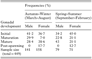
Table 1. Frequencies of Cyphocarax gilbert in different gonadal development stages at middle Itabapoana River
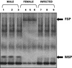
Fig. 1. Plasma protein profiles ([male ], [female] and infected fish) by non-denaturing PAGE. (5–15%). Lanes 1, 2, 3=non-infected male plasma of 3 different fish showing testes in stage 2; Lanes 4, 5, 6=non-infected female plasma of 3 different fish showing ovaries in stage 2; Lanes 7, 8, 9=infected fish plasma of 3 different fish showing atrophic gonads. The gel was stained with Coomassie Blue. Arrows indicated the sex-specific proteins: MSP, male-specific protein; FSP, female-specific protein.
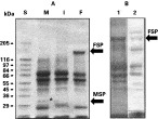
Fig. 2. Plasma protein profiles ([male ], [female] and infected fish) by SDS-PAGE (5–15%). (A) Coomassie Blue-stained gel: S, molecular weight markers; M, non-infected male plasma; I, infected fish; F, non-infected female plasma; B, non-infected female plasma; 1, Coomassie Blue-stained gel; 2, PAS-Schiff reagent-stained gel.
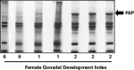
Fig. 3. Plasma protein profiles at 3 gonadal development stages. SDS-PAGE (5–15%). 0, Females presenting post-spawing gonads; 1, females presenting gonads in initial development; 2, females presenting gonads in maturation or mature. Arrow indicates FSP.
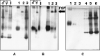
Fig. 4. Non-denaturing PAGE analysis of the KBr gradient fractions (A) Coomassie Blue-stained gel; (B) Sudan Black-stained gel. [male ], total male plasma; [female], total female plasma; I, total infected fish plasma. 1, Top of the gradient – plasma female; 2, medium of the gradient-plasma female; 3, bottom of the gradient – plasma female. (C) Coomassie Blue- stained gel of the infected fishes fractions: 1 and 2, top of the gradient; 3, medium of the gradient; 4, 5 and 6, bottom of the gradient.
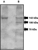
Fig. 5. SDS-PAGE analysis of the FSP isolated by KBr gradient ultracentrifugation. The band of the non-denaturing PAGE corresponding to FSP was detached and submitted to SDS-PAGE. (A) The 143 kDa subunit of FSP was detached from another SDS-PAGE gel and re-submitted to SDS-PAGE. (B) FSP was detached from a non-denaturing PAGE and analysed by SDS-PAGE. Arrows indicate the molecular weight of FSP subunits.
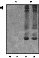
Fig. 6. Identification of phosphoproteins from Cyphocharax gilbert plasma. Non-denaturing PAGE of the fish plasma was stained with methyl green. (A) Non-denaturing PAGE stained with methyl green. (B) Non-denaturing PAGE stained with Coommassie Blue. M, plasma males; F, plasma females. Arrow indicates FSP.
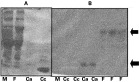
Fig. 7. Binding of 45Ca to FSP and calmodulin. Fish plasma samples were subjected to SDS-PAGE (5–15%) and then transferred to nitrocellulose membranes. In (A) the gel was stained with Coomassie Blue. (B) Autoradiogram of the nitrocellulose membrane incubated for 5 min with 45Ca, as described in the Materials and Methods section. M, Male plasma; F, female plasma; Ca, calmodulin as a positive control; Cc, cytochrome C as a negative control.
 You have
Access
- 7
- Cited by