Introduction
The control of primordial follicle activation involves two-way communication between the oocyte and its surrounding somatic cells (Cortvrindt & Smitz, Reference Cortvrindt and Smitz2001), but the factors and mechanisms responsible for the activation and growth of these follicles have not been fully elucidated (Kerr et al., Reference Kerr, Myers and Anderson2013). The development of an in vitro culture system able to promote the growth of primordial follicles is extremely important to optimize female reproductive potential, as well as to a better understanding of early folliculogenesis.
Several substances and growth factors have been tested in in vitro studies, including bone morphogenetic protein 4 (BMP4). This protein binds to heterogeneous complexes of transmembrane serine/threonine (Ser/Thr) kinase receptors, known as the BMP type IA and IB receptors (BMPR-IA and BMPR-IB) (Chen et al., Reference Chen, Zhao and Mundy2004). BMP4 is derived from the thecal tissue and has been observed to increase the proliferation of ruminant granulosa cells in vitro (Glister et al., Reference Glister, Richards and Knight2004; Juengel et al., Reference Juengel, Reader, Bibby, Lun, Ross, Haydon and McNatty2006) and to regulate the action of FSH on progesterone and oestradiol production (Shimasaki et al., Reference Shimasaki, Zachow, Li, Kim, Iemura, Ueno, Sampath, Chang and Erickson1999; Pierre et al., Reference Pierre, Pisselet, Dupont, Mandon-Pépin, Monniaux, Monget and Fabre2004). In bovine species, the presence of BMP4 was demonstrated in theca cells of antral follicles and the expression of BMPRII was found in primordial, primary and secondary follicles both in granulosa cells and oocytes (Fatehi et al., Reference Fatehi, Van Den Hurk, Colenbrander, Daemen, Van Tol H, Monteiro, Roelen and Bevers2005). In addition, BMP4 mRNA and its receptors (BMPR-IA and BMPR-IB) have been observed in goat and sheep preantral follicles, suggesting that BMP4 mediates development during this stage of folliculogenesis (Costa et al., Reference Costa, Passos, Leitão, Vasconcelos, Saraiva, Figueiredo, van den Hurk and Silva2012; Bertoldo et al., Reference Bertoldo, Duffard, Bernard, Frapsauce, Calais, Rico, Mermillod and Locatelli2014). In mono-ovulatory species, culture in vitro of granulosa cells demonstrated that BMP-4 and other members of the BMP family have a major role in modulating proliferative and differentiative responses (Campbell et al., Reference Campbell, Souza, Skinner, Webb and Baird2006). Rossi et al. (Reference Rossi, Portela, Passos, Cunha, Silva, Costa, Saraiva, Donato, Peixoto, Van Der Huck and Silva2015) reported that BMP-4 contributes to preserve the ultrastructure of bovine secondary follicles cultured in vitro and that, in combination with FSH, BMP4 increases the expression of mRNA for BMP15. Despite the role BMP4 on primordial germ cell formation (humans: Park et al., Reference Park, Woods and Tilly2013; buffalo: Shah et al., Reference Shah, Saini, Ashraf, Singh, Manik, Singla, Palta and Chauhan2015; goat: Singhal et al., Reference Singhal, Singhal, Malik, Singh, Kumar, Kaushik, Mohanty and Malakar2015) and secondary follicles (Rossi et al., Reference Rossi, Portela, Passos, Cunha, Silva, Costa, Saraiva, Donato, Peixoto, Van Der Huck and Silva2015), has already been reported, it is still not known if BMP4 regulates primordial follicle activation and development in bovine species.
During follicle development, expression of oocyte-secreted factors, like growth and differentiation factor 9 (GDF9, Carabatsos et al., Reference Carabatsos, Elvin, Matzuk and Albertini1998) and BMP-15 (Otsuka et al., Reference Otsuka, Suda, Li, Matsumoto and Watanabe2000), is an important event that contributes to the slow maturation process observed in domestic species (Van den Hurk & Zhao, Reference Van Den Hurk and Zhao2005). GDF9 play a role during early follicle development and maturation (Carabatsos et al., Reference Carabatsos, Elvin, Matzuk and Albertini1998) and treatment with GDF9 enhances primary and preantral follicular growth in vitro and in vivo (Hayashi et al., Reference Hayashi, Mcgee, Min, Klein, Rose, Van Duin and Hsueh1999; Vitt et al., Reference Vitt, Mcgee, Hayashi and Hsueh2000). Furthermore, BMP15 contributes to the growth during the different phases of folliculogenesis, including the process of follicular activation (Juengel et al., Reference Juengel, Hudson, Whitinig and Mcnatty2004). In sheep, immunization against BMP-15 resulted in a blockage of follicular growth (Juengel et al., Reference Juengel, Hudson, Heath, Smith, Reader, Lawrence, O'Connell, Laitinen, Cranfield, Groome, Ritvos and Mcnatty2002). Cuboidal granulosa cells from growing follicles express proliferating nuclear antigen (PCNA), which is a nuclear protein essential for follicular growth and thus is considered to be a marker of proliferating granulosa cells (Wandji et al., Reference Wandji, Srsen, Voss, Eppig and Fortune1996). Bax and Bcl2 are pro-apoptotic and anti-apoptotic genes, respectively, which are involved in growth regulation and follicular apoptosis (Choi et al., Reference Choi, Hwang, Lee, Yoon, Yoon and Bae2004). However, it is still not known if BMP4 influences the expression of all these factors in ovarian cortical tissue cultured in vitro.
The aim of this study was to evaluate the effects of different concentrations of BMP4 on activation and survival of bovine follicles after culture of ovarian cortical tissue. Moreover, the influence of different concentrations of BMP4 on mRNA expression of GDF9, BMP15, PCNA, Bax and Bcl2 in follicles cultured in vitro was evaluated.
Materials and methods
Chemicals
Unless mentioned otherwise, the culture media, BMP4 and other chemicals used in the present study were purchased from Sigma Chemical (St Louis, MO, USA).
Source of ovaries
Bovine ovaries (n = 20) were collected from females obtained from a local slaughterhouse. Immediately postmortem, the surrounding fat tissue and ligaments were removed and the ovaries were washed in 70% alcohol followed by two washes in sterile saline solution. The ovaries were placed into tubes containing 20 ml alpha minimum essential medium (α-MEM), supplemented with 100 IU/ml penicillin and 150 mg/ml streptomycin and then transported to the laboratory at 4°C within 1 h.
Experimental protocol
Briefly, ovarian tissue samples from each ovarian pair were cut into slices (3 × 3 × 1 mm) using a needle and scalpel under sterile conditions. The tissue pieces were then either directly fixed for histological and ultrastructural analysis (fresh control) or placed in culture for 6 days. Bovine tissues were transferred to 24-well culture dishes containing 1 ml culture medium. Culture was performed at 39°C in 5% CO2 in air in a humidified incubator and all media were incubated for 1 h before use. The basic culture medium (cultured control) was called α-MEM+ and consisted of α-MEM (pH 7.2–7.4) supplemented with ITS (insulin 6.25 ng/ml, transferrin 6.25 ng/ml and selenium 6.25 ng/ml), 2 mM glutamine, 2 mM hypoxanthine, 1.25 mg/ml bovine serum albumin (BSA), 100 IU/ml penicillin and 150 mg/ml streptomycin. Different concentrations of BMP4 (0, 10, 50 or 100 ng/ml) were added to the MEM+ to test the effects of this growth factor. Each treatment was repeated four times and the culture medium was replenished every other day.
Morphological analysis and assessment of in vitro follicular growth
Before culture (fresh control) and after 6 days of culture, the pieces of ovarian tissue were fixed overnight at room temperature in 4% paraformaldehyde in phosphate-buffered saline (PBS; pH 7.4) for histological studies. After fixation, the ovarian fragments were dehydrated in a graded series of ethanol, clarified with xylene, and embedded in paraffin wax. For each piece of ovarian cortex, 7-µm sections were mounted on slides and stained with hematoxylin and eosin. Coded anonymized slides were examined under a microscope (Nikon, Tokyo, Japan) at ×100 and ×400 magnification. The developmental stages of follicles were classified as primordial (one layer of flattened or flattened and cuboidal granulosa cells around the oocyte) or growing follicles (primary: one layer of cuboidal granulosa cells, and secondary: two or more layers of cuboidal granulosa cells around the oocyte). These follicles were classified further individually as histologically normal when an intact oocyte was present, surrounded by granulosa cells that are well organized in one or more layers, and have no pyknotic nucleus. Degenerated follicles were defined as those with a retracted oocyte that has a pyknotic nucleus and/or is surrounded by disorganized granulosa cells, which are detached from the basement membrane. Overall, from 140–212 follicles were evaluated for each treatment. The percentages of healthy primordial and developing follicles were calculated before (fresh control) and after culture in a particular medium. Follicle and oocyte diameters were only measured in healthy follicles. Follicle diameter was recorded from one edge of the granulosa cell membrane to the other edge, or from the outside edge of the theca cell layer when present. Oocyte diameter was recorded from edge to edge of the oocyte membrane. Two perpendicular diameters were recorded for each and the average was reported as the follicle and oocyte diameters, respectively.
Expression of mRNA for GDF9, BMP15, PCNA, Bax and Bcl2 in bovine ovarian cortical tissue
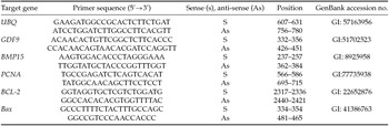
For mRNA isolation, bovine ovarian cortex from fresh control, as well as after culture in the different treatments were collected and stored at −80°C until RNA extraction. Total RNA extraction was performed using a Trizol ® purification kit (Invitrogen, São Paulo, Brazil). In accordance with the manufacturer's instructions, 800 µl of Trizol solution was added to each frozen samples and the lysate was aspirated through a 20-gauge needle before centrifugation at 10,000 g for 3 min at room temperature. Thereafter, all lysates were diluted 1:1 with 70% ethanol and subjected to a mini-column. After binding of the RNA to the column, DNA digestion was performed using RNase-free DNase (340 Kunitz units/ml) for 15 min at room temperature. After washing the column three times, the RNA was eluted with 30 µl RNase-free water. The RNA concentration was estimated by reading the absorbance at 260 nm and was checked for purity at 280 nm in a spectrophotometer (Amersham Biosciences, Cambridge, UK) and 2 µl of total RNA was used for reverse transcription. Before the reverse transcription reaction, samples of RNA were incubated for 5 min at 70°C and then cooled in ice. Reverse transcription was performed in a total volume of 20 µL, which was comprised of 10 µl of sample RNA, 4 µl 5× reverse transcriptase buffer (Invitrogen, São Paulo, Brazil), 8 units RNase out, 150 units Superscript III reverse transcriptase, 0.036 U random primers (Invitrogen, São Paulo, Brazil), 10 mM DTT, and 0.5 mM of each dNTP. The mixture was incubated for 1 h at 42°C, for 5 min at 80°C, and then stored at −20°C. Negative controls were prepared under the same conditions, but without the inclusion of the reverse transcriptase. Quantification of mRNA was performed using SYBR Green. PCR reactions were composed of 1 µl cDNA as a template in 7.5 µl of SYBR Green Master Mix (PE Applied Biosystems, Foster City, CA,USA), 5.5 µl of ultra-pure water, and 0.5 µM of each primer. The primers were designed by using the PrimerQuestSM program (http://www.idtdna.com) to perform amplification of GDF9, BMP15, Bcl2, Bax and the housekeeping gene Ubiquitin (UBQ) (Table 1). This housekeeping gene has shown highest stability in bovine preantral follicles (Rebouças et al., Reference Rebouças, Costa, Passos, Passos, van den Hurk and Silva2013) and, thus, was used to normalize expression of target genes. The specificity of each primer pair was confirmed by melting curve analysis of PCR products. The efficiency amplification for all genes was verified according to Pfaffl et al. (Reference Pfaffl, Lange, Daxenberger and Meyer2001). The thermal cycling profile for the first round of PCR was: initial denaturation and activation of the polymerase for 10 min at 95°C, followed by 40 cycles of 15 s at 95°C, 30 s at 58°C, and 30 s at 72°C. The final extension was for 10 min at 72°C. All reactions were performed in a real-time PCR Realplex (Eppendorf, Germany). The ΔCt method was used to transform the Ct values into normalized relative expression levels (Livak & Schmittgen, Reference Livak and Schmittgen2001).
Primer pairs used for real-time PCR

Statistical analysis
Data of follicular development were subjected to potential transformation (x3,9), while data of degeneration received logarithmic transformation (log10 (x)) and then, evaluated by ANOVA. The mean values of degeneration were contrasted with fresh control means, before culture, by Dunnett's test (P < 0.05). The mean values of follicular activation were compared by Student−Newman−Keuls (SNK, P < 0.05).The diameters of oocytes and follicles under the various treatments were subjected to ANOVA followed by SNK. Levels of mRNA for GDF9, BMP15, Bcl2 and Bax in cultured fragments were analyzed by using the non-parametric Kruskal−Wallis test (P < 0.05). Data were expressed as mean ± standard error of the mean (SEM). Differences were considered to be significant when the P-value was < 0.05.
Results
Effect of BMP4 concentration on follicular survival
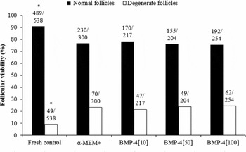
Histological analysis showed that degenerated and normal follicles were found in non-cultured and cultured ovarian cortical pieces. Degenerated follicles show a pyknotic nucleus, shrunken oocyte or unorganized granulosa cells. In total, 1513 follicles were counted to evaluate follicular morphology, activation and growth. After 6 days of culture, there is an increase of degenerated follicles cultured in all treatments compared with fresh control. However, the percentage of viable follicles was not influenced by treatments (Fig. 1).
Percentages of normal and degenerated follicles in uncultured tissue (fresh control) and in tissues cultured for 6 days in α-MEM+ alone or supplemented with various concentrations of BMP4. *P < 0.05, significantly different from cultured ovarian cortex tissue (fresh control).

Effect of BMP4 concentration on follicular activation and development
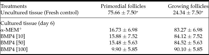
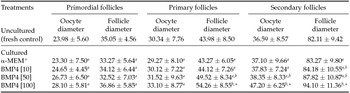
After 6 days of culture, a decrease in primordial follicles and increase in primary and secondary follicles was observed in cultured tissues when compared to fresh control (Table 2, P < 0.05). However, no significant differences were found among tissues cultured in the different treatments. Follicle and oocyte diameters at different follicular categories before and after in vitro culture are shows in Table 3. At day 6 of culture, follicular and oocyte diameters of primordial follicles cultured in α-MEM+ alone or supplemented with BMP4 (10, 50 or 100 ng/ml) had no differences in size when compared with fresh control. In addition, no differences among treatments were seen (P < 0.05). Conversely, primary follicles showed a significant increase in their diameter after culture in presence of 100 ng/ml BMP4, when compared with follicles from uncultured control, or cultured in α-MEM+ alone or supplemented with 10 ng/ml BMP4. In addition, an increase in the diameters of secondary follicles was observed in ovarian tissues cultured with 100 ng/ml when compared with follicles from uncultured control or cultured in α-MEM+ (P < 0.05). Furthermore, oocytes from these follicles increased their diameters after culture in the presence of 100 ng/ml BMP4, when compared with follicles cultured in α-MEM+ alone or added with 10 ng/ml BMP4.
Percentages (mean ± S.E.M.) of primordial and growing follicles (primary and secondary) in uncultured tissues and tissues cultured for 6 days in α-MEM+(control medium) and α-MEM+ supplemented with various concentrations of BMP4

*P < 0.05, significantly different from uncultured ovarian cortex tissue (fresh control).
Follicle and oocyte diameter (µm, mean ± SD) in uncultured (fresh control) tissues and tissues cultured for 6 days in α-MEM+ (control medium) and α-MEM+ supplemented with various concentrations of BMP4

*P < 0.05, significantly different from uncultured ovarian cortex tissue (fresh control).
a,b Values within columns with different letters among treatments are significantly different (P < 0.05).
Expression of mRNA for PCNA, GDF9, BMP15, Bax and Bcl2 in bovine preantral follicles
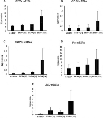
The levels of mRNA for PCNA, GDF9, BMP15, Bax and Bcl2 in tissues cultured for 6 days in α-MEM+ alone or supplemented with different concentrations of BMP4 are shown in Fig. 2(A−E). Tissues cultured in the presence of BMP-4 at concentration of 100 ng/ml had an increase in the levels of PCNA, GDF9, BMP15, Bax and Bcl2 mRNA, compared with those cultured in medium supplemented with α-MEM+ alone. However, the differences were not statistically significant (P > 0.05).
Discussion
The present study demonstrates that BMP4 does not influence the transition from primordial to developing follicles in bovine species, but stimulates the growth of primary and secondary follicles in vitro. Various studies have shown that the transition from resting primordial follicles to growth stages occurs spontaneously when cortical ovarian tissue is cultured in vitro (Cushman et al., Reference Cushman, Wahl and Fortune2002). It has been suggested that ovarian fragmentation increases actin polymerization and stops the Hippo signaling pathway, which leads to increased expression of growth factors, including connective tissue growth factor (CTGF or CCN2) and nephroblastoma overexpressed (NOV or CCN3) (Kawamura et al., Reference Kawamura, Cheng, Suzuki, Deguchi, Sato, Takae, Ho, Kawamura, Tamura, Hashimoto, Sugishita, Morimoto, Hosoi, Yoshioka, Ishizuka and Hsueh2013). Hsueh et al. (Reference Hsueh, Kawamura, Cheng and Fauser2015) reported that secretion of CCN2 and related factors promotes the growth of primordial follicles in vitro. In ovine species, BMP4 also does not influence the activation of primordial follicles in vitro (Bertoldo et al., Reference Bertoldo, Duffard, Bernard, Frapsauce, Calais, Rico, Mermillod and Locatelli2014). Conversely, BMP4 promoted an increase in primordial to primary follicle transition in mouse (Ding et al., Reference Ding, Zhang, Mu, Li and Hao2013) and rat cultured ovaries (Nilsson & Skinner, 2003). In mouse ovary, BMP4 protein was detected in all stages of follicular development including primordial follicles, suggesting that BMP4 might act in paracrine/autocrine manner to affect the transition of primordial to primary follicles (Tanwar et al., Reference Tanwar, O'Shea and McFarlane2008; Tanwar & McFarlane, Reference Tanwar and McFarlane2011).
Regarding follicular diameter, after in vitro culture, BMP4 increased the diameters of primary and secondary follicles after culture of ovarian tissue, suggesting the existence of functional BMP4 signaling in the preantral follicle. A study in sheep demonstrated that BMP4 increases the diameter of follicles cultured in ovarian tissue fragments (Bertoldo et al., Reference Bertoldo, Duffard, Bernard, Frapsauce, Calais, Rico, Mermillod and Locatelli2014). Nilsson & Skinner (2003) reported an increase in the number of developing follicles compared with controls when rat ovaries were treated with BMP4. Park et al. (Reference Park, Woods and Tilly2013) reported a direct effect of BMP4 on mice oogonial stem cell differentiation into oocyte, sustaining hypothesis of functional BMP signaling in early steps of oogenesis/folliculogenesis. In vivo, BMP4 is synthesized by the theca cells and acts on nearby granulosa cells and oocytes in a paracrine manner (Young & McNeilly, Reference Young and McNeilly2010). Possibly BMP4 acts by increasing the capacity of the granulosa cells to secrete factors of which the oocyte is a target, promoting the increase in diameter of primary and secondary follicles.
This study shows that follicles enclosed in ovarian tissues cultured in presence of BMP4 had their morphology preserved after 6 days of culture. A previous study suggested that BMP4 may be associated with the survival of oocytes and the development of primordial follicles in neonatal pig ovaries (Shimizu et al., Reference Shimizu, Yokoo, Miyake, Sasada and Sato2004). It was also demonstrated that treatment of neonatal rat ovaries with anti-BMP4 antibody resulted in the apoptosis of ovarian stromal-interstitial cells, as well as apoptosis of follicular granulosa and oocytes, which indicate the role of BMP4 as a cell survival factor (Nilsson & Skinner, 2003). Ding et al. (Reference Ding, Zhang, Mu, Li and Hao2013) demonstrated that BMP4 enhanced the phosphorylation of SMAD1/5/8 and prevented oocyte apoptosis via up-regulation of Sohlh2 and c-kit in primordial follicles. Childs et al. (Reference Childs, Kinnell, Collins, Hogg, Bayne, Green, Mcneilly and Anderson2010) reported that BMP4 reduces apoptosis levels in human granulosa cells cultured in vitro. BMP4 is also associated with the inhibition of apoptosis in bovine granulosa cells through the PI3K/PDK-1/PKC pathway (Shimizu et al., Reference Shimizu, Kayamori, Murayama and Miyamoto2012). Spicer et al. (Reference Spicer, Aad, Allen, Mazerbourg and Hsueh2006) reported that BMP4 has no effect on granulosa cell proliferation, but prevented premature differentiation of the granulosa cells during growth of follicles. In addition, Fabre et al. (Reference Fabre, Pierre, Pisselet, Mulsant, Lecerf, Pohl, Monget and Monniaux2003) reported that BMP4 have no effect on ovine granulosa cell proliferation while significantly affecting steroidogenesis in vitro.
In this study, exogenous addition of BMP4 did not affect mRNA expression for GDF9, BMP15, PCNA, Bax and Bcl2, perhaps a longer in vitro culture period is necessary to have a more pronounced increase in gene expression. Sadeu & Smitz (Reference Sadeu and Smitz2008) observed follicular activation and increased expression of GDF9 after 28 days of culture of ovarian cortex in humans. It has been proposed that the addition of BMP4 to culture medium promotes a balance between various factors involved in the mechanisms of folliculogenesis (Pierre et al., Reference Pierre, Pisselet, Dupont, Mandon-Pépin, Monniaux, Monget and Fabre2004). GDF9 expression has been found in oocytes from bovine follicles at early stages of follicular development (Bodensteiner et al., Reference Bodensteiner, Clay, Moeller and Sawyer1999). It is known that GDF9 promotes follicular activation after 7 days of in vitro culture. GDF9 also stimulates the transition from primary to secondary follicles while maintaining their ultrastructural integrity (Martins et al., Reference Martins, Celestino, Saraiva, Matos, Bruno, Rocha-Junior, Lima-Verde, Lucci, Báo and Figueiredo2008). In goats, high levels of mRNA for BMP15 were found during the transition from primary to secondary follicle stages (Celestino, et al., Reference Celestino, Bruno, Saraiva, Rocha, Brito, Duarte, Araújo, Silva, Matos, Campello, Silva and Figueiredo2011). PCNA performs the essential function of providing replicative polymerases with the high processivity required to duplicate the entire genome and has been used as a marker of granulosa cell proliferation (Maga & Hubscher, Reference Maga and Hubscher2003; Muskhelishvili et al., Reference Muskhelishvili, Wingard and Latendresse2005). Bax is a pro-apoptotic protein involved in granulosa cell apoptosis and is an important regulator of follicle growth (Tilly et al., Reference Tilly, Tilly, Kenton and Johnson1995). Bcl2 expression is found in the granulosa cells of both fetal and adult ovaries (Hussein, Reference Hussein2005; Hussein et al., Reference Hussein, Bedaiwy and Falcon2006).
In conclusion, 100 ng/ml BMP4 promotes an increase in follicular and oocyte diameters of primary and secondary follicles after 6 days of in vitro culture. Furthermore, BMP4 is able to promote the maintenance of follicular viability.
Levels of mRNA for PCNA (A), GDF9 (B), BMP15 (C), Bax (D) and Bcl2 (E) in tissues cultured for 6 days in α-MEM+ alone or supplemented with various concentrations of BMP4.

Acknowledgements
The authors acknowledge the members of Biotechnology Graduation School (Renorbio) of Federal University of Ceara, Brazil.
Financial support
This research was supported by grants from the National Council for Scientific and Technological Development (CNPq, Brazil) and Coordination for the Improvement of Higher Education Personnel (CAPES), grant number 23038.007469/2011–1027. J.R.V. Silva is an investigator for CNPq. E.V. Cunha is the recipient of a PhD scholarship from the Cearense Foundation for the Support of Scientific and Technological Development (FUNCAP), Brazil.
Statement of interest
None. The authors declare that there is no conflict of interest that would prejudice the impartiality of this scientific work.