Introduction
Amyloidosis is an idiopathic disorder characterised by the extracellular deposition of non-soluble fibrillar proteins (amyloid) that deposit in organs and tissues, which can cause organ failure and death.Reference Sipe, Benson, Buxbaum, Ikeda, Merlini and Saraiva 1 , Reference Gertz 2 It may manifest as systemic disease or be localised to certain organs, most commonly the kidney, heart and liver (in order of decreasing incidence).Reference Merlini and Westermark 3
Amyloidosis is a very rare disease with an incidence approaching 5–10 per million per year, with up to 20 per cent of cases involving the head and neck.Reference Sipe, Benson, Buxbaum, Ikeda, Merlini and Saraiva 1 , Reference Passerotti, Caniello, Hachiya, Santoro, Imamura and Tsuji 4 The larynx is the most common site of amyloid deposition in the head and neck, representing 0.2–1.2 per cent of all benign laryngeal tumours, and is rarely associated with systemic amyloidosis.Reference Thompson, Derringer and Wenig 5 , Reference Lebowitz and Morris 6 It is typically primary; it is occasionally associated with a plasma cell dyscrasia and very rarely systemic disease.Reference Lebowitz and Morris 6 Amyloidosis primarily affects those aged 40–60 years, with a peak incidence in the fifth decade of life. It is more prevalent in males, with a 3:1 male:female ratio.Reference Pribitkin, Friedman, O'Hara, Cunnane, Levi and Rosen 7 It rarely presents during childhood; only 10 cases have been reported in the literature, with the youngest being an 8-year-old female.Reference Balbani, Formigoni, Sennes, Jacob, Miniti and de Carvalho 8 – Reference Clevens, Wiatrak and Myers 14
Symptoms are dependent on size and specific location of the deposit. Symptoms may include dysphonia, stridor, dysphagia or exertional dyspnoea.Reference Wierzbicka, Budzyński, Piwowarczyk, Bartochowska, Marszałek and Szyfter 15 The ‘gold standard’ methods for diagnosis are laryngoscopy and tissue biopsy. Amyloidosis presents as subepithelial extracellular deposits of acellular, amorphous, eosinophilic material within the stroma on haematoxylin and eosin stain. Diagnosis is confirmed with Congo red stain positive for amyloid with apple-green birefringence when viewed with polarisation microscopy.Reference Sipe, Benson, Buxbaum, Ikeda, Merlini and Saraiva 1
This paper aims to highlight the diversity of presentations, increase awareness of paediatric amyloidosis and update the reader on current management.
Case series
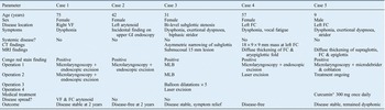
See Table I for patient demographics, disease locations, symptoms, investigations, treatments and outcomes.
Case series patient summary

*BioCeuticals® Theracurmin BioActive. VF = vocal fold; FC = false cord; GI = gastrointestinal; CT = computed tomography; MRI = magnetic resonance imaging; MLB = microlaryngoscopy and bronchoscopy
Case one
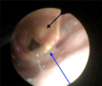
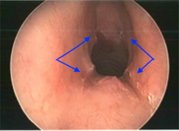
A 75-year-old female presented with an insidious onset of dysphonia, which had gradually progressed over many years, with no other ENT symptoms. Fibre-optic nasopharyngeal examination demonstrated a tiny mid-right vocal fold nodule, for which no treatment was prescribed. Repeat examination conducted 15 months later for persistent dysphonia demonstrated unusual submucosal thickening of her right vocal fold. She underwent endoscopic resection on two separate occasions over the next six months for progressive disease. On the last review, over two years after the initial diagnosis, the disease appeared stable and involved the right vocal fold, right arytenoid and especially the right false cord (Figure 1).
On fibre-optic nasopharyngeal examination of case one at the last review, over two years after initial diagnosis, the disease appeared stable, and involved the right vocal fold, right arytenoid (blue arrow) and especially the right false cord (black arrow).

Case two
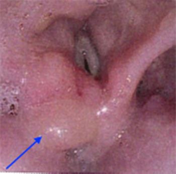
A 42-year-old female presented for examination of an abnormal laryngeal appearance observed on upper gastrointestinal endoscopy conducted for an investigation of iron deficiency. She did not report any cough, dysphonia, dyspnoea or dysphagia at the time of presentation. Hence, amyloidosis was an incidental finding on upper gastrointestinal endoscopy. Microlaryngoscopy demonstrated a left arytenoid swelling, with soft, cyst-like contents but no obvious external wall; the contents were immediately submucosal, with no deep wall or margin (Figure 2). There was no clear demarcation between pathological and normal tissue.
Microlaryngoscopy of case two demonstrated left arytenoid swelling with soft, cyst-like contents but no obvious external wall; the contents were immediately submucosal, with no deep wall or margin (blue arrow). There was no clear demarcation between pathological and normal tissue.

Case three
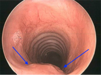
A 31-year-old female presented with an 18-month history of dysphonia and recent episodic stridor. On examination, she had soft biphasic stridor. Fibre-optic nasopharyngeal examination demonstrated stenosis in the subglottis, with mobile vocal folds. Computed tomography of the neck with contrast demonstrated mild-to-moderate asymmetric narrowing in the subglottic trachea, with thickening of the lateral and posterior mucosal surfaces, but with no underlying cause evident. Microlaryngoscopy demonstrated two areas of stenosis. The superior area of stenosis commenced 7 mm below the vocal folds and extended for 26 mm (Figure 3). Inferior stenosis proceeded along the posterior trachea for a distance of 40 mm. Subsequent dilatations allowed passage of a size 4.0–6.0 endotracheal tube (Figure 4). Carbon dioxide (1W) laser cruciate incisions were made to the circumferential subglottic stenosis, with subsequent balloon dilatation to 19 mm (Figure 5).
Microlaryngoscopy of case three demonstrated two areas of stenosis. The superior area of stenosis commenced 7 mm below the vocal folds and extended for 26 mm.

In case three, inferior stenosis was observed proceeding along the posterior trachea for a distance of 40 mm (blue arrows). Subsequent dilatations allowed passage of a size 4.0–6.0 endotracheal tube.

Carbon dioxide (1W) laser cruciate incisions in case three to the circumferential subglottic stenosis (blue arrows), with subsequent balloon dilatation to 19 mm.

Case four
A 57-year-old female schoolteacher presented with persistent dysphonia with vocal fatigue, which she had experienced for over six months. The lesion was evaluated with computed tomography of the larynx and neck, which demonstrated an 18 × 9 × 9 mm mass along the left false vocal fold and in the vestibule, extending from the anterior commissure to the arytenoid. Microlaryngoscopy and debulking of the lesion including a biopsy was performed two weeks later. Histopathology confirmed a diagnosis of laryngeal amyloidosis.
Case five
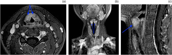
A nine-year-old boy presented with a six-month history of dysphonia and exertional dyspnoea, which had increased significantly in severity during the previous three months. At the time of examination, he was stridulous whilst asleep, despite being otherwise medically well. Fibre-optic nasopharyngeal examination demonstrated a cystic lesion arising from the left ventricular fold, causing 70 per cent obstruction of his airway. Vocal fold movement was intact. Microlaryngoscopy and biopsy was performed and confirmed amyloid. Amyloid deposits were observed to occupy the supraglottis bilaterally on magnetic resonance imaging, filling the false cords and base of the epiglottis (Figure 6a–c).
In case five, amyloid deposits were observed, on (a) axial, (b) coronal and (c) sagittal magnetic resonance imaging scans, to occupy the supraglottis bilaterally, filling the false cords and base of epiglottis (blue arrows).

Discussion
Amyloidosis is characterised by the extracellular deposition of pathological, insoluble fibrils in various tissues and organs. The fibrils have a typical B-pleated sheet configuration and result from misfolded proteins. The current classification is based on the main fibril protein, which allows a specific designation of amyloid disease. The amyloid protein is a designated protein ‘A’ plus a suffix (which specifies the protein).Reference Sipe, Benson, Buxbaum, Ikeda, Merlini and Saraiva 1 At present, there are over 36 human biochemical forms. Type AL (light chain) amyloidosis accounts for 90 per cent of cases; it is derived from plasma cells and contains kappa or lambda immunoglobulin light chains.Reference Gertz 2 It may be localised or systemic.
Localised AL amyloidosis results from the secretion of light chains from plasma cells within a specific tissue, but without systemic dissemination of these proteins. In comparison, in systemic AL amyloidosis, the plasma cell dyscrasia occurs in the bone marrow, spreads systemically and deposits within organs. Systemic amyloidosis is typically associated with higher mortality and morbidity, depending on the organs affected and the disease load.
Amyloidosis affects the head and neck in up to 20 per cent of cases, with disease having been described occurring in the orbit, nasopharynx, lips, floor of mouth, tongue, larynx, and tracheobronchial tree.Reference Simpson, Skinner, Strong and Cohen 16 The larynx is the most common site, with localisation most commonly to the vestibular folds (false cords) or ventricle, followed by the vocal folds (true cords), aryepiglottic folds and subglottis (in order of decreasing frequency).Reference Mitrani and Biller 12 , Reference Barnes and Zafar 17 , Reference Lewis, Olsen, Kurtin and Kyle 18 Synchronous multiple laryngeal sites have been reported.Reference Passerotti, Caniello, Hachiya, Santoro, Imamura and Tsuji 4 , Reference Thompson, Derringer and Wenig 5 Symptoms are determined by the relative size and anatomical location of lesions. Hoarseness and progressive dyspnoea are the two most common symptoms, whilst dysphagia, cough, haemoptysis are frequently reported.Reference Lewis, Olsen, Kurtin and Kyle 18 – Reference Aydin, Üstündaǧ, İşeri, Özkarakaş and Oǧuz 20
Appearance on laryngoscopy varied significantly between cases, ranging from nodules, to soft, cystic swelling to diffuse tissue mass. On direct laryngoscopy, amyloid has previously presented as firm, non-ulcerated, yellow, red or white lesions.Reference Barnes and Zafar 17 Investigation with computed tomography prior to surgery was performed in the third and fourth cases, and typically demonstrates marked thickening of laryngeal soft tissues with a high-density appearance. Magnetic resonance imaging is the technique of choice to demonstrate the most specific features of amyloidosis. Magnetic resonance imaging demonstrates intermediate T1-weighted signal intensity and low T2-weighted signal intensity, as evident in case five (Figures 6a–c).Reference Aydin, Üstündaǧ, İşeri, Özkarakaş and Oǧuz 20
Initial biopsy confirmed the diagnosis of amyloidosis in four of the five cases, with case three returning positive results on repeat biopsy four months later. For all cases, on histological examination, special Congo red staining was positive and demonstrated the characteristic apple-green birefringence when viewed under polarised light, which is pathognomonic of the disease.
After diagnosis, all patients underwent investigation for evidence of systemic disease; the findings were negative in all cases. First-line investigations should consist of general blood examination, serum free light chain testing, and urinary examination including proteinuria and light chain testing.Reference Bartels, Dikkers, van der Wal, Lokhorst and Hazenberg 21 Injected radio-labelled 123iodine serum amyloid P (‘SAP’) scanning localises rapidly and specifically to amyloid deposits in proportion to the amount of amyloid present. It has a reported sensitivity of 90 per cent in AL and AA amyloidosis, and is considered the best modality in clinical use for assessing the extent and distribution of all amyloidosis types.Reference Sachchithanantham and Wechalekar 22 – Reference Hawkins, Richardson and Vigushin 24 The serum amyloid P scan should be performed in all patients to rule out systemic disease, given the vastly higher morbidity and mortality compared with localised disease.
The current regimen for the treatment of systemic disease is high-dose melphalan followed by autologous stem cell transplantation, which carries a high risk of mortality. Median overall survival has improved and has been reported as up to 8.2 years, with 5-year and 10-year survival rates of 63.9 per cent and 43.4 per cent respectively.Reference Rosengren, Mellqvist, Nahi, Forsberg, Lenhoff and Strömberg 25
The goal of treatment for localised laryngeal amyloidosis is to maintain an adequately patent airway with as few procedures as possible, whilst definitive treatment involves excision of the affected tissue. Management strategies range from observation to balloon dilatation, endoscopic excision and laryngectomy.Reference Thompson, Derringer and Wenig 5 , Reference Celenk, Durucu, Baysal, Karatas, Polat and Bakir 26 Whilst laser (carbon dioxide and potassium titanyl phosphate) excision was considered effective in several studies,Reference Deviprasad, Pujary, Balakrishnan and Nayak 27 – Reference Yiotakis, Georgolios, Charalabopoulos, Hatzipantelis, Golias and Charalabopoulos 30 there is evidence that cold endoscopic excision for small laryngeal and localised glottic lesions is appropriate.Reference Bartels, Dikkers, van der Wal, Lokhorst and Hazenberg 21 The paediatric case in the current study employed the use of a microdebrider and coblation wands, the first such case in the literature. Laryngeal amyloidosis is rarely fatal; however, it can have a significant impact on quality of life, and voice quality is commonly affected as a result of disease burden and aggressive surgery. Treatment modalities must be balanced against the potential morbidity of any therapy. This said, tracheostomy has been performed in light of airway concerns.Reference Lewis, Olsen, Kurtin and Kyle 18 , Reference Gilad, Milillo and Som 31
-
• Amyloidosis is an idiopathic, localised or systemic disorder characterised by extracellular deposition of non-soluble fibrillar proteins (amyloid)
-
• Laryngeal amyloidosis represents 1 per cent of all benign laryngeal lesions, and causes variable symptoms depending on location and size
-
• Diagnosis is confirmed on histology; positive Congo red stain and apple-green birefringence under polarised light indicate disease
-
• A serum amyloid P scan should be performed to rule out systemic disease associated with higher morbidity and mortality
-
• Treatment ranges from observation to balloon dilatation, endoscopic, laser or microdebrider and coblation wand excision, and laryngectomy
-
• Annual follow up for at least 10 years is recommended given the long latency period
The evidence for medical treatment of localised disease is limited. The paediatric patient in the current study was commenced on curcumin (300 mg, daily) for persisting disease within his false cords and epiglottis. There is modest evidence demonstrating clinical benefit in patients with plasma cell dyscrasias.Reference Golombick, Diamond, Manoharan and Ramakrishna 32 No patient underwent radiation therapy and the evidence in the literature is mixed.Reference Neuner, Badros, Meyer, Nanaji and Regine 33 , Reference Truong, Kachnic, Grillone, Bohrs, Lee and Sakai 34 Whilst chemotherapy gives the best chance of remission in systemic disease, it not advocated for localised deposits as it is not regarded as effective.Reference Thompson, Derringer and Wenig 5 , Reference Pribitkin, Friedman, O'Hara, Cunnane, Levi and Rosen 7 , Reference Ghosh, Allgar and Elliott 35
Laryngeal amyloidosis can have long latency, with recurrence reported after 8–14 years, whilst lesions have remained unchanged for up to 17 years.Reference Bartels, Dikkers, van der Wal, Lokhorst and Hazenberg 21 , Reference Graamans and Lubsen 36 Recurrence is heavily reported in the literature, with one study reporting that almost half of its cases required subsequent surgery because of localised recurrent or large lesions.Reference Ma, Bandarchi, Sasaki, Levine and Choi 37 There appears to be no consistent area most vulnerable to recurrence; however, supraglottic disease had a higher incidence of recurrence in a limited case series, potentially attributable to surgical techniques.Reference Bartels, Dikkers, van der Wal, Lokhorst and Hazenberg 21 Annual follow up for at least 10 years is recommended.
Conclusion
Laryngeal amyloidosis is a rare and benign condition for which it is uncommon to observe progression to systemic amyloidosis; however, localised recurrence is frequently reported. This case series demonstrates that lesion size and anatomical location are the primary determinants of symptoms, and these largely dictate surgical management. Paediatric amyloidosis is exceedingly rare, with only 10 cases reported in the literature. This paper presents the second youngest case of laryngeal amyloidosis, and the first to be treated with a microdebrider and coblation wands.
Clinicians should consider the diagnosis of amyloidosis for unusual appearing submucosal laryngeal lesions with poorly defined borders, in both adult and paediatric patients. The serum amyloid P scan should be performed in all patients, with referral to a haematologist, to rule out systemic disease. Treatment consists of observation, balloon dilatation, endoscopic, laser or coblation wand excision, and rarely laryngectomy, with some cases requiring tracheostomy. Long-term follow up for 10 years is essential given the slowly progressive nature of the disease and risk of systemic amyloidosis development.