I. INTRODUCTION
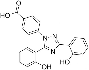
Deferasirox (Exjade®, ICL670, Figure 1), chemical name 4-[3,5-bis (2-hydroxyphenyl)-1,2,4- triazol-1-yl] benzoic acid, is an oral, once-daily iron chelator widely approved for the treatment of transfusional chronic iron overload (Yang et al., Reference Yang, Keam and Keating2007), and also as salvage therapy for a patient with progressive rhinocerebral mucormycosis (Reed et al., Reference Reed, Ibrahim, Edwards, Walot and Spellberg2006). Deferasirox was the first oral iron chelator approved by the Food and Drug Administration (FDA). Compared with other iron chelators, deferasirox can rapid access to intracellular iron pools (Neufeld, Reference Neufeld2006), and with less side effects. Therefore, deferasirox has become one of the most promising iron chelator drugs.
Structural formula of deferasirox.

The single-crystal structure of deferasirox [a = 8.900(2) Å, b = 26.946(5) Å, c = 7.558(2) Å, β = 94.77(3)°, unit-cell volume V = 1806.3(7) Å3, Z = 4, and space group P21/c] was first reported by Steinhauser et al. (Reference Steinhauser, Heinz, Bartholomä, Weyhermüller, Nick and Hegetschweiler2004). But detailed X-ray powder diffraction data for deferasirox have not been reported. In this report, the X-ray powder diffraction data of deferasirox were collected and analyzed.
II. EXPERIMENTAL
A. Sample preparation
The title compound, deferasirox (≥98% purity), in powder form was obtained from Wuhan E-ternity Technologies Co., Ltd. (Shanghai, China), and was characterized by FT-IR measurements (Thermo Fisher Scientific, Madison, USA, online Figure S1). The melting point and measured density of deferasirox are 262.48 °C and 1.40 g cm−3. The compound was re-crystallized by slow evaporation of a methanol solution at room temperature. Then the crystallized product with a needle-like shape was dried and ground into powder.
B. Powder diffraction data collection and reduction
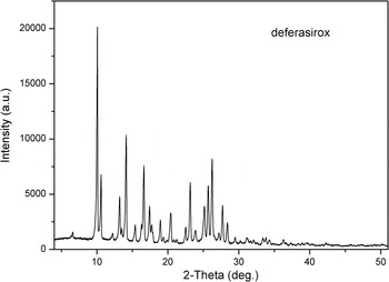
X-ray powder diffraction measurement was performed at room temperature using an X'Pert PRO diffractometer (PANalytical Co., Ltd., Netherlands) with a PIXcel 1D detector and CuKα radiation (generator setting: 40 kV and 40 mA). The diffraction data were collected over the angular range from 4° to 51°2θ with a step size of 0.013 13°2θ and a counting time of 50 ms step−1. Figure 2 shows the powder X-ray diffraction (XRD) pattern of the compound.
Powder XRD pattern of deferasirox.

The software package Material Studio 8.0 (Accelrys Co., Ltd., CA, USA) was used to process the data in the Analytical & Testing Center (Sichuan University, Chengdu, China). The powder XRD pattern was pretreated by subtracting the background, smoothing, and stripping off the Kα 2 component (Wang et al., Reference Wang, Sun, Li, Guo and Li2016). Automatic indexing results were obtained by the DICVOL91 method (Boultif and Louër, Reference Boultif and Louër1991). The indexing results were then refined with the type of Pawley (Reference Pawley1981), which involves assigning the Miller indices (h, k, l) to each observed peak in the experimental powder XRD pattern (Xu et al., Reference Xu, Liang, Wu, Zhang, Tang and Li2014).
III. RESULTS
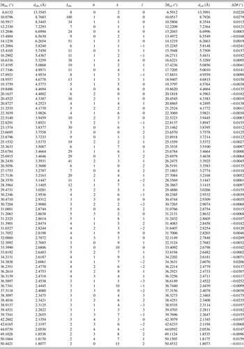
Pawley refinement results confirmed that deferasirox is monoclinic with space group P21/c and unit-cell parameters: a = 8.821(7) Å, b = 26.798(2) Å, c = 7.540(4) Å, α = 90°, β = 94.655(2)°, γ = 90°, unit-cell volume V = 1776.7(3) Å3, Z = 4, and ρ cal = 1.396 g cm−3. The values of 2θ obs, d obs, I obs, h, k, l, 2θ cal, d cal, and Δ2θ are listed in Table I. The figures of merit were achieved: F 24 = 47.5 (0.0071, 71) (Smith and Snyder, Reference Smith and Snyder1979) and M 24 = 22.9 (de Wolff, Reference de Wolff1968). The comparison of this pattern with the calculated pattern of single-crystal data reported by Steinhauser et al. (Reference Steinhauser, Heinz, Bartholomä, Weyhermüller, Nick and Hegetschweiler2004) is shown in online Figure S2. The results showed that both single-crystal and powder diffraction methods can get similar structure data.
X-ray powder diffraction data for deferasirox, C21H15N3O4. The d-values were calculated using CuKα 1 radiation (λ = 1.540 56 Å).

ACKNOWLEDGEMENTS
This work was supported by the National Development and Reform Commission and Education of China (Grant No. 2014BW011) and the Large-scale Science Instrument Shareable Platform Construction of Sichuan Province (Grant No. 2015JCPT0005-15010102).
SUPPLEMENTARY MATERIAL
The supplementary material for this article can be found at http://dx.doi.org/10.1017/S0885715616000476