Microbial adhesion to biological surfaces is an important criterion for selection of promising probiotic strains. This phenomenon guides the survival and colonisation of probiotics into the gut lumen, exclusion of pathogens as well as communication of the bacteria with host cells for the execution of its beneficial effects. Microbial interactions are generally mediated by the weak non-specific interaction (physicochemical) between biological surfaces and specific interaction between legend/adhesin-receptors of both interactive systems (Sengupta et al. Reference Sengupta, Altermann, Anderson, McNabb, Moughan and Roy2013; Beaussart et al. Reference Beaussart, El-Kirat-Chatel, Sullan, Alsteens, Herman, Derclaye and Dufrêne2014). For this, cell surface properties of a candidate probiotic strain are considered as an important contributing factor for the assessment of its probable beneficial effects on the host. Among nonspecific interactions, hydrophobic interaction and aggregating interaction between surface-macromolecules have been considered as primary markers for the evaluation of colonisation potential of probiotics (Andriantsoanirina et al. Reference Andriantsoanirina, Teolis, Xin, Butel and Aires2014; Nishiyama et al. Reference Nishiyama, Nakazato, Ueno, Seto, Kakuda, Takai, Yamamoto and Mukai2015; Shahbazi et al. Reference Shahbazi, Nateghi and Aghababyan2016). Similarly, the specific adhesin-receptor mediated attachment between host tissue and microbes play an important role in the accomplishment of probiotic attributes (Turpin et al. Reference Turpin, Humblot, Noordine, Thomas and Guyot2012). Therefore, microbial cell surface hydrophobicity, auto-aggregation and adhesion to biological surface models are ubiquitously accepted as determinant factors in the assessment of colonisation potential of probiotic strains.
The initial non-specific physico-chemical interactions of microbes are mediated by electrostatic, van der Walls, hydrogen bonding and hydrophobic interactions (Beaussart et al. Reference Beaussart, El-Kirat-Chatel, Sullan, Alsteens, Herman, Derclaye and Dufrêne2014). These physico-chemical features are largely predisposed by the composition of bacterial surface covering components. Mainly, exopolysaccharide, glycoproteins, lipoproteins, lipotechoic acids, moonlightining proteins, S layer proteins and surface appendages influence the behaviour of the cell. The cell surface hydrophobicity could be measured at cell-cell, cell-solid surface or cell-liquid interactions (Benoit et al. Reference Benoit, Gabriel, Gerisch and Gaub2000; Wilson et al. Reference Wilson, Wade, Holman and Champlin2001; Tsuneda et al. Reference Tsuneda, Aikawa, Hayashi, Yuasa and Hirata2003; Salerno et al. Reference Salerno, Logan and Velegol2004). Among them, bacterial adhesion to hydrocarbons (BATH) is a simple method which measures the hydrophobic behaviour of candidate probiotic strains in the presence of aliphatic or aromatic hydrocarbons in a liquid-liquid interference system.
Similarly, cellular aggregation implicates the ability of precipitation of similar kinds of cells. Auto-aggregation of probiotic cells is reported to be mediated by the cell surface proteins and few soluble proteins (Miljkovic et al. Reference Miljkovic, Strahinic, Tolinacki, Zivkovic, Kojic, Golic and Kojic2015; Nishiyama et al. Reference Nishiyama, Nakazato, Ueno, Seto, Kakuda, Takai, Yamamoto and Mukai2015). This form of interaction is advantageous for the effective colonisation and communication of probiotics with the host tissue. Additionally, it strengthens the antimicrobial potential of probiotics and helps them to compete and exclude the pathogens from the intestinal mucosa (Nishiyama et al. Reference Nishiyama, Nakazato, Ueno, Seto, Kakuda, Takai, Yamamoto and Mukai2015). Both hydrophobicity and auto-aggregation are also known to affect the specific attachment of probiotics with host epithelial mucosa (Garcia-Cayuela et al. Reference García-Cayuela, Korany, Bustos, de Cadiñanos, Requena, Peláez and Martínez-Cuesta2014). For attachment to the host epithelium, specific interaction of adhesive cell surface appendages and complimentary structures of opposite surface is required. This system can be mimicked quite simply by the formation of in vitro models of extracellular collagen matrix. The specific interaction of probiotic with collagen surface is mediated by the collagen binding moieties on bacterial surface. Indeed, the presence of different types of binding proteins determines the colonisation advantage of a probiotic strain over the other competitors of commensal microflora including pathogenic strains.
Collectively, cell surface hydrophobicity, auto-aggregation and adhesion are the basic essential characteristics of a probiotic which potentially empowers their colonisation into the intestine. In this manner, we aimed to investigate the cell surface properties of indigenous candidate probiotic Lactobacillus strains of human milk origin.
While it is well known that human milk is a significant source of potential commensal microflora to the infant gut (Jost et al. Reference Jost, Lacroix, Braegger, Rochat and Chassard2014; Moles et al. Reference Moles, Escribano, de Andrés, Montes, Rodríguez, Jiménez, Sáenz de Pipaón and Espinosa-Martos2015; Damaceno et al. Reference Damaceno, Souza, Nicoli, Paula, Assis, Figueiredo, Azevedo and Martins2017); attempts were made to explore the rich source of valuable bacteria to find the potential probiotic lactobacilli for human use. Thereby, the study aimed to explore the cell surface properties of lactobacilli isolates of human milk origin.
Material and methods
Sample collection and isolation of strains
Human milk samples (2–5 ml per volunteer; 40 samples) from healthy volunteers of the age group of 25–30 years were collected from hospitals and local vicinity of Guru Angad Dev Veterinary and Animal Sciences University (GADVASU), Ludhiana. Samples were collected in sterile containers (screw capped tubes; 15 ml) and transported to the laboratory under refrigerated (5 °C) conditions. Lactic acid bacterial flora from human milk were isolated by standard serial plate dilution method over de Man Rogosa Sharpe (MRS) agar medium.
Bacterial strains and growth conditions
Eleven native Lactobacillus strains were isolated and identified from human milk collected from volunteers from Ludhiana, Punjab region of India. Isolates were initially identified by genus-specific PCR and further characterised by 16S rRNA based sequencing. These strains were earlier evaluated for their antimicrobial activities and antibiotic susceptibility pattern (Sharma et al. Reference Sharma, Singh, Thakur, Gulati, Gupta, Mishra and Panwar2017a, Reference Sharma, Gulati, Thakur, Singh, Gupta, Kaur, Mishra, Puniya, Gill and Panwarb). Reference Lactobacillus strains viz. L. plantarum (NCDC 20), L. bulgaricus (NCDC 26), L. fermentum (NCDC 214); and non-probiotic control, Lactococcus lactis subsp. Lactis (NCDC 91) were procured from National Collection of Dairy cultures (NCDC), ICAR-NDRI, Karnal, India. Reference probiotic, L. rhamnosus GG (ATCC 53103) and L. casei (ATCC 393) were procured from American Type Culture Collection (ATCC). Besides, two enteropathogens viz. E. coli (MTCC 723) and Salmonella enteric serovar Typhi (MTCC 733) were obtained from Microbial Type Culture Collection (MTCC, India). Lactobacillus isolates and reference cultures were maintained and propagated in de Man Rogosa Sharpe (MRS) medium (HiMedia, India); Lactococci in M17 (HiMedia, India); and pathogens in Brain Heart Infusion (BHI) medium. All the strains were preserved at −80 °C as glycerol stocks.
Cell surface hydrophobicity (CSH)
Cell surface hydrophobicity of all the test strains was assessed using bacterial adhesion to hydrocarbons (BATH) assay as defined by Rosenberg et al. (Reference Rosenberg, Judes and Weiss1983), with some modifications. Briefly, log phase bacterial cultures were harvested by centrifugation at 8000 g for 10 min. The cell pellets were washed twice and re-suspended in phosphate urea magnesium sulphate (PUM) buffer (22·2 g K2HPO4·3H2O; 7·26 g KH2PO4; 1·80 g Urea; 0·20 g MgSO4; pH: 7·1) to OD600 of 1·0 ± 0·05. Initially, 3 ml of cell suspension was transferred in a glass test-tube, followed by addition of 1 ml of hydrocarbon. In this study, five hydrocarbons viz. Toluene (monopolar, aromatic solvent), Xylene (apolar, aromatic solvent), Chloroform (monopolar, aliphatic solvent), n-Hexane (apolar, aliphatic solvent), and n-Octane (apolar, aliphatic solvent) were tested. Tubes were incubated at 37 °C for 10 min for temperature equilibrium, followed by vortex for 15 s. Cell suspensions were kept undisturbed at 37 °C for 20 min for phase separation. Lower aqueous phase was collected carefully and OD600 was recorded. Per cent hydrophobicity was calculated by using the following formula: % Hydrophobicity = (ODi – ODt/ODi) × 100; Wherein, ODi is the initial OD600 of cell suspension, and ODt represents the OD600 of aqueous phase recorded at the time t.
Auto-aggregation
Test bacterial strains were harvested at log phase by centrifugation at 8000 g for 10 m and washed twice with phosphate buffer saline (PBS, pH7·4). Cell pellets were re-suspended in same buffer and OD600 of 1·0 ± 0·05 was adjusted. Cell suspension (4 ml) was taken in a clean glass test tube and incubated undisturbed at 37 °C for 1, 3, 5, 7, 9, 11 and 24 h. Upper fraction (1 ml) was collected at different time intervals and per cent auto-aggregation was calculated as per Kos et al. (Reference Kos, Suskovic, Vukovic, Simpraga, Frece and Matosic2003) with the help of the following formula: % Auto-aggregation = (ODi – ODt/ODi) × 100; Wherein, ODi is the initial OD600 of cell suspension , and ODt is the OD600 of cell suspension at time t.
Adhesion to collagen
The adhesive activity of test strains on collagen surface was determined as reported by Yadav et al. (Reference Yadav, Tyagi, Kaushik, Saklani, Grover and Batish2013) with minor modifications. Briefly, the wells of 96 well microtiter plate were treated with 200 µl of collagen solution (50 µg/ml) prepared in PBS, pH5·5. The plate was incubated overnight at 4 °C and subsequently washed twice with PBS. Coated wells were blocked with 200 µl solution of 0·1% Tween 20 prepared in PBS and further incubated for 1 h at 37 °C. In the meantime, log phase bacterial cultures were harvested at 8000 g for 10 min. Cells were washed twice with PBS (pH7·4) and re-suspended in the same buffer to obtain an OD600 of 0·1 ± 0·05. To enumerate the bacterial cell number in prepared solution, ten fold serial dilutions were prepared in normal saline and appropriate dilution were plated in respective agar medium.
After 1 h of incubation with blocking buffer, solution was aspirated and 100 µl of prepared bacterial cell suspension was added and incubated in the wells for 2 h at 37 °C. Control wells contained buffer only. Following incubation, un-attached cells were removed by washing the wells thrice with PBS. The adherent bacteria were detached by adding 100 µl of 0·05% Triton × 100 and plated on agar plates. After 18–24 h of incubation at 37 °C, bacterial colonies were counted and per cent adhesion calculated using following formula: % Attachment to collagen = (N t/N 0) × 100; Wherein, N t is the number of attached cells after incubation, and N 0 is the initial number of cell added to the wells.
Maintenance and growth of HT-29 cell line
Human colon-carcinoma cell line, HT-29 was procured from National Center for Cell Science (NCCS), Pune. Cells were maintained in Dulbecco's Modified Eagles Medium (DMEM, Hi-Media) containing 5 mm glucose, 4 mm L-glutamine and 2 g/l sodium bicarbonate, 10% (v/v) heat inactivated fetal bovine serum (Cell Clone), 25 mm HEPES, 100 U/ml penicillin, 100 µg/ml streptomycin and 1% non-essential amino acids in 25 cm2 culture flask at 5% CO2/37 °C in a humidified atmosphere. Cells were preserved in freezing medium containing dimethyl sulphoxide (DMSO) (1X) (Hi-Media). An aliquot of 1·5 ml cell suspension in freezing medium was stored in liquid nitrogen at −196 °C.
Bacterial adhesion to HT-29 cells
The adhesion potential of selected candidate probiotic strains was carried out as per the method described by Duary et al. (Reference Duary, Rajput, Batish and Grover2011). HT-29 cells were cultured to develop a mature semi-confluent cells monolayer in a 12 well cluster plate and incubated until 80% confluence reached. Prior to the experiment, cells were supplemented with serum and antibiotic free DMEM for overnight. Cell monolayer was washed twice with 1XPBS and challenged at multiplicity of infection (MOI) of 10 : 1 bacteria per well and incubated for 2 h at 37 °C/5%CO2. After incubation, cells were washed thrice followed by treatment with 1 ml of a 0·05% (v/v) triton X-100 solution to lyse the cells and detach the bound bacteria. Cell suspension was aspirated, diluted in sterile 1XPBS and plated onto MRS agar. After 24–48 h of incubation at 37 °C, bacterial colonies were counted. Per cent adhesion was calculated using following formula: % Adhesion = (N t/N 0) × 100; Wherein, N t is number of attached bacteria after 2 h incubation period, and N 0 represents number of bacteria added at 0 h.
Statistical analysis
Per cent values of cell surface and adhesion properties were calculated using MS Excel. Results were expressed as mean ± se value of at least three separate duplicate experiments. One way analysis of variance (ANOVA) (for collagen and HT-29 adhesion assay) and two way ANOVA (for CSH and auto-aggregation assay) were applied to the mean data of positive control and test samples at P value of <0·05. For cell surface hydrophobicity and auto-aggregation data, Bonferroni post test and for collagen attachment assay, Dunnett post test were applied to determine the statistical significance of mean values using Prism 5·0 software (Graph Pad Prism, USA).
Results and discussion
Cell surface hydrophobicity
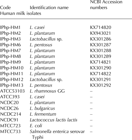
The eleven Lactobacillus isolates along with five reference strains of Lactobacillus, one Lactococcus and two pathogenic strains (Table 1) were investigated for the cell surface hydrophobicity using BATH assay. Among eleven tested candidate probiotic lactobacilli, three strains viz. HM1, HM9 and HM12 exhibited constantly higher hydrophobicity with all the five hydrocarbons used in this study. Notably, the hydrophobic disposition of HM1 (L. casei) was recorded highest among all the bacterial strains including reference cultures (Fig. 1). The per cent hydrophobic value of HM1 and LGG with chloroform was 97·10 ± 3·35% and 98·92 ± 1·24% respectively, which indicates almost complete removal of the culture from the aqueous phase. Additionally, adhesion of HM1 with n-Hexane (70·49 ± 5·64%) and toluene (77·59 ± 3·17%), HM12 with chloroform (75·11 ± 4·42%) and L. plantarum NCDC20 with chloroform (72·67 ± 5·93%) was also quite promising. On the other hand, negligible hydrophobic interaction was observed between n-Octane: L. fermentum NCDC214 (1·15 ± 3·80%), E. coli (0·91 ± 0·00%), S. Typhi (−2·00 ± 0·00%); Xylene: L. fermentum NCDC214 (−1·73 ± 5·80%); Toluene: L. fermentum NCDC214 (−2·57 ± 3·05%) and S. Typhi (−0·76 ± 5·31%). To our surprise, reference culture L. bulgaricus NCDC26 exhibited lowest hydrophobicity (−7·97 ± 5·04%) with toluene, which was even lower than that recorded with S. Typhi.
Cell surface hydrophobicity of Lactobacillus and reference strains using three aliphatic (Chloroform, n-Hexane and n-Octane) and two aromatic (Toluene and Xylene) solvents. Error bars indicate ± mean sd (n = 3).

List of Lactobacillus isolates and other strains used in this study

The hydrophobic interactions of bacteria have key roles in adhesion and biofilm formation. The behaviour of bacteria as a hydrophobic particle is allied with the composition of surrounding medium, growth stage of bacteria and structure of cell surface components (Wu & Shah, Reference Wu and Shah2014; Nivoliez et al. Reference Nivoliez, Veisseire, Alaterre, Dausset, Baptiste, Camarès, Paquet-Gachinat, Bonnet, Forestier and Bornes2015). In line with our results, strain dependent adhesiveness with hydrocarbons has also been documented by several earlier reports (Xu et al. Reference Xu, Jeong, Lee and Ahn2009; Garcia-Cayuela et al. Reference García-Cayuela, Korany, Bustos, de Cadiñanos, Requena, Peláez and Martínez-Cuesta2014; Piwat et al. Reference Piwat, Sophatha and Teanpaisan2015).
It has been verified that binding to hydrophobic surfaces is a time independent interaction, which may include multiple small length forces of unfolded microbial cell surface proteins (Beaussart et al. Reference Beaussart, El-Kirat-Chatel, Herman, Alsteens, Mahillon, Hols and Dufrêne2013). The microbial adhesion chemistry may contain combination of electrostatic and other short range hydrophobic contacts between the two surfaces. Therefore, in present study, affinity for solvents of different physicochemical properties (aliphatic/aromatic/apolar/polar/acidic/basic) was assessed to express the hydrophobic/ hydrophilic/ acidic/ basic nature of cell surface components of tested bacteria. The highest affinity of Lactobacillus strains towards chloroform and toluene (monopolar aliphatic and aromatic solvents respectively) indicates the presence of low electron acceptor/acidic surface components on cell surface of most of the strains. Similar kinds of observations were recorded by Xu et al. (Reference Xu, Jeong, Lee and Ahn2009) with xylene (apolar), chloroform (polar, acidic) and ethyl acetate (polar, basic) solvents. Affinities of the strains of Bifidobacterium, Lactobacillus, Luconostoc, Pediococcus and food pathogens to ethyl acetate were relatively low, when compared to xylene and chloroform.
Interestingly, among the apolar aliphatic (n-Hexane, n-Octane) and aromatic (Xylene) solvents, Xylene and n-Hexane removed microorganisms more efficiently then n-Octane. Around 68·42 and 73·68% tested bacterial strains exhibited moderate to high hydrophobicity with n-Hexane and xylene respectively. The same level of hydrophobic interaction with n-Octane was recorded with 52·63% strains only. Comparative less hydrophobic affinity with n-Octane may be due to stearic hindrance and absence of short range hydrogen bonding between long hydrocarbon and unfolded surface proteins of the microbes. A more in-depth study in this line by Van Der Mei et al. (Reference Van der Mei, Van de Belt-Gritter and Busscher1995) has described that attractive forces between hydrocarbons and microorganisms are predominantly long range van der Waals forces and short-range interactions. Authors stated that removal by hydrocarbon of the microorganisms increases towards the isoelectric points of the organisms or towards solution pH at which the hydrocarbons appear uncharged. Moreover, these interactions are compromised in long aliphatic hydrocarbons due to steric hindrance.
Auto-aggregation kinetics
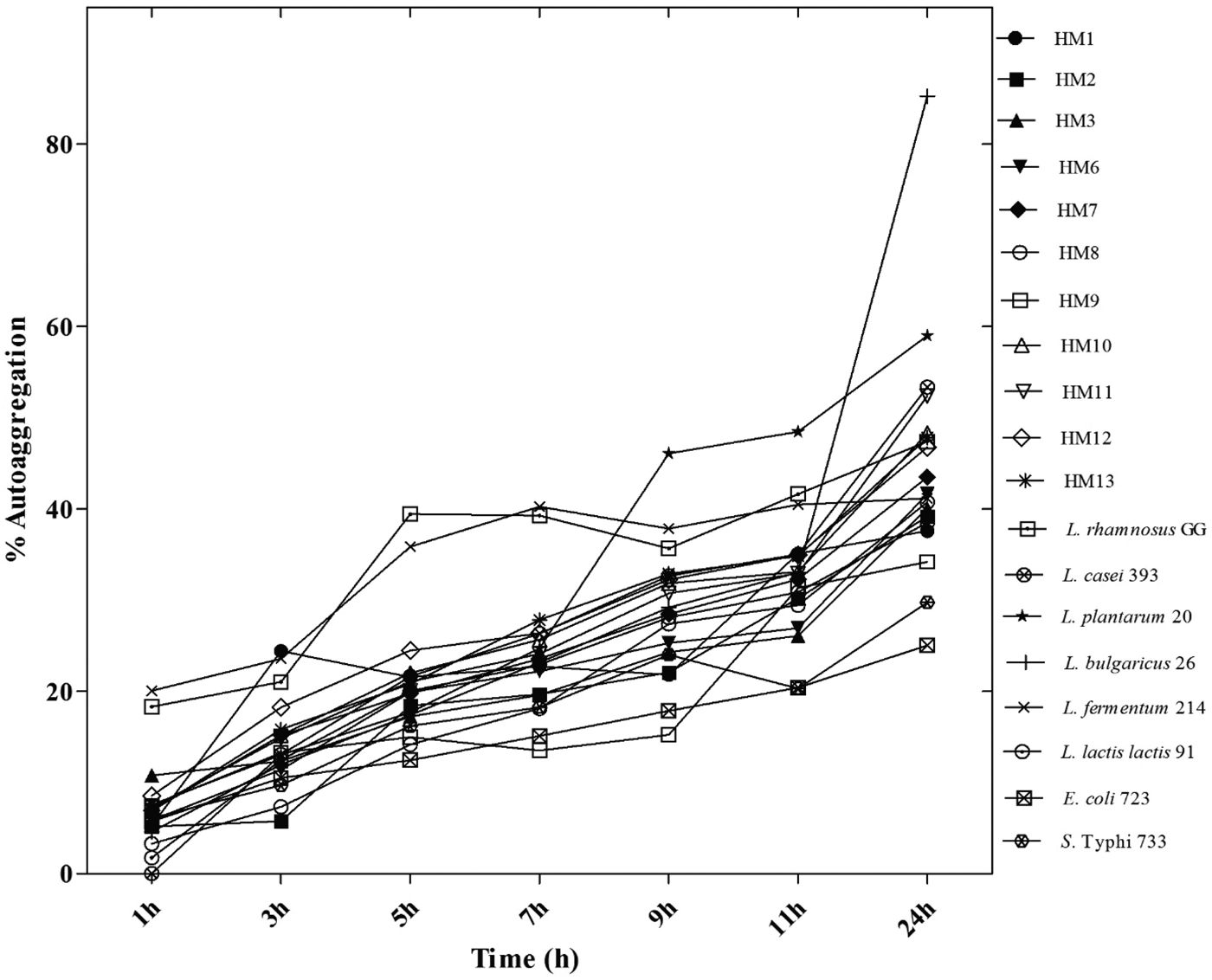
In contrast to the results logged with BATH assay, a moderate auto-aggregation attribute was observed in all of our Lactobacillus isolates. Only HM10, HM12 and HM13 exhibited comparatively enhanced precipitation rate after 7 h of incubation period (Fig. 2). Overall, the rate of sedimentation was slow during initial hours of incubation and slightly enhanced with increasing time, except L. plantarum NCDC20 and L. fermentum NCDC214. These two reference cultures aggregated at augmented rate from the beginning and maintained the pattern throughout the experimental period (Fig. 2). After completion of 24 h incubation period, the per cent auto-aggregation values of all the Lactobacillus strains tapered between the ranges of 29·05 to 57%, except L. casei ATCC393, for which significantly higher (P < 0·01) amount of precipitation (85·25 ± 10·84%) was recorded. Besides, HM7 and two pathogenic strains, E. coli and S. Typhi could aggregate at lowest rate which indicates minor intercellular physicochemical interaction among the cells. The mechanism of cellular aggregation involves the complex interaction of surface and/or secreted components of the cells (Malik et al. Reference Malik, Petrova, Claes, Verhoeven, Busschaert, Vaneechoutte, Lievens, Lambrichts, Siezen, Balzarini and Vanderleyden2013; Wang et al. Reference Wang, Qin, Yang, Zhai, Zhou, Sun, Pan, Ji, Wang, Gu and Feng2015). Overall, the auto-aggregation of similar kind of cells is strain and environment specific (Li et al. Reference Li, Liu, Dong, Zhou and Wang2015). Likewise, the time and strain dependent difference in aggregation phenotype in bacteria has also been revealed in several earlier reports (Rokana et al. Reference Rokana, Mallappa, Batish and Grover2016; Vanzieleghem et al. Reference Vanzieleghem, Couniot, Herman-Bausier, Flandre, Dufrêne and Mahillon2016).
Auto-aggregation kinetics of Lactobacillus and reference strains at different time intervals.

Attachment to collagen surface
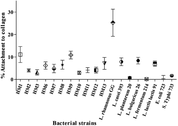
The binding potential of tested bacteria with extracellular matrix was evaluated using an in vitro model of immobilised collagen protein. The results presented in Fig. 3 indicates that L. rhamnosus GG could bind to the collagen surface at significantly high (P < 0·001) rate (26·94 ± 5·83%) and was followed by HM1 (11·07 ± 3·54%) and HM9 (10·85 ± 1·74%). Additionally, L. bulgaricus NCDC26 and L. casei ATCC393 exhibited moderate binding property by showing 8·36 ± 1·47% and 7·85 ± 1·72% attachment to the collagen surface. The lowest attachment potential was observed in L. plantarum NCDC20 (0·73 ± 0·52%) and L. fermentum NCDC214 (0·15 ± 0·03%), which was lower than two pathogens E. coli (1·03 ± 0·74%) and S. Typhi (1·67 ± 0·31%). The above average attachment efficiency of HM1 and HM9 could be correlated with their high hydrophobic attribute. In this line, similar type of correlation have been observed in various previous reports (Piwat et al. Reference Piwat, Sophatha and Teanpaisan2015; Rokana et al. Reference Rokana, Mallappa, Batish and Grover2016)
Per cent Attachment to collagen matrix of Lactobacillus and reference strains. Error bars indicate ± mean sd (n = 3). L. rhamnosus GG exhibited highest attachment attribute which was significantly higher (P < 0·001) from all the tested strains.

Contrary to this, auto-aggregation and collagen binding attributes have not demonstrated any correlation with each other in our study, although a positive relation between these two properties has been observed by some researchers (Collado et al. Reference Collado, Surono, Meriluoto and Salminen2007; Terraf et al. Reference Terraf, Mendoza, Tomás, Silva and Nader-Macías2014). After critical evaluation of the results obtained with both the assays in our study, it could be deciphered that cell to cell aggregation phenotype may not be an essential contributing factor in attachment traits of the bacteria. Thus, in the absence of appropriate binding potential, the possibility of washing out of the highly precipitated bacteria cannot be ruled out. A previous study by Lukic et al. (Reference Lukic, Strahinic, Milenkovic, Nikolic, Tolinacki, Kojic and Begovic2014) made similar observations by testing the role of aggregation factor (AggL) and mucin binding protein (MbpL) from Lactobacillus sp. in adhesion to gastrointestinal mucosa. In this study, expression of AggL had negative effect on binding of L. salivarius BGHO1 to gut mucosa. However, AggL did not decrease the adhesion in intestinal fragments in ex vivo condition. Authors suggested that intestinal motility could be the crucial force for eliminating aggregation factor bearing bacteria. Moreover, microbial adhesion to host epithelium is a multifactorial process which includes various elements and simultaneous forces from the both sides of a biological system (Schaer-Zammaretti & Ubbink, Reference Schaer-Zammaretti and Ubbink2003; Bron et al. Reference Bron, Tomita, Mercenier and Kleerebezem2013; Dufrêne, Reference Dufrêne2015).
Bacterial adhesion to HT-29 cells
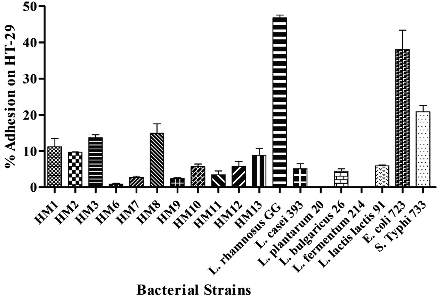
The range of per cent adhesion potential of our isolates was recorded from 0·85 ± 0·27% (L. pentosus HM6) to 14·99 ± 3·61% (L. plantarum HM8) (Fig. 4). However, the reference standard culture, LGG exhibited significantly higher (P < 0·001) adhesion value (46·81 ± 1·07%) in comparison to our isolates. On similar pattern, Pisano et al. (Reference Pisano, Viale, Conti, Fadda, Deplano, Melis, Deiana and Cosentino2014) have evaluated the adhesion potential of Lactobacillus strains of dairy origin on Caco2 cell line. The per cent adhesion property of the strains evaluated in this study ranged from 3% to 20% which is in accordance with the results obtained with our isolates. However, the percentage adhesion of L. rhamnosus GG used as a positive control was recorded at very low level (<5%) by Pisano et al. (Reference Pisano, Viale, Conti, Fadda, Deplano, Melis, Deiana and Cosentino2014), which is much lower than the value recorded in our study. A study by Mahmoudi et al. (Reference Mahmoudi, Moussa, Khaldi, Kebouchi, Soligot, Le Roux and Hassouna2016) could explain the differential adhesion feature of bacterial cultures under in vitro conditions. Mahmoudi et al. (Reference Mahmoudi, Moussa, Khaldi, Kebouchi, Soligot, Le Roux and Hassouna2016) reported different levels of adhesion of Lactobacillus isolates of camel milk origin on two different cell lines viz. Caco-2 and HT29-MTX. Previous observations and results obtained from our work confirm that the adhesion property of lactobacilli is controlled by several factors including environmental, treatment conditions and strain specificity of the organism. Here we also noticed that strains like HM6 and HM9 showing high or moderate attachment potential on collagen surface failed to adhere to HT-29 monolayer. Whereas, few isolates i.e. HM2, HM3 and HM8, which exhibited very low attachment affinity with collagen were able to adhere on HT-29 at substantially higher rate. In this context, Kainulainen & Korhonen, (Reference Kainulainen and Korhonen2014) has explained the possible role of moonlighting proteins in adhesion and functionality of bacteria. Authors have described that change in environmental factors, such as pH alter the position of these proteins from cell bound to detached form, which could inhibit the other interactions with human cells. However, the complete functionality of probiotics is also mediated by the combination of cell surface anchored and free proteins bound with electrostatic forces on the surface of bacteria. Thus, here we speculate that variable adhesion properties of our human milk isolates as well as standard cultures on different types of biological surfaces is a cumulative effect of the composition of cell surface components and surrounding conditions during interaction with epithelial cells.
Per cent adhesion of human milk isolates, non-milk origin standard and pathogen strains on HT-29 cells.

The outcomes of our study describe that the cell surface properties and adhesion prospective of probiotic bacteria are undoubtedly strain dependent. Nonetheless, the source of a probiotic strain could also add significant advantages to their survival, colonisation and beneficial attributes. The comparison of human milk origin candidate probiotic Lactobacillus strains with reference probiotic, non-probiotic and pathogenic strains under the reference of cell surface properties was quite interesting, in that HM1, HM2, HM3, HM8 and HM13 exhibited a marked difference from reference Lactobacillus strains. Our observations are supported by an earlier study by Pérez-Cano et al. (Reference Pérez-Cano, Dong and Yaqoob2010), who also recorded a better immunomodulatory activity of two human milk origin lactobacilli over reference probiotic strains.
Conclusions
The data presented in this study will help to understand the mechanism of non-specific and specific attachment properties of a candidate probiotic strain. In future, more precise transcriptomic and interactomic based studies would help to identify the mechanical factors behind the specific phenotypic effects of probiotic species. Additionally, it will also provide important information regarding the strain level metamorphoses of lactic acid bacteria. Besides, the outcomes of our study will pave the way for the establishment of indigenous probiotics of human milk origin for the promotion of the nutraceutical market and human health.
Authors would like to acknowledge the financial grant from Science and Engineering Research Board – Ministry of Food Processing and Industries (SERB/MoFPI/026/2015), Government of India for carrying out this research work.