




No CrossRef data available.
Article contents
Excretion of fluorescent substrates of mammalian multidrug resistance-associated protein (MRP) in the Schistosoma mansoni excretory system
Published online by Cambridge University Press: 19 January 2004
Abstract
The protonephridium of platyhelminths including Schistosoma mansoni plays a pivotal role in their survival by excretion of metabolic wastes as well as xenobiotics, and can be revealed in the living adult parasite by certain fluorescent compounds which are concentrated in excretory tubules and collecting ducts. To determine the presence of the multidrug resistance-associated protein (MRP) as a possible transporter in protonephridial epithelium, adult schistosomes were exposed to a fluorescent Ca2+ indicator, fluo-3 acetyloxymethyl ester, which is a potential substrate of mammalian MRP. Specific fluorescence related to fluo-3/Ca2+ chelate delineated the whole length of the protonephridial system. Simultaneously, a fluorescent substance was accumulated in the posterior part of collecting ducts and the excretory bladder. Similarly, when other fluorogenic substrates for mammalian MRP such as monoclorobimane, fluorescein diacetate, and 5(6)-carboxyfluorescein diacetate were applied to adult schistosomes, these fluorescent markers were observed in the excretory tubules through to the excretory bladder. The excretory system of mechanically-transformed schistosomula was not labelled with any of these 4 fluorescent markers. These findings suggest that the protonephridial epithelium of adult schistosomes, but not schistosomula, might express the homologue of the mammalian MRP transporting organic anionic conjugates with glutathione, glucuronate or sulphate as well as unconjugated amphiphilic organic anions.
Keywords
Information
- Type
- Research Article
- Information
- Copyright
- 2004 Cambridge University Press
INTRODUCTION
The protonephridium of platyhelminths including Schistosoma mansoni plays a pivotal role in their survival by excretion of metabolic wastes as well as xenobiotics (Wilson & Webster, 1974). The physiology and biochemical mechanisms of the excretory process are poorly understood. Recently, the P-glycoprotein (P-gp) has been suggested as one of the expressed transport proteins in the protonephridial epithelium of platyhelminths such as S. mansoni and Fasciola hepatica (Bosch et al. 1994; Reed et al. 1998; Sato, Kusel & Thornhill, 2002).
The P-gp and the multidrug resistance-associated protein (MRP) are energy-dependent transport proteins of the ATP-binding cassette (ABC) superfamily, pumping out normal cell-metabolites and xenobiotics (Keppler & König, 1997; Borst et al. 1999; König et al. 1999). Their over-expression confers broad drug resistance, called ‘multidrug resistance’, upon neoplastic mammalian cells (Endicott & Ling, 1989; Higgins, 1992; Ambudkar et al. 1999; Borst et al. 1999; Klein, Sarkadi & Váradi, 1999). The expression of the P-gp and MRP homologues, or their over-expression in organisms exhibiting drug resistance, has been documented in several protozoan (reviewed by Upcroft, 1994; Ullman, 1995) as well as metazoan parasites such as Haemonchus contortus (Xu et al. 1998; Sangster et al. 1999), Onchocerca volvulus (Huang & Prichard, 1999), S. mansoni (Bosch et al. 1994) and F. hepatica (Reed et al. 1998). In S. mansoni, our previous study (Sato, Kusel & Thornhill, 2002) focused on the excretion of a fluorescent P-gp substrate, resorufin, by the protonephridium and its interruption by a variety of modulators of the P-gp activity, strongly suggesting the physiological role of the P-gp in the excretory process.
In contrast to P-gp that uses a broad range of neutral or cationic molecules as substrates, members of the MRP family transport anionic conjugates of lipophilic substances with glutathione (GSH), glucuronate, or sulphate as well as unconjugated amphiphilic anions (Keppler & König, 1997; Borst et al. 1999; Hipfner, Deeley & Cole, 1999; König et al. 1999). Although 7 members designated MRP1 through MRP7 have been identified in human beings, only MRP2, previously also termed the hepatocyte canalicular multidrug resistance protein (cMRP) or canalicular multispecific organic anion transporter (cMOAT), is localized in the surface plasma membrane of polarized cells i.e., the brush border membrane of intestinal cells, the biliary canalicular membrane of hepatocytes, and the luminal membrane of the epithelium of the proximal urinary tubules (Schaub et al. 1997, 1999; Borst et al. 1999; König et al. 1999; Hopper et al. 2001). This surface localization of MRP2 like P-gp is strongly associated with the efflux of a variety of substrates with an affinity to this transporter from polarized cells to the external environment.
This study was conducted to assess the possible expression of the MRP homologue in the protonephridial epithelium of S. mansoni using fluorescent microscopy after incubation of the parasites with fluorescent markers that are specific substrates of mammalian MRP. Potential substrates examined for schistosome MRP included a fluorescent Ca2+ indicator, fluo-3/acetyloxymethyl ester (fluo-3/AM), monoclorobimane (mBCl), fluorescein diacetate (FDA), and 5(6)-carboxyfluorescein diacetate (CFDA).
MATERIALS AND METHODS
Parasites
A Puerto Rican strain of S. mansoni was maintained using TO mice and Biomphalaria glabrata in our facilities. Adult worms were collected by retro-grade hepatic perfusion of TO mice, according to the method of Smithers & Terry (1965), after 6–7 weeks p.i. Medium used for perfusion was Glasgow minimum essential medium (Sigma Chemical Co., St Louis, MO, USA) with a few drops of heparin. After perfusion, parasites were immediately immersed, washed and kept in the medium containing 5% heat-inactivated newborn calf serum (Sigma Chemical Co.) throughout the experiment. Schistosomula were prepared by so-called ‘mechanical transformation’ of cercariae, and these fresh schistosomula were placed in the medium supplemented with 5% newborn calf serum for 2–3 h at 37 °C before use.
Fluorescent substrates for MRP and P-gp
Rhodamine 123 (Rh123), calcein/AM, fluo-3/AM, indo-1/AM, and mBCl were purchased from Molecular Probes Europe BV (Rijnsburgeweg, Leiden, The Netherlands). Resorufin, FDA, and CFDA were obtained from Sigma Chemical Co. The acetyloxymethyl ester (AM) derivatives of fluorescent markers and chelators mentioned below can permeate cell membranes of live cells efficiently, being cleaved by non-specific esterases to become a charged form that leaks out of cells far more slowly than its parent compound (Homolya et al. 1993). Rh123 and resorufin are originally fluorescent. Calcein/AM, FDA and CFDA are non-fluorescent until hydrolysis. Fluo-3/AM and indo-1/AM are non-fluorescent, becoming fluorogenic intracellularly after hydolysis and forming a chelate with Ca2+. MBCl is non-fluorescent, but the conjugate with GSH, glutathione bimane, is fluorescent. For each compound, a stock solution was prepared in dimethyl sulphoxide (DMSO) at a concentration of 5 mg/ml.
Exposure of parasites with fluorescent substrates
A 24-well culture plate was used for all processing. In each well containing 0·5 ml of the medium, at least 3–6 pairs or males of S. mansoni were placed. The well was then replaced with medium containing appropriate concentrations of substrates mentioned above. In preliminary experiments, the plate was incubated at 37 °C for 20, 40 and 60 min. From the results of this time-course experiment, it was concluded that optimal fluorescence could be obtained by incubation for between 40 and 60 min. The plate was therefore incubated for 60 min at 37 °C in subsequent experiments, unless otherwise described. Incubations were also carried out at 4 °C in a refrigerator, to examine the energy dependence of labelling. As fluorescent dyes unrelated to the MRP membraneous transport, 10 μg/ml monodansylcadaverine (Sigma Chemical Co.) specific for autophagosomes (Niemann et al. 2001) (stock solution 10 mg/ml DMSO) and 1 μg/ml Nile Red (Sigma Chemical Co.) specific for neutral lipids (Haugland, 1996) (stock solution 10 mg/ml ethanol) were also tested. Labelled schistosomes were washed 5 times by replacing the well with 1 ml of medium. For parasite relaxation, 10 μl of saline containing carbachol (carbamylcholine chloride; Sigma Chemical Co.) was added to each well at a final concentration of 200 μg/ml. Worms were then transferred into a silicone grease-surrounded chamber on a glass slide immediately prior to observation under a Leitz Laborlux S microscope with 3-1 PLEOMOPAK fluorescence illuminator and appropriate filters (excitation/emission max. of Rh123=510 nm/534 nm; calcein=494 nm/517 nm; fluo-3=490–510 nm/526 nm; indo-1=345–360 nm/400 nm [Ca2+-bound], 485 nm [Ca2+-free]; glutathione bimane=395 nm/490 nm; resorufin=571 nm/585 nm; fluorescein and 5(6)-carboxyfluorescein=495 nm/520 nm, monodansylcadaverine=335 nm/518 nm; and nile red=485–530 nm/525–605 nm).
Ca2+ chelators and Ca2+-uptake inhibitors
To examine the interaction between fluo-3 and intracellular Ca2+, the following compounds known to interact with Ca2+ were examined: BAPTA/AM (Molecular Probes Europe BV) to deplete cytoplasmic Ca2+ by forming a chelate; thapsigargin to increase cytoplasmic Ca2+ by inhibiting Ca2+-uptake to the intracellular store (Genazzani & Galione, 1996; Treiman, Caspersen & Christensen, 1998); and nicotinic acid adenine dinucleotide phosphate (NAADP) sodium salt to increase cytoplasmic Ca2+ by releasing Ca2+ from the intracellular store (Genazzani & Galione, 1996). The latter two compounds were purchased from Sigma Chemical Co. For each compound, a stock solution was prepared in DMSO. In addition, effects of exhaustion of Ca2+ in the medium upon the fluorescent labelling of schistosomes by fluo-3 by ethylenediamine-tetraacetic acid (EDTA) was examined.
GSH depletion
To deplete GSH in the parasites, schistosomes were immersed in medium containing 50 μM diamide (Sigma Chemical Co.) for 5 min before substrate exposure (Holló et al. 1996).
ATP depletion
To deplete ATP by disturbing the mitochondrial electron transport or oxidative phosphorylation, schistosomes were pre-treated with sodium azide (NaN3), iodoacetic acid, or carbonyl cyanide m-chlorophenylhydrazne (CCCP) (all from Sigma Chemical Co.), for 1 h before substrate exposure (Abrahamse & Rechkemmer, 2001).
Assessment of effects of various compounds on the excretion of fluorogenic substrates in the protonephridium
Exposure of S. mansoni to Ca2+ chelators, Ca2+-uptake inhibitors, and ATP-depleting compounds was commenced 1 h prior to substrate exposure, unless otherwise described. Repeated washings were made using the medium to which each compound was added at the appropriate concentration. All observations were repeated 4 times as an independent experiment to demonstrate the reproducibility of the results.
RESULTS
Excretion of fluorogenic substrates for mammalian MRP in the protonephridium of S. mansoni
Adult schistosomes and mechanically-transformed schistosomula were exposed to various fluorogenic compounds known to be substrates for mammalian MRP and P-gp (Table 1). After simple diffusion into the schistosome body, remarkable accumulation of several fluorescent substances was observed in the protonephridium of live adults kept at 37 °C (Figs 1 and 2). Worms kept at a lower temperature in the refrigerator showed no accumulation of the fluorescence in the excretory system, although the substance had been processed intracellularly to become fluorescent. Excreted compounds included mammalian-MRP substrates such as fluo-3/Ca2+ chelate, fluorescein, 5(6)-carboxyfluorescein, and GSH bimane (a conjugate of mBCl with GSH), in addition to resorufin, a potential schistosome P-gp substrate (Sato et al. 2002). The fluorescence originating from these compounds was distinct from autofluorescence, and was fine and thin in the excretory tubules, accumulating remarkably in the posterior part of the collecting ducts and the excretory bladder. To see whether increased exposure time of adult schistosomes to fluo-3/AM and mBCl affected the excretion of fluorescent substances, the observation was carried out after the incubation of schistosomes for 1, 2, 3 and 4 h. Neither increased exposure time nor increased concentration (compared at 0·5, 5 and 50 μg/ml) of fluo-3/AM and mBCl enhanced the excretion of fluorescent substances in the protonephridium.

Fig. 1. Accumulation of fluorescent glutathione bimane (after incubation of parasites with monochlorobimane) in the protonephridium of adult male and female Schistosoma mansoni with its excretion from the nephridiopore. (a and c) Males, showing excretion of the fluorescent dye from the nephridiopore after accumulation in the excretory system. (b and d) Accumulation and excretion of the fluorescent dye from females. (e) Male, pre-incubated in 20 μM indomethacin, an inhibitor of mammalian MRP, showing absence of any accumulation of fluorescent dye in the protonephridium. (f and h) Males, with accumulation of the fluorescent dye in collecting ducts in the anterior end at 37 °C (f) but not at 4 °C (h). (g) Male incubated in monodansylcadaverine, a fluorescent dye not related to the transport mediated by MRP. Excretory bladder and collecting ducts can be seen in dark relief. Abbreviations: CD, collecting duct; EB, excretory bladder; ET, excretory tubules; EX, excreted fluorescent dye through the nephridiopore; NP, nephridiopore; and OS, oral sucker. Scale bar shown on (h)=750 μm for (a) and (c); 150 μm for (b) and (d); and 300 μm for (e)–(h).

Fig. 2. Accumulation of fluorescent dyes after incubation of parasites with fluo-3/AM (a–e) and CFDA (f and g) in the protonephridium of adult male and female Schistosoma mansoni. (a–c, g) Accumulation of fluorescence (fluo-3/Ca2+ chelate) delineating the whole length of the excretory system. Male (a–c) and female (g) adults are shown. (d) Male pre-treated with diamide (which depletes glutathione, and affects accumulation of glutathione bimane, but has no effect on fluo-3/Ca2+ chelate accumulation). (e) Males pre-treated with 10 μM carboxylcyanide m-chlorophenylhydrazine (CCCP), and a much reduced accumulation is seen as a fine and thin lining of the excretory tubules and collecting ducts. (f) Male exposed to 5(6)-carboxyfluorescein diacetate (CFDA), showing accumulation of the fluorescent dye in the excretory system and its excretion from a nephridiopore. Abbreviations: CD, collecting duct; ET, excretory tubules; EX, excreted fluorescent dye through the nephridiopore; NP, nephridiopore; OS, oral sucker; and VS, ventral sucker.

Schistosomula exposed to fluo-3/AM, mBCl, and calcein/AM showed no fluorescence localized in the protonephridium, although the whole body was labelled weakly or strongly by fluo-3 and calcein, respectively, and specific fluorescence due to GSH-bimane localized in the acetabular gland and its ducts.
Labelling of the protonephridium with fluo-3/Ca2+ chelate
All observations under the fluorescence microsope of the excretion of fluorogenic MRP substrates in the protonephridium contrasted sharply with the robust tubular fluorescence during resorufin excretion, suggesting that the excretion of MRP substrates through the protonephridial epithelium, was quantitavely less than resorufin. Although excretion of fluorescent fluo-3/Ca2+ chelate in the protonephridium was not as robust as observed previously with resorufin (Sato et al. 2002), the specific fluorescence delineated the whole length of the protonephridium (Fig. 2).
Specificity of labelling with fluorescent MRP substrates
(1) The entry of the fluorescent MRP substrates into the excretory tubules was shown to be energy dependent by incubating the schistosomes with substrates at 4 °C. The pattern seen after such incubations is shown in Fig. 1(h) and indicates no accumulation of the dye in the excretory tubules, even though the fluorescent substrate is present in the parenchyma and intercellular spaces. Thus ATP is required by MRP transporters for their activity. This is also seen in Table 2 and Fig. 2(e) where the mitochondrial uncoupling agent CCCP inhibits transport of fluo-3/Ca2+ chelate.
Table 2. Effects of Ca2+, GSH and ATP depletion upon excretion of fluo-3/Ca2+ chelate and GSH bimane in the protonephridium (Effects are expressed as follows: ++, more than untreated control; +, the same level as untreated control; and −, negative. N.E., Not examined.)

(2) Fluorescent MRP substrates did not enter the excretory tubules via the nephridiopore, distinct from the demonstrated entry of macromolecules after membrane damage of schistosomula or adult schistosomes (Tan et al. 2003; Wippersteg et al. 2003). Undamaged adult worms accumulated all 4 MRP substrates into the excretory tubules and collecting ducts, but did not show simultaneous uptake of fluorescent bovine serum albumin via the nephridiopore (data not shown).
(3) The entry of the fluorescent MRP substrates was accompanied by rapid efflux through the nephridiopore which could be observed under the fluorescent microscope as shown in Fig. 1(a), (c), (d) and Fig. 2(g). Discrete fluorescent clouds were emitted from the nephridiopore at regular intervals due to muscular activity of the tissue musculature and of the nephridiopore sphincter. These observations, taken together, demonstrated that the excretory system is a very active system in removing the 4 MRP substrates from the worm tissues.
(4) Additional evidence for the specificity of labelling of the protonephridium by the 4 MRP substrates was provided by the distribution of the fluorescent dyes monodansylcadaverine and Nile Red that are not MRP substrates. As seen in Fig. 1(g) these dyes were not concentrated in the excretory tubules; indeed the excretory system can be observed as a dark, unstained tubular system, against a fluorescent background (Nile Red not shown).
(5) A specific inhibitor of MRP transport (indomethacin at 20 μM, the concentration used in mammalian systems) prevented accumulation of GSH bimane in the protonephridium as shown in Fig. 1 (e).
The evidence mentioned above in points (1)–(5) strongly suggests the specificity of the accumulation of MRP substrates in the protonephridium of S. mansoni.
Effects of Ca2+, GSH and ATP depletion upon the labelling/excretion of fluo-3/Ca2+ chelate and GSH bimane
Specific fluorescence of fluo-3/Ca2+ chelate in the protonephridium could not be seen in adult schistosomes pre-treated with BAPTA/AM at a minimum concentration of 0·5 μM, whereas the same pre-treatment had no effect upon GSH bimane excretion (Table 2). Ca2+ depletion of the medium by EDTA had no effect upon excretion of both dyes. An increase of cytoplasmic Ca2+ concentration by pre-treatment of adult schistosomes with thapsigargin at a concentration of 10 μM greatly enhanced the staining of the fine tubules and collecting ducts by fluo-3/Ca2+ chelate. Little or reduced fluorescence of fluo-3/Ca2+ chelate and GSH bimane was observed in adult schistosomes pre-treated with 1 mM iodoacetic acid or 10–40 μM CCCP (Fig. 2(e)), suggesting that energy is required for the accumulation of fluorescence in the excretory system.
DISCUSSION
The substrate of MRP is represented by amphiphilic anions like conjugates of lipophilic compounds with GSH, glucuronate, or sulphate (Keppler & König, 1997; Hipfner et al. 1999; König et al. 1999). With reduced GSH as a co-substrate, MRP transports also unmodified neutral or cationic substances that are usually transported by P-gp without GSH. Furthermore, MRP directly transports several amphiphilic organic anions which are not conjugated with GSH (Nies et al. 1998; Keppler et al. 1999). According to König et al. (1999), major fluorescent substrates used for the functional study of MRP in intact polarized cells are divided into 2 categories as follows: (1) fluorescent anions such as carboxyfluorescein (van der Kolk et al. 1998), carboxy-2′,7′- dichlorofluorescein (Courtois et al. 1999; Laupeze et al. 1999), calcein (Feller et al. 1995; Holló et al. 1996), and fluo-3 (Lohoff et al. 1998; Nies et al. 1998; Cantz et al. 2000); and (2) fluorescent GSH S-conjugates such as GSH-bimane (Oude Elferink et al. 1993; Roelofsen et al. 1995), and GSH-methylfluorescein (Roelofsen et al. 1998). The original compounds of the latter two are mBCl and 5-chloromethylfluorescein diacetate, respectively.
As shown in this study, excretion of a limited but significant amount of fluorescent substances was observed in the protonephridium of adult schistosomes, but not schistosomula, following exposure to fluo-3/AM, mBCl, CFDA and FDA (not shown). Since all these compounds become fluorogenic after intracellular processes, the accumulation of fluorescence in the protonephridium should be mediated by the excretory process, and not ascribed to the uptake of fluorescent dyes in the culture medium into the protonephridium via the nephridiopore. The latter phenomenon has been noted by some groups of researchers using fluorogenic macromolecules that are originally fluorescent in their cultures of schistosomes, and Tan et al. (2003) and Wippersteg et al. (2003) clearly showed that excretory uptake of fluorescent macromolecules via the nephridiopore occurs after non-lethal damage to the parasite. Furthermore, by maintenance of adult schistosomes at a lower temperature in the refrigerator during exposure to the fluorogenic substrates, or pre-treatment of adult schistosomes with ATP-depleting compounds such as iodoacetic acid or CCCP, the accumulation of the fluorescence in the protonephridium disappeared or reduced remarkably, indicating that the process is energy dependent. Localization of specific fluorescence due to GSH-bimane in the acetabular gland and its ducts of schistosomula has been reported previously (Ribeiro et al. 1998). Our previous work demonstrated remarkable excretion of a fluorescent substrate of P-gp, resorufin, in the protonephridium of adult schistosomes, but not of schistosomula (Sato et al. 2002). The reason is unknown why the excretory system of different developmental stages of schistosomes showed such differences following exposure to fluorescent substances such as resorufin and the 4 compounds studied here. Distinct expression of ABC transporter in the different developmental stages of S. mansoni and F. hepatica has been indicated by Bosch et al. (1994) and Reed et al. (1998). Further research is needed to explain the significance of these findings for the development of the parasite in the host.
Intensity of fluo-3-related fluorescence depends on the Ca2+ concentration (Kao, Harootunian & Tsien, 1989; Minta, Kao & Tsien, 1989), so that this fluorogenic chemical is used as a cytoplasmic or cytosolic Ca2+ indicator. The finding that specific fluorescence following fluo-3/AM exposure delineated the protonephridium of adult schistosomes indicates that the protonephridial tubule is rich in free Ca2+. In confirmation of this, pre-treatment of adult schistosomes with BAPTA/AM, a Ca2+-chelating compound, completely abolished fluorescence delineating the protonephridium. To increase cytoplasmic Ca2+ concentration in the parenchyma, schistosomes were pre-treated with thapsigargin and NAADP (Genazzani & Galione, 1996) before fluo-3/AM exposure. Pre-treatment with thapsigargin at a concentration of 10 μM either induced marked increases in excretion of fluo-3/Ca2+ chelate in the protonephridium or increased Ca2+ concentration in the tubules as it enhanced the staining of the protonephridium by fluo-3. Although the physiological significance of a high concentration of cytoplasmic Ca2+ in the protonephridial tubule is uncertain and further work will determine which of the alternatives mentioned above is correct, it has a practical significance since it enables the whole tubular system of the protonephridia of adult S. mansoni to be visualized clearly.
In this study, we have not provided data showing effects of modulators of mammalian MRP and P-gp upon the excretion of 4 fluorogenic MRP substrates by adult schistosomes, except for the effects of indomethacin on mBCl excretion. Our preliminary observations suggested that modulators of MRP such as probenecid, sulfinpyrazone and indomethacin appeared to reduce the excretion, but it was difficult to assess the effects clearly due to fine and thin fluorescence of any 4 MRP substrates accumulated in the protonephridium. This contrasted sharply with the robust tubular fluorescence during resorufin excretion (Sato et al. 2002). Confocal microscopy can be used in further work to quantify the effects of these drugs.
Recent studies have implicated MRP homologues in the resistance to heavy metal; cadmium resistance in yeast (Szczypka et al. 1994), arsenite resistance in Leishmania tarentolae (Papadopoulou et al. 1994), and cadmium and arsenite resistance in Caenorhabditis elegans (Broeks et al. 1996). Although final identification of the transporter that mediated the excretion of fluorogenic MRP substrates in adult S. mansoni needs further research including molecular studies, this study suggests that the protonephridial epithelium of adult schistosomes, but not schistosomula, might express the homologue of the mammalian MRP transporting organic anionic conjugates with glutathione, glucuronate or sulphate as well as unconjugated amphiphilic organic anions. Further exploration of biochemical mechanisms of the protonephridial excretion is important for understanding the basic physiology of the schistosome (Pearce & MacDonald, 2002). In addition, expression of possible homologues of P-gp or MRP on the protonephridial epithelium should make us aware of their possible role in removing drugs from the parasite. This knowledge may influence our thinking about the process of development of novel schistosomicides that are needed to combat praziquantel-resistant strains (Fallon et al. 1996; Kusel & Hagan, 1999).
We would like to acknowledge the Japan Health Sciences Foundation for a scholarship to H. Sato, and express sincere thanks to Drs Donato Cioli, Jane A. Plumb, Alessandro Rossi, and Cristiana Valle and Professor Godfrey Smith for valuable discussion. This work was supported in part by a Grant-in-Aid (No. 15390134) from the Japan Society for the Promotion of Science.
References
REFERENCES
ABRAHAMSE, S. L. &
RECHKEMMER, G.
(2001).
Identification of an organic anion transport system in the human colon carcinoma cell line HT29 clone 19A.
Pflügers Archiv: European Journal of Physiology 441, 529–537.CrossRefGoogle Scholar
AMBUDKAR, S. V.,
DEY, S.,
HRYCYNA, C. A.,
RAMACHANDRA, M.,
PASTAN, I. &
GOTTESMAN, M. M.
(1999).
Biochemical, cellular, and pharmacological aspects of the multidrug transporter.
Annual Review of Pharmacology and Toxicology 39, 361–398.CrossRefGoogle Scholar
BORST, P.,
EVERS, R.,
KOOL, M. &
WIJNHOLDS, J.
(1999).
The multidrug resistance protein family.
Biochimica et Biophysica Acta 1461, 347–357.CrossRefGoogle Scholar
BOSCH, I. B.,
WANG, Z.-X.,
TAO, L.-F. &
SHOEMAKER, C. B.
(1994).
Two Schistosoma mansoni cDNAs encoding ATP-binding cassette (ABC) family proteins.
Molecular and Biochemical Parasitology 65, 351–356.CrossRefGoogle Scholar
BROEKS, A.,
GERRARD, B.,
ALLIKMETS, R.,
DEAN, M. &
PLASTERK, R. H. A.
(1996).
Homologues of the human multidrug resistance genes MRP and MDR contribute to heavy metal resistance in the soil nematode Caenorhabditis elegans.
EMBO Journal 15, 6132–6143.Google Scholar
CANTZ, T.,
NIES, A. T.,
BROM, M.,
HOFMANN, A. F. &
KEPPLER, D.
(2000).
MRP2, a human conjugate export pump, is present and transports fluo-3 into apical vacuoles of Hep G2 cells.
American Journal of Physiology: Gastrointestinal and Liver Physiology 278, G522–G531.Google Scholar
COURTOIS, A.,
PAYEN, L.,
LAGADIC, D.,
GUILLOUZO, A. &
FARDEL, O.
(1999).
Evidence for a multidrug resistance-associated protein 1 (MRP1)-related transport system in cultured rat liver biliary epithelial cells.
Life Sciences 64, 763–774.CrossRefGoogle Scholar
ENDICOTT, J. A. &
LING, V.
(1989).
The biochemistry of P-glycoprotein-mediated multidrug resistance.
Annual Review of Biochemistry 58, 137–171.CrossRefGoogle Scholar
FALLON, P. G.,
TAO, L.-F.,
ISMAIL, M. M. &
BENNETT, J. L.
(1996).
Schistosome resistance to praziquantel: fact or artifact?
Parasitology Today 12, 316–320.Google Scholar
FELLER, N.,
BROXTERMAN, H. J.,
WÄHRER, D. C. R. &
PINEDO, H. M.
(1995).
ATP-dependent efflux of calcein by the multidrug resistance protein (MRP): no inhibition by intracellular glutathione depletion.
FEBS Letters 368, 385–388.CrossRefGoogle Scholar
GENAZZANI, A. A. &
GALIONE, A.
(1996).
Nicotinic acid-adenine dinucleotide phosphate mobilizes Ca2+ from a thapsigargin-insensitive pool.
The Biochemical Journal 315, 721–725.CrossRefGoogle Scholar
HAUGLAND, R. P.
(1996).
Handbook of Fluorescent Probes and Research Chemicals, 6th Edn. Molecular Probes Inc., Eugene, Oregon.
HIGGINS, C. F.
(1992).
ABC transporters: from microorganisms to man.
Annual Review of Cell Biology 8, 67–113.CrossRefGoogle Scholar
HIPFNER, D. R.,
DEELEY, R. G. &
COLE, S. P. C.
(1999).
Structural, mechanistic and clinical aspects of MRP1.
Biochimica et Biophysica Acta 1461, 359–376.CrossRefGoogle Scholar
HOLLÓ, Z.,
HOMOLYA, L.,
HEGEDÜS, T. &
SARKADI, B.
(1996).
Transport properties of the multidrug resistance-associated protein (MRP) in human tumour cells.
FEBS Letters 383, 99–104.CrossRefGoogle Scholar
HOMOLYA, L.,
HOLLÓ, Z.,
GERMANN, U. A.,
PASTAN, I.,
GOTTESMAN, M. M. &
SARKADI, B.
(1993).
Fluorescent cellular indicators are extruded by the multidrug resistance protein.
Journal of Biological Chemistry 268, 21493–21496.Google Scholar
HOPPER, E.,
BELINSKY, M. G.,
ZENG, H.,
TOSOLINI, A.,
TESTA, J. R. &
KRUH, G. D.
(2001).
Analysis of the structure and expression pattern of MRP7 (ABCC10), a new member of the MRP subfamily.
Cancer Letters 162, 181–191.CrossRefGoogle Scholar
HUANG, Y.-J. &
PRICHARD, R. K.
(1999).
Identification and stage-specific expression of two putative P-glycoprotein coding genes in Onchocerca volvulus.
Molecular and Biochemical Parasitology 102, 273–281.CrossRefGoogle Scholar
KAO, J. P. Y.,
HAROOTUNIAN, A. T. &
TSIEN, R. Y.
(1989).
Photochemically generated cytosolic calcium pulses and their detection by Fluo-3.
Journal of Biological Chemistry 264, 8179–8184.Google Scholar
KEPPLER, D. &
KÖNIG, J.
(1997).
Expression and localization of the conjugate export pump encoded by the MRP2 (cMRP/cMOAT) gene in liver.
FASEB Journal 11, 509–516.CrossRefGoogle Scholar
KEPPLER, D.,
CUI, Y.,
KÖNING, J.,
LEIER, I. &
NIES, A.
(1999).
Export pumps for anionic conjugates encoded by MRP genes.
Advances in Enzyme Regulation 39, 237–246.CrossRefGoogle Scholar
KLEIN, I.,
SARKADI, B. &
VÁRADI, A.
(1999).
An inventory of the human ABC proteins.
Biochimica et Biophysica Acta 1461, 237–262.CrossRefGoogle Scholar
KÖNIG, J.,
NIES, A. T.,
CUI, Y.,
LEIER, I. &
KEPPLER, D.
(1999).
Conjugate export pumps of the multidrug resistance protein (MRP) family: localization, substrate specificity, and MRP2-mediated drug resistance.
Biochimica et Biophysica Acta 1461, 377–394.CrossRefGoogle Scholar
KUSEL, J. R. &
HAGAN, P.
(1999).
Praziquantel: its use, cost and possible development of resistance.
Parasitology Today 15, 352–354.CrossRefGoogle Scholar
LAUPEZE, B.,
AMIOT, L.,
COURTOIS, A.,
VERNHET, L.,
DRENOU, B.,
FAUCHET, R. &
FARDEL, O.
(1999).
Use of the anionic dye carboxy-2′,7′-dichlorofluorescein for sensitive flow cytometric detection of multidrug resistance-associated protein activity.
International Journal of Oncology 15, 571–576.CrossRefGoogle Scholar
LOHOFF, M.,
PRECHTL, S.,
SOMMER, F.,
ROELLINGHOFF, M.,
SCHMITT, E.,
GRADEHANDT, G.,
ROHWER, P.,
STRIDE, B. D.,
COLE, S. P. C. &
DEELEY, R. G.
(1998).
A multidrug-resistance protein (MRP)-like transmembrane pump is highly expressed by resting murine T helper(Th)2, but not Th1 cells, and is induced to equal expression levels in Th1 and Th2 cells after antigenic stimulation in vivo.
Journal of Clinical Investigation 101, 703–710.CrossRefGoogle Scholar
MINTA, A.,
KAO, J. P. Y. &
TSIEN, R. Y.
(1989).
Fluorescent indicators for cytosolic calcium based on rhodamine and fluorescein chromophores.
Journal of Biological Chemistry 264, 8171–8178.Google Scholar
NIEMANN, A.,
BALTES, J. &
ELSÄSSER, H.-P.
(2001).
Fluorescence properties and staining behaviour of monodansylpentane, a structural homologue of the lysosomotropic agent monodansylcadaverine.
Journal of Histochemistry and Cytochemistry 49, 177–185.CrossRefGoogle Scholar
NIES, A. T.,
CANTZ, T.,
BROM, M.,
LEIER, I. &
KEPPLER, D.
(1998).
Expression of the apical conjugate export pump, Mrp2, in the polarized hepatoma cell line, WIF-B.
Hepatology 28, 1332–1340.CrossRefGoogle Scholar
OUDE ELFERINK, R. P. J.,
BAKKER, C. T. M.,
ROELOFSEN, H.,
MIDDELKOOP, E.,
OTTENHOFF, R.,
HEIJN, M. &
JANSEN, P. L. M.
(1993).
Accumulation of organic anion in intracellular vesicles of cultured rat hepatocytes is mediated by the canalicular multispecific organic anion transporter.
Hepatology 17, 434–444.CrossRefGoogle Scholar
PAPADOPOULOU, B.,
ROY, G.,
DEY, S.,
ROSEN, B. P. &
OUELLETTE, M.
(1994).
Contribution of the Leishmania P-glycoprotein-related gene ltpgpA to oxyanion resistance.
Journal of Biological Chemistry 269, 11980–11986.Google Scholar
PEARCE, E. J. &
MacDONALD, A. S.
(2002).
The immunobiology of schistosomiasis.
Nature Reviews 2, 499–511.CrossRefGoogle Scholar
REED, M. B.,
PANACCIO, M.,
STRUGNELL, R. A. &
SPITHILL, T. W.
(1998).
Developmental expression of a Fasciola hepatica sequence homologous to ABC transporters.
International Journal for Parasitology 28, 1375–1381.CrossRefGoogle Scholar
RIBEIRO, F.,
COELHO, P. M. Z.,
VIEIRA, L. Q.,
WATSON, D. G. &
KUSEL, J. R.
(1998).
The effect of praziquantel treatment on glutathione concentration in Schistosoma mansoni.
Parasitology 116, 229–236.CrossRefGoogle Scholar
ROELOFSEN, H.,
BAKKER, C. T. M.,
SCHOEMAKER, B.,
HEIJN, M.,
JANSEN, P. L. M. &
OUDE ELFERINK, R. P. J.
(1995).
Redistribution of canalicular organic anion transport activity in isolated and cultured rat hepatocytes.
Hepatology 21, 1649–1657.Google Scholar
ROELOFSEN, H.,
SOROKA, C. J.,
KEPPLER, D. &
BOYER, J. L.
(1998).
Cyclic AMP stimulates sorting of the canalicular organic anion transporter (Mrp2/cMoat) to the apical domain in hepatocyte couplets.
Journal of Cell Science 111, 1137–1145.Google Scholar
SANGSTER, N. C.,
BANNAN, S. C.,
WEISS, A. S.,
NULF, S. C.,
KLEIN, R. D. &
GEARY, T. G.
(1999).
Haemonchus contortus: sequence heterogeneity of internucleotide binding domains from P-glycoproteins and an association with avermectin/milbemycin resistance.
Experimental Parasitology 91, 250–257.CrossRefGoogle Scholar
SATO, H.,
KUSEL, J. R. &
THORNHILL, J.
(2002).
Functional visualization of the excretory system of adult Schistosoma mansoni by the fluorescent marker resorufin.
Parasitology 125, 527–535.CrossRefGoogle Scholar
SCHAUB, T. P.,
KARTENBECK, J.,
KÖNIG, J.,
SPRING, H.,
DÖRSAM, J.,
STAEHLER, G.,
STÖRKEL, S.,
THON, W. F. &
KEPPLER, D.
(1999).
Expression of the MRP2 gene-encoded conjugate export pump in human kidney proximal tubules and in renal-cell carcinoma.
Journal of the American Society of Nephrology 10, 1159–1169.Google Scholar
SCHAUB, T. P.,
KARTENBECK, J.,
KÖNIG, J.,
VOGEL, O.,
WITZGALL, R.,
KRIZ, W. &
KEPPLER, D.
(1997).
Expression of the conjugate export pump encoded by the mrp2 gene in the apical membrane of kidney proximal tubules.
Journal of the American Society of Nephrology 8, 1213–1221.Google Scholar
SMITHERS, S. R. &
TERRY, R. J.
(1965).
The infection of laboratory hosts with cercariae of Schistosoma mansoni and the recovery of adult worms.
Parasitology 55, 695–700.CrossRefGoogle Scholar
SZCZYPKA, M. S.,
WEMMIE, J. A.,
MOYE-ROWLEY, W. S. &
THIELE, D. J.
(1994).
A yeast metal resistance protein similar to human cystic fibrosis transmembrane conductance regulator (CFTR) and multidrug resistance-associated protein.
Journal of Biological Chemistry 269, 22853–22857.Google Scholar
TAN, H. H. C.,
THORNHILL, J. A.,
AL-ADHAMI, B. H.,
AKHKHA, A. A. &
KUSEL, J. R.
(2003).
A study of the effect of surface damage on the uptake of Texas Red-BSA by schistosomula of Schistosoma mansoni.
Parasitology 126, 235–240.CrossRefGoogle Scholar
TREIMAN, M.,
CASPERSEN, C. &
CHRISTENSEN, S. B.
(1998).
A tool coming of age: thapsigargin as an inhibitor of sarcoendeplasmic reticulum Ca2+-ATPases.
Trends in Pharmacological Sciences 19, 131–135.CrossRefGoogle Scholar
ULLMAN, B.
(1995).
Multidrug resistance and P-glycoproteins in parasitic protozoa.
Journal of Bioenergetics and Biomembranes 27, 77–84.CrossRefGoogle Scholar
UPCROFT, P.
(1994).
Multiple drug resistance in the pathogenic protozoa.
Acta Tropica 56, 195–212.CrossRefGoogle Scholar
VAN DER KOLK, D. M.,
DE VRIES, E. G. E.,
KONING, J. A.,
VAN DEN BERG, E.,
MÜLLER, M. &
VELLENGA, E.
(1998).
Activity and expression of the multidrug resistance proteins MRP1 and MRP2 in acute myeloid leukemia cells, tumor cell lines, and normal hematopoietic CD34+ peripheral blood cells.
Clinical Cancer Research 4, 1727–1736.Google Scholar
WILSON, R. A. &
WEBSTER, L. A.
(1974).
Protonephridia.
Biological Reviews 49, 127–160.CrossRefGoogle Scholar
WIPPERSTEG, V.,
RIBEIRO, F.,
LIEDTKE, S.,
KUSEL, J. R. &
GREVELDING, C. G.
(2003).
The uptake of Texas Red BSA in the excretory system of schistosomes and its colocalisation with ER60 promoter induced GFP in transiently transformed adult males.
International Journal for Parasitology 33, 1139–1143.CrossRefGoogle Scholar
XU, M.,
MOLENTO, M.,
BLACKHALL, W.,
RIBEIRO, P.,
BEECH, R. &
PRICHARD, R.
(1998).
Ivermectin resistance in nematodes may be caused by alteration of P-glycoprotein homolog.
Molecular and Biochemical Parasitology 91, 327–335.CrossRefGoogle Scholar

Fig. 1. Accumulation of fluorescent glutathione bimane (after incubation of parasites with monochlorobimane) in the protonephridium of adult male and female Schistosoma mansoni with its excretion from the nephridiopore. (a and c) Males, showing excretion of the fluorescent dye from the nephridiopore after accumulation in the excretory system. (b and d) Accumulation and excretion of the fluorescent dye from females. (e) Male, pre-incubated in 20 μM indomethacin, an inhibitor of mammalian MRP, showing absence of any accumulation of fluorescent dye in the protonephridium. (f and h) Males, with accumulation of the fluorescent dye in collecting ducts in the anterior end at 37 °C (f) but not at 4 °C (h). (g) Male incubated in monodansylcadaverine, a fluorescent dye not related to the transport mediated by MRP. Excretory bladder and collecting ducts can be seen in dark relief. Abbreviations: CD, collecting duct; EB, excretory bladder; ET, excretory tubules; EX, excreted fluorescent dye through the nephridiopore; NP, nephridiopore; and OS, oral sucker. Scale bar shown on (h)=750 μm for (a) and (c); 150 μm for (b) and (d); and 300 μm for (e)–(h).
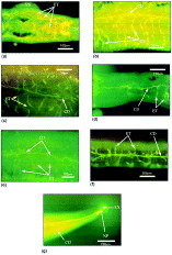
Fig. 2. Accumulation of fluorescent dyes after incubation of parasites with fluo-3/AM (a–e) and CFDA (f and g) in the protonephridium of adult male and female Schistosoma mansoni. (a–c, g) Accumulation of fluorescence (fluo-3/Ca2+ chelate) delineating the whole length of the excretory system. Male (a–c) and female (g) adults are shown. (d) Male pre-treated with diamide (which depletes glutathione, and affects accumulation of glutathione bimane, but has no effect on fluo-3/Ca2+ chelate accumulation). (e) Males pre-treated with 10 μM carboxylcyanide m-chlorophenylhydrazine (CCCP), and a much reduced accumulation is seen as a fine and thin lining of the excretory tubules and collecting ducts. (f) Male exposed to 5(6)-carboxyfluorescein diacetate (CFDA), showing accumulation of the fluorescent dye in the excretory system and its excretion from a nephridiopore. Abbreviations: CD, collecting duct; ET, excretory tubules; EX, excreted fluorescent dye through the nephridiopore; NP, nephridiopore; OS, oral sucker; and VS, ventral sucker.

Table 1. Fluorogenic substrates for mammalian MRP and P-gp examined to assess the interaction with the schistosome protonephridium
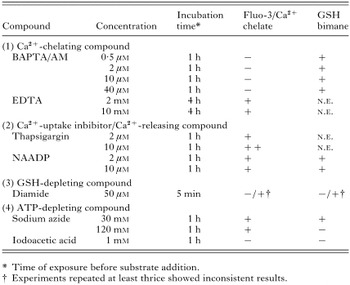
Table 2. Effects of Ca2+, GSH and ATP depletion upon excretion of fluo-3/Ca2+ chelate and GSH bimane in the protonephridium
 You have
Access
- 36
- Cited by