Coronary artery disease can have an atherosclerotic or non-atherosclerotic aetiology, the latter including rare systemic diseases with coronary involvement such as fibromuscular dysplasia, connective tissue disorders, and systemic inflammation. Fibromuscular dysplasia is a non-inflammatory, non-atherosclerotic arteriopathy, and involves numerous histopathological types of fibromuscular tissue accumulation (e.g., medial fibroplasia, medial hyperplasia, and intimal fibroplasia). Reference Olin, Gornik and Bacharach1 We present a rare case of fibromuscular dysplasia with first clinical manifestation in childhood and rapid disease progression.
Case presentation
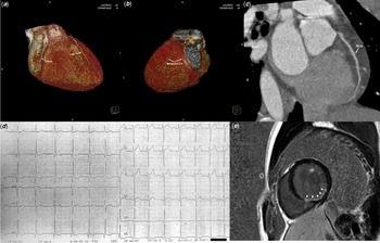
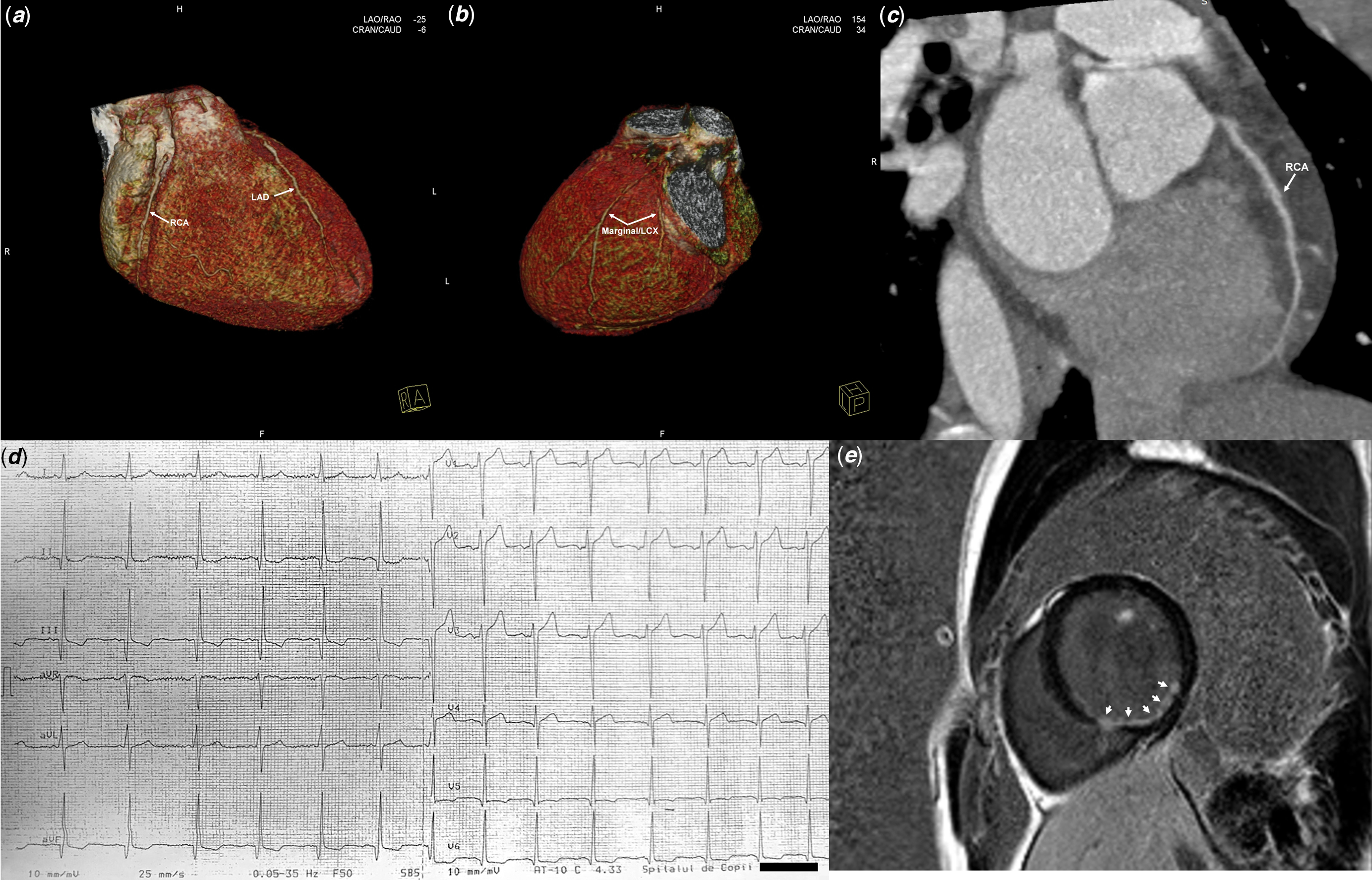
A 22-year-old male suffered a cardiac arrest by ventricular fibrillation preceded by chest pain at the age of 14 years. At that time, the patient was investigated for arrhythmic, inflammatory, infiltrative, or other structural causes of the event and found no obvious cause, thus a primary idiopathic ventricular fibrillation diagnosis was considered. Although genetic testing for myocardial diseases was recommended as part of the routine investigations after the cardiac arrest, no genetic analysis was performed. The patient had no obvious cardiovascular risk factors and no familial history of sudden cardiac death or coronary artery disease. However, rest electrocardiography revealed normal sinus rhythm with pathological Q waves in inferior leads, high serum levels of cardiac enzymes, inferior and lateral left ventricular wall ischaemia at cardiac magnetic resonance, and a normal coronary CT angiogram (Fig 1, Supplemental Video No. 1 - QuickTime codec). At that time, the legal tutor refused invasive coronary angiography. An internal cardiac defibrillator was implanted for secondary prevention of sudden cardiac death, and no appropriate therapy has been administered since that event.
Investigations performed in 2012, at 6 weeks after the cardiac arrest: coronary computed tomography angiography showing apparently healthy epicardial arteries ( a , b – three-dimensional reconstruction images, c – two-dimensional visualization of the right coronary artery); electrocardiogram ( d ) with pathological Q waves and subsequent ST-segment depression and T-wave changes in leads DII, DIII, aVF, and V5-V6; late gadolinium enhancement sequence of cardiac magnetic resonance imaging ( e ) showing subendocardial accumulation of contrast media, corresponding to a possible ischemic lesion of the infero-lateral wall and the inferior interventricular septum (arrows). LAD – left anterior descending coronary artery; LCX – left circumflex coronary artery; Marginal – obtuse marginal branch; RCA – right coronary artery.
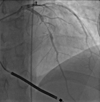
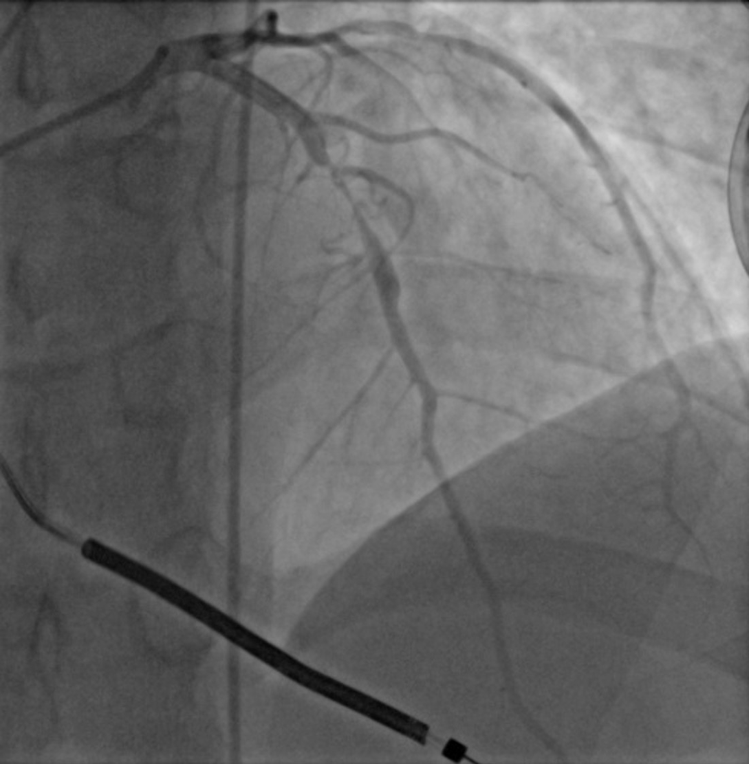
After 8 years of evolution, the patient was referred to our clinic for measurement of pulmonary arterial resistances and coronary angiography in preparation for cardiac transplantation due to recent and aggressive deterioration of left ventricular function. Transthoracic echocardiography revealed a dilated left ventricle with posterior and inferior wall hypokinesia, as well as severe motion abnormalities at the level of the interventricular septum, anterior and lateral walls (indexed left ventricular diastolic volume of 98 ml/m2 and left ventricular ejection fraction of 25%, Supplemental Video No. 2). Cardiac catheterisation revealed normal pulmonary arterial resistance of 0.4 Wood Units. Coronary angiography evidenced right dominance and severe three-vessel disease with unaffected left main coronary artery: left anterior descending artery with subtotal occlusion in the second segment with ipsilateral bridging collaterals, followed by an ectasia distally and smooth narrowing of septal and diagonal branches; left circumflex artery with distal subocclusive stenosis and an obtuse marginal artery with long and diffuse stenosis of intermediate severity; and right coronary artery with tortuous, “cork-screw”-like side-branches and chronic occlusion in the distal segment with large bridging ipsilateral collateralisation (Fig 2 and Supplemental Video No. 3). Given the severely diseased coronary arteries, and especially the occluded right coronary artery, a thrombophilia screening was performed. However, the analyses were negative for antinuclear antibodies, mutation of factor V (G1691A) and factor II (G20210A), antithrombin-, protein C- and protein S-deficiency.
Coronary angiogram showing left anterior descending artery subtotal occlusion.
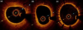
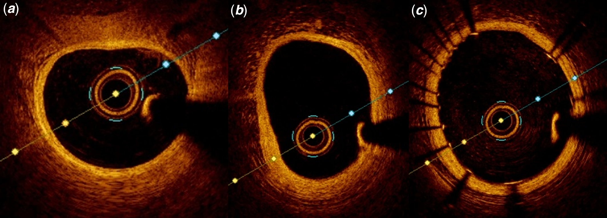
Percutaneous coronary revascularisation was decided by the heart team with patient’s consent. The left anterior descending artery lesion was approached first, pre-dilation was performed with a NC Trek® 2.5/15 mm balloon, Abbott, IL, USA, multiple inflations at 12–14–16 atmospheres, followed by drug- eluting stent implantation (Resolute Onyx® 3.5/22 mm, Medtronic, CA, USA) and post-dilation with NC Solarice® 4.0/8 mm balloon (Medtronic, MN, USA) up to 18 atmospheres. Optical coherence tomography) was performed with Dragonfly OPTIS® Imaging Catheter (St. Jude Medical, MN, USA), to confirm appropriate stent apposition and to reveal vessel wall characteristics. Left anterior descending presented a focal high-reflectivity (fibrotic) intimal and medial thickening (Fig 3).
Optical coherence tomography of the left anterior descending artery showing ( a ) normal vessel wall structure in the first segment, ( b ) focal intimal and medial thickening in the second segment and ( c ) correct stent apposition.
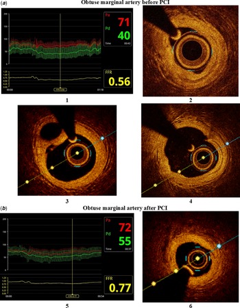
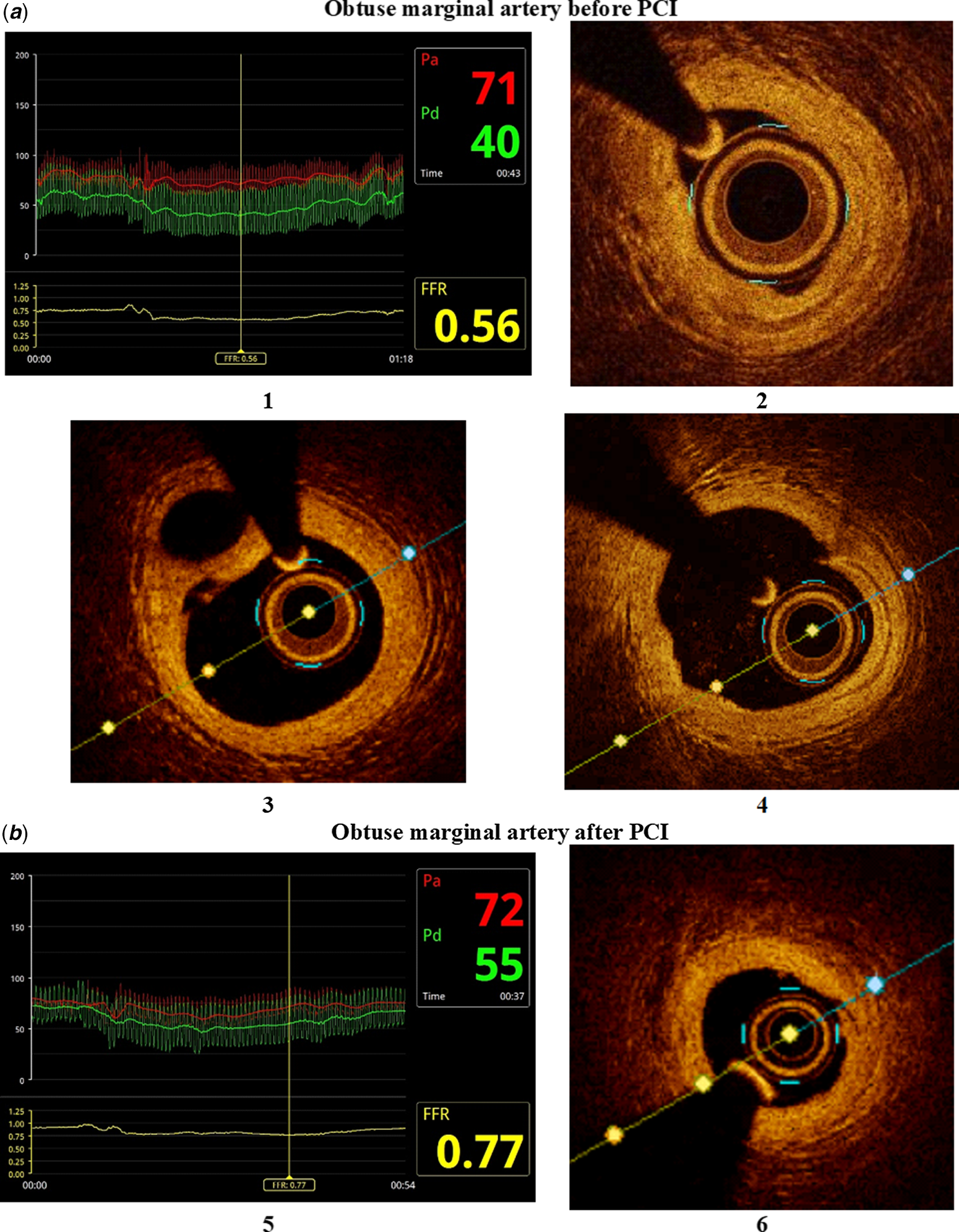
Obtuse marginal artery was approached next. Baseline fractional flow reserve was measured with PressureWire X®, St. Jude Medical, MN, USA, showing a value of 0.56 after intracoronary glycerin trinitrate and adenosine administration. Baseline optical coherence tomography of the vessel was performed showing diffuse fibrotic intimal and medial thickening with cavitation changes and neovascularisation (Fig 4a). Angiographic and optical coherence tomography co-registration is available as Supplemental Video No. 4. Pre-dilation of the vessel was performed. However, in the medial segment of the vessel, we found a not fully dilatable stenosis even with semi-compliant, non-compliant, and scoring balloons. This stenosis corresponded with the minimum lumen area and maximum amount of vessel wall thickening on optical coherence tomography imaging. After multiple dilatations, performed with successively upsized balloons inflated at high pressures (NC Trek® 2.5/8 mm, 2.5/15 mm, and 3.0/20 mm balloons, Abbott IL, USA, inflated at up to 24 atmospheres and AngioSculpt® 3.0/10 mm scoring balloon, Biotronik, Bülach, Switzerland, inflated at 18 atmospheres), an acceptable angiographic result was obtained. Final fractional flow reserve value was found to be 0.77 after adenosine administration. Final optical coherence tomography of the vessel was performed showing an increase of the minimum lumen area to 1.45 mm2 (Fig 4b). Noteworthy, only minimum wall dissection was found even after high pressure balloon inflation. Concurrently, intramural cavities were remitted after balloon dilation (Supplemental Video No. 5). Given the acceptable haemodynamic and optical coherence tomography result, and the presence of a not fully dilatable lesion, no stent was implanted in the marginal branch. The distal left circumflex artery artery was treated with plain balloon angioplasty because of its small lumen size (Mini Trek® 1.5/15 mm balloon, Abbott, IL, USA, inflated at 12 atmospheres). The patient received chronic treatment with ramipril, carvedilol, and spironolactone along with acetylsalicylic acid and clopidogrel.
Optical coherence tomography showing ( a ) obtuse marginal artery before angioplasty with (1) baseline FFR measurement, (2) minimal lumen area of 0.79 mm2 with diffuse thick fibrous intimal and media in form of Saturn’s ring, (3) cavitation of the vessel wall, (4) subclinical intimal tear into fibrotic lesion (healed spontaneous arterial wall dissection) and ( b ) obtuse marginal artery after angioplasty with (5) final FFR measurement, (6) minimal lumen area increased to 1.45 mm2 with minimum dissection.
Six months follow-up revealed similar left ventricular ejection fraction with improved symptoms and exercise tolerance. Brain-to-pelvis CT angiogram showed no extracoronary arteriopathies.
Discussion
Coronary fibromuscular dysplasia is a non-atherosclerotic, non-inflammatory, and rare cause of coronary artery disease. Although initially was thought to be associated invariably with spontaneous coronary artery dissection, it was later found that only about 40% of cases present with this complication, and numerous other hallmarks are present. Angiographically, both irregular and smooth stenosis of different severity; coronary ectasia <1.5 times adjacent segment; coronary “cork-screw”-like tortuosity; and smooth coronary narrowing should raise suspicion of fibromuscular dysplasia, although these criteria cannot differentiate entirely from atherosclerotic lesions. Reference Saw, Bezerra, Gornik, Machan and Mancini2,Reference Michelis, Olin, Kadian-Dodov, d’Escamard and Kovacic3 Our young patient presented all the above-mentioned angiographic criteria, which raised the suspicion of fibromuscular dysplasia.
Recently, optical coherence tomography characteristics of fibromuscular dysplasia have been described, including intima-media focal or tubular thickening with high reflectivity due to fibrous and/or muscular tissue accumulation, intramural haematoma, coronary dissection, subclinical intimal tear into the fibrotic plaque, and cavitation in ectatic segments. Reference Saw, Bezerra, Gornik, Machan and Mancini2,Reference Saw, Poulter and Fung4 Our case presented most of the above-mentioned optical coherence tomography criteria (1, 3, 4, and 5) and no criteria for atherosclerotic disease, thus confirming fibromuscular dysplasia. In our opinion, the absence of intramural haematoma optical coherence tomography criteria reflects the chronic stage of the disease at the moment of coronary angiography, as this complication of FMD would have been associated with an acute coronary syndrome.
Clinically, fibromuscular dysplasia manifestation may include sudden cardiac death, acute myocardial infarction, and left ventricular dysfunction. In our patient, the acute myocardial infarction complicated with ventricular fibrillation at the age of 14 years was retrospectively diagnosed, given the appearance of chest pain before cardiac arrest, Q-waves in inferior leads, high serum levels of cardiac enzymes, and segmental left ventricular ischaemia at cardiac MRI, in addition to chronic occlusion of right coronary artery. Furthermore, acute myocardial infarction caused by spontaneous right coronary artery dissection was considered, knowing that coronary CT angiography can be normal and may miss the diagnosis of spontaneous coronary artery dissection. Reference Eleid, Tweet, Young, Williamson, Hayes and Gulati5 Interestingly, in this case, no extracoronary arteriopathy was detected, as it is known that remote arteriopathies are infrequently associated with coronary fibromuscular dysplasia. Reference Persu, Lopez-Sublet and Al-Hussaini6
Conclusion
In conclusion, we illustrated a case of fibromuscular dysplasia with clinical presentation of acute coronary syndrome, left ventricular dysfunction, and aborted cardiac arrest in a young male with first manifestation at the age of 14 years and progression to ischaemic dilated cardiomyopathy by the age of 22 years. To our knowledge, such case of fibromuscular dysplasia with early onset and aggressive evolution was never reported before.
Supplementary material
To view supplementary material for this article, please visit https://doi.org/10.1017/S1047951122000865
Acknowledgements
None.
Financial support
This work was funded by the Romanian Academy of Medical Sciences and European Regional Development Fund, MySMIS 107124: Funding Contract 2/Axa 1/31.07.2017/ 107124 SMIS.