Introduction
Cryopreserved semen samples have been widely used in the breeding industry for artificial insemination (AI). This approach enables long-term storage of the semen before AI such that semen can be transported over long distances, quarantine of the semen can occur or samples can be used as a genetic bank to store commercially desirable traits. Advances in freezing techniques have improved the availability of superior genetic material, and various thawing procedures have also been employed to optimize maximum recovery of viable frozen spermatozoa at insemination (Córdova-Izquierdo et al., Reference Córdova-Izquierdo, Oliva, Lleó, Garcia-Artiga, Corcuera and Pérez-Gutiérrez2006; Aamdal & Andersen, Reference Aamdal and Andersen2007).
Thawing reverses the freezing process by forcing the cell back to its original path after various environmental changes (Anel et al., Reference Anel, Gomes-Alves, Alvarez, Borragan, Anel, Nicolas, Martinez-Pastor and de Paz2010). Other factors that contribute to the determination of suitable thawing rates include extenders, freezing rate, glycerol concentration and the packaging method (Robbins et al., Reference Robbins, Saacke and Chandler1976; Li et al., Reference Li, Saenz, Godke and Devireddy2006). According to the bifactorial theory of cryoinjury, ideal cell survival requires that the freezing rate and thawing rate be evaluated together (Mazur & Schmidt, Reference Mazur and Schmidt1968).
Various methods of thawing frozen bovine semen have been tried in order to achieve maximum recovery of viable spermatozoa. This approach includes experiments ranging from thawing cryopreserved semen in iced water (1–3°C) to thawing cells at 75°C for a minimum of 30 s or 12 s respectively (Pickett et al., Reference Pickett, Berndtson and Sullivan1978; Pace et al., Reference Pace, Sullivan, Elliott, Graham and Coulter1981). Studies by Pace et al. (Reference Pace, Sullivan, Elliott, Graham and Coulter1981) indicated that thawing at 37°C for a minimum of 30 s is an acceptable method for gaining superior post-thaw sperm quality and fertility in the field. In addition to the thawing procedure used, it is important to use a suitable semen preparation technique.
The most commonly used sperm preparation technique is sperm washing. It is believed that this technique is able to remove seminal plasma, and reduce the occurrence of free radicals through the process of replacing seminal plasma in cultured medium (Mortimer & Mortimer, Reference Mortimer and Mortimer1990). At the present time, the sperm washing technique has been found to have unacceptable limitations. Its ability to separate damaged sperm from healthy sperm is questionable. A revision of the swim-up technique has been developed. This technique is based on the assumption that phosphatidylserine (PS) is expressed when spermatozoa enter an early phase of apoptosis. Identifying spermatozoa that express PS and excluding them from the semen is postulated to increase the likelihood of successful fertilization (Vermes et al., Reference Vermes, Haanen, Steffens-Nakken and Reutellingsperger1995). The system described here uses microbeads bound with annexin V, which is a 35–36 kDa phospholipid-binding protein. When PS is exposed, these microbeads bind to apoptotic spermatozoa that have membrane deterioration, and therefore can be easily detected and separated from other spermatozoa. This system is known as the magnetic-activated cell sorting (MACS) system (Grunewald et al., Reference Grunewald, Paasch, Said, Rasch, Agarwal and Glander2006). Previous studies have shown that MACS can yield motile, viable, morphologically normal spermatozoa that display higher cryosurvival rates with a higher fertilization potential in human sperm (Grunewald et al., Reference Grunewald, Paasch and Glander2001). Hence, this study had been replicated in animals to evaluate the effects of this technique on viability and capacitation status, especially in bovine spermatozoa.
According to Watson (Reference Watson1995), cryopreservation modifies sperm membranes to a partially capacitated state and makes them more reactive to the environment. Besides causing an apoptotic-like mechanism by the induction of PS translocation, the cryopreservation and thawing process induces many changes in sperm cells, including membrane disorder and cell death. Diminished motility and membrane changes, including sperm capacitation or acrosomal reaction, are some of the main forms of damage brought about by cryopreservation (Martin et al., Reference Martin, Finucane, Amarante-Mendes, O'Brien and Green1996).
Past research has focused more towards the effects of AnMACS solely on human sperm, not on bovine sperm. Therefore, the focal points of this research were to study the effects of AnMACS on kinematics, viability and capacitation status of Bos taurus sperm when subjected to various thawing temperatures.
Materials and methods
Semen collection
Fresh semen samples were collected from three adult Piedmontese breed bulls using an artificial vagina at the Institut Bioteknologi Veterinar Kebangsaan, (IBVK), Jerantut, Pahang, Malaysia. Three ejaculates were obtained; average yield per ejaculation was about 5 ml.
Semen preparation
The sperm suspension was divided into two separate fractions. The first fraction was subjected to AnMACS followed by cryopreservation and thawing, whereas the second fraction was cryopreserved without AnMACS and was designated the control group. Semen was separated by binding annexin V microbeads to PS, which was externalized on the sperm membrane, and using a MiniMacs kit (Miltenyi Biotec, GmbH, Germany). Semen were centrifuged for 10 min at 300 relative centrifugal force (rcf). Supernatant was then removed completely and the cell pellet was re-suspended in 80 μl of annexin V buffer per 107 cells. Next, 20 μl of MACS annexin V microbeads were added per 107 total cells. The mixture was incubated for 15 min in the dark at 10°C. The sperm were then washed with 1–2 ml buffer per 107 cells and resuspend in 500 μl buffer per 108 total cells. The cells were added to the separation column, which was placed in a magnet such that the annexin V microbead-labelled spermatozoa (ANMB-positive) were retained in the separation column. The fraction with intact membranes that passed through the column was labelled as the ANMB-negative fraction. This fraction was eluted and collected from the column (Said et al., Reference Said, Grunewald, Paasch, Rasch, Agarwal and Glander2005; Said et al., Reference Said, Agarwal, Zborowski, Grunewald, Glander and Paasch2008).
Cryopreservation-thawing protocol
The control group and ANMB-negative fraction was cryopreserved using Bioxcell® extender (IMV, France). The concentration of semen mixture was determined using a SpermaCue photometer (Miltenyi Biotec Asia Pacific Pte Ltd, Singapore) and adjusted to 20 million spermatozoa per 0.25 ml straw. The mixture was placed in chiller at 4°C for at least 3 h. Samples were then transferred to 0.25 ml straws and put in liquid nitrogen vapour (–70°C) for 9 min. Finally, the straws were plunged into liquid nitrogen (–196°C) and stored in liquid nitrogen tanks. The direct plunging cryopreservation technique was used.
Straws were then thawed in water baths at 20°C for 13 s, 37°C for 30 s, 40°C for 7 s, 60°C for 6 s and 80°C for 5 s respectively.
Eosin–nigrosin staining technique
The viability of spermatozoa was assessed by the eosin–nigrosin staining method. For eosin–nigrosin staining, the final composition of the stain was eosin-Y (1.67 g), nigrosin (10 g), and sodium citrate (2.9 g), dissolved in 100 ml distilled water. Sperm suspension smears were prepared by mixing a drop of sperm sample with two drops of stain on a warm slide and spreading the stain with a second slide. The smear was allowed to dry. Viability was assessed by counting 200 spermatozoa under a bright-field microscope at ×400 magnification. Sperm that displayed partial or complete purple staining were considered to be non-viable whilst only sperm that showed strict exclusion of stain were counted as viable (Kim et al., Reference Kim, Yu and Kim2009). Images were captured using phase-contrast inverted microscope with an analyzer system (Olympus 1 × 51, and Image Pro Plus 5.1 software).
Chlorotetracycline staining assay
A modified chlorotetracycline (CTC) staining protocol was used to determine the sperm cell capacitation status (Ward & Storey, Reference Ward and Storey1984). Hundreds of 50 ml of washed sperm were mixed with 3 ml propidium iodide (PI) and 3 ml Hoechst342 stain (40 μg/ml) and incubated for 15 min at 37°C in the dark. One drop of the mixture was added to one drop of CTC solution (805 μmol/l CTC and 5 mmol/l cysteine in CTC buffer that contained 130 mmol/l NaCl and 20 mmol/l Tris (Trizma base, pH 7.8) and fixed immediately with one drop of 12.5% (w/v) paraformaldehyde in 1 mol/l Tris–HCl, pH 8.0. Next, a drop of 0.22 M 1,4-diazabicyclo[2.2.2]octane (DABCO) was added to the mixture to prevent fading of fluorescence. Two slides were prepared per treatment samples and analyzed within 2 h of preparation.
Chlorotetracycline staining was assessed on live spermatozoa under blue-violet illumination (excitation at 330–380 nm, emission at 420 nm). The staining patterns observed was similar to those described by Wang et al. (Reference Wang, Abeydeera, Fraser and Niwa1995), namely: F, full fluorescence (uncapacitated); B, a fluorescence-free band in the post-acrosomal domain (capacitated); and AR, low fluorescence over the entire head with a band of bright fluorescence across the equatorial segment (acrosome reacted cells).
Analysis of sperm parameters
Sperm count and motility were performed using a CEROS sperm analyser (Hamilton Thorne Inc, Beverly, MA, USA). Five straws were thawed per treatment in triplicate and analyzed for computer-assisted sperm analysis (CASA) parameters of sperm velocity and sperm progression. Briefly, 10 μl of fresh semen were diluted at a 1:9 ratio in 3% sodium citrate to access CASA parameters.
For sperm velocity, three parameters were considered, these were average path velocity (VAP) μm/s, straight line velocity (VSL) μm/s and curvilinear velocity (VCL) μm/s. Sperm velocity is a measure of how fast sperm are moving along their trajectory. Curvilinear velocity is defined as the time-average velocity of a sperm head along its actual curvilinear path. Straight line velocity is the average time-velocity of a sperm head in a straight line between its first and last spotted point. Average path velocity is the time-average velocity of sperm along its average path (Ibrahim et al., Reference Ibrahim, Jaffar, Osman and Syed2011). Sperm progression is the movement pattern along its path. Wobble (WOB %), describes the sperm head movement side to side, linearity (LIN %) explains the trajectory straightness of the sperm's path and STR (%) illustrates the average distance of the sperm from its origin on the average path in all frames (Ibrahim et al., Reference Ibrahim, Osman, Das III, Othman, Majid and Rahman IV2008).
Statistical analysis
Two-way analysis of variance (ANOVA) was used to determine the effects of the thawing procedures on the control and treatment groups assessed for sperm kinematics, viability and capacitation status. Results were reported as mean ± standard error of the mean (SEM). A P-value of 0.05 was considered to be significant.
Results
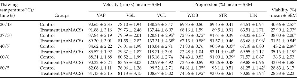
Following the cryopreservation–thawing process, AnMACS positively selected sperm differed significantly only in velocity (VCL μm/s) and sperm straightness (STR %), which displayed significantly higher values compared with the control group at a P-value <0.05 (Table 1). The effect of AnMACS on sperm was mostly apparent on sperm velocity, especially when samples wre thawed at 37°C for 30 s. For sperm progression the effect of MACS was significant when cells were thawed at 40°C for 7 s or at 60°C for 6 s, P < 0.05.
Motility characteristics (mean ± SEM) for sperm velocity, progression and viability in thawed bull semen after magnetic separation (n > 200) a

aThe study was done in triplicate.
b The study significance level was set at P < 0.05 between control and treatment groups. SEM, standard error of the mean.
LIN, linearity; STR, straightness; VAP, average path velocity; VCL, curvilinear velocity; VSL, straight line velocity; WOB, wobble.
Furthermore, in sperm progression, two-way ANOVA showed that WOB (%), LIN (%) and STR (%) were all affected by inclusion in control or treatment group, thawing temperature and time (P < 0.05). Only STR (%) in the treatment group showed a significantly higher value compared with the control group when cells were thawed at 40°C for 7 s or 60°C for 6 s.
In the AnMACS-treated group, sperm viability was mostly reduced compared with the control group. The highest per cent viability (43.2 ± 2.89%) was found in the control group following thawing at 40°C for 7 s. Nevertheless, the CTC staining pattern revealed that AnMACS-treated sperms showed a significantly higher number of uncapacitated sperms compared with the control group after thawing 37°C for 30 s, 40°C for 7 s or 60°C for 6 s.
Most characteristics of sperm motility were not changed, except for VCL (μm/s) and STR (%), which were higher in the treatment group. Hence, the reliability of this system was shown only for sperm VCL (μm/s), STR (%) and uncapacitated sperm.
Spermatozoa viability assessment through eosin–nigrosin staining technique
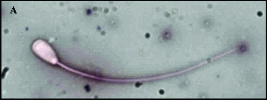
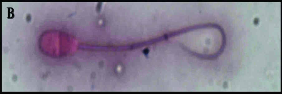
The commonly used method for spermatozoa viability is eosin–nigrosin staining. Eosin is an orangish pink to red dye stains the cytoplasmic material. It penetrates in non-viable cells, which then appear red. Nigrosin gives a dark background, facilitating the detection of viable, non-stained cells (Figs. 1 and 2).
Live spermatozoa stained with eosin–nigrosin dye (×400 magnification).

Dead spermatozoa stained with eosin–nigrosin dye (×400 magnification).

Statistical analysis revealed that thawing at 20°C for 13 s, 37°C for 30 s or 40°C for 7 s yielded significantly lower values for sperm viability P = 0.005, P = 0.02 and P = 0.03 respectively compared with the control group (Table 1).
Cyro-capacitation status
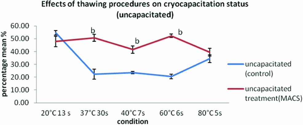
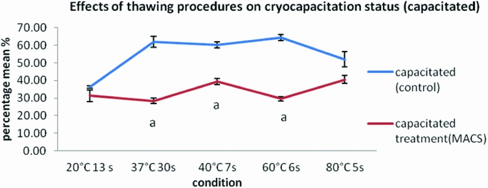
After thawing at 37°C for 30 s, 40°C for 7 s or 60°C for 6 s, there was a significantly higher number of sperm in an uncapacitated state in the MACS-treated sample. Meanwhile, at the same temperatures, fewer capacitated sperms were found in the MACS-treated sample compared with the control group, P < 0.05.
Discussion
At present, one of the main important parameters used to evaluate semen quality is sperm motility. CASA software is one option used to represent motility and viability of sperm. Parameters of sperm motility have been divided into two categories, namely sperm velocity (VCL, VSL, VAP; μm/s) and sperm progression (WOB, LIN, STR; %). The thawing temperatures and time selected for this study were based on results from previous studies (Nur et al., Reference Nur, Dogan, Soylu and Ak2003; Muiño et al., Reference Muiño, Rivera, Rigau, Rodriguez-Gil and Peña2008; Calamera et al., Reference Calamera, Buffone, Doncel, Brugo-Olmedo, de Vincentiis, Calamera, Storey and Alvarez2010). Methodologies were combined and the best thawing temperatures and times were chosen to suit the goals for our study.
In this study, the most significant changes on sperm motility could be seen for the AnMACS-treated samples, for VCL (μm/s) and STR (%). According to Muiño et al. (Reference Muiño, Rivera, Rigau, Rodriguez-Gil and Peña2008) spermatozoa with relatively low velocity (medium VCL and VAP) and with high progressiveness (high LIN %, STR % and WOB %) could be classified as subpopulation 1, comprised of relatively slow but progressive spermatozoa. This slow progression was a result of post-thaw thermal stress, as some of the spermatozoa may reduce their flagella activity, probably due to sublethal cryoinjury, although their movement is still forward. This type of subpopulation was observed when thawing was carried out at 80°C for both control and treatment groups. Furthermore, spermatozoa that exhibited highly active but non-progressive movement with high values of VCL μm/s, low LIN % and low STR % were observed when thawing was performed at 20°C for 13 s. These spermatozoa can be classified as subpopulation 2 and could be considered as having ‘hyperactivated-like’ movement. This pattern of movement is due to the accumulation of intracellular Ca2+ above physiological concentrations (Yanagimachi, Reference Yanagimachi, Knobil and Neill1994).
Our study indicated that sperm move most rapidly and progressively after AnMACS and when thawed at 37°C for 30 s, presumably at this temperature energy production and cell respiration occur at their maximum levels. As we can see from the results, there is a crossover of results at certain temperatures between control and treatment samples. For the average path velocity (VAP μm/s) the crossover was at 70°C, whilst for curve line velocity (VCL μm/s) the intercepts were at 37°C and 70°C. This phenomenon might be due to spermatozoa changes in VAP μm/s and VCL μm/s at these temperatures, especially for AnMACS-treated spermatozoa. After incubation at 70°C, separated spermatozoa that have undergone AnMACS are not tolerant to heat stress and hence decrease their velocity. However, different phenomena occurred for progression parameters (WOB %, LIN % and STR %). Results obtained indicated that spermatozoa progression was increased as the temperature increased and that progression was increased by decreasing velocity. These types of motility, with low velocity and high progression, are not suitable for sperm to achieve fertilization with the egg. Hence, a compromise between velocity and progression is needed in order for the sperm to successfully penetrate the egg.
Survival of the spermatozoa after the freezing–thawing process depends on the plasma membrane, as this is the most crucial region and regarded as the primary site of cryoinjury (Söderquist et al., Reference Söderquist, Madrid-Bury and Rodriguez-Martinez1997). The AnMACS preparation technique was believed to yield motile, viable, morphologically normal spermatozoa that displayed higher cryosurvival rates as well as higher fertilization potential (Grunewald et al., Reference Grunewald, Paasch and Glander2001; Said et al., Reference Said, Grunewald, Paasch, Rasch, Agarwal and Glander2005). Moreover, non-apoptotic sperm separated by AnMACS prior to cryopreservation had significantly higher motility following cryopreservation thawing than sperm that were not separated by MACS (Said et al., Reference Said, Agarwal, Zborowski, Grunewald, Glander and Paasch2008).
Furthermore, it is believed that membranes are more intact in the AnMACS-processed samples as there is no phosphatidylserine to be exposed. AnMACS is an excellent method for separating cells of interest out of a mixed cell population. The principle underlying this tool is the utilization of magnetic micro-particles conjugated with specific antibodies. Untagged cells are not influenced by the magnetic field and can be used directly for insemination (Kang & Park, Reference Kang and Park2004). Additionally, AnMACS usage allows the enrichment of cells with and without PS exposure on their surface. Externalization of plasma membrane PS is part of an oxidative signalling pathway, identified as one of the early and prominent features of apoptosis. In cells, PS exposure on the outer leaflet on the plasma membrane has several functions, one of which is recognition and removal of apoptotic cells by phagocytes (Kagan et al., Reference Kagan, Fabisiak, Shvedova, Tyurina, Tyurin, Schor and Kawai2000). In the present study, sperm were labelled magnetically with annexin V microbeads and passed through a MACS column, which was placed in the magnetic field of a MACS separator. Cells with externalized PS were tagged, separated from the normal healthy cells and then flushed out from the system. As a result, only healthy viable cells were collected for cryopreservation. Removal of abnormal spermatozoa and spermatozoa undergoing apoptosis can decrease the oxidative stress among vital healthy sperm (Dirican et al., Reference Dirican, Özgün, Akarsu, Akın, Ercan, Uğurlu, Çamsarı, Kanyılmaz, Kaya and Ünsal2008).
The results found in this study might relate to decreased oxidative stress in the ejaculate after magnetic separation. In other words, AnMACS acts on sperm at the molecular level and this technique can be considered as an effective method in sperm preparation to enhance sperm quality and function. AnMACS is also a simple, reliable and rapid sorting system for separating sperms in large numbers (Said et al., Reference Said, Grunewald, Paasch, Rasch, Agarwal and Glander2005).
It is clear that fertility is reduced in half the number of sperm that have undergone the cryopreservation process compared with fresh semen. It is now recognized that the surviving population of spermatozoa is compromised as a result of capacitation-like changes (Watson, Reference Watson1995; Bailey et al., Reference Bailey, Bilodeau and Cormier2000). In this study, the capacitation status of AnMACS-treated spermatozoa and control spermatozoa were assessed using the CTC assay. Results indicated that AnMACS-treated samples produced more uncapacitated spermatozoa compared with the control group (Figs. 3 and 4), even after different thawing procedures were applied. Kurz et al. (Reference Kurz, Viertel, Herrmann and Müller2005) reported that PS is localized in the cytoplasmic leaflet of the plasma membrane as well as on the outer acrosomal membrane. During the capacitation and acrosome reaction, PS was not exposed on the surface of the viable sperm cells. This point clarifies and adds weight to our result that more uncapacitated cells were reveled during the capacitation and acrosome reaction as a consequence of magnetic separation. This point furthermore explains the fact that good quality sperms did not expose PS on the outer leaflet even though they were undergoing an extreme event such as cryopreservation, which is believed to trigger cryo-capacitation-like damage.
Line graph shows the effects of the magnetic-activated sperm system on capacitation status of cryopreserved spermatozoa with the uncapacitated status. bHigher significantly compared with control group at P < 0.05.

Line graph shows the effects of the magnetic-activated sperm system on capacitation status of cryopreserved spermatozoa with capacitated status. aLower significantly compared with control group at P < 0.05.

Results further showed that thawing at 37°C, 40°C or 60°C respectively yielded significant results for AnMACS-processed samples rather than for the control group (P < 0.05). Here we postulated that using AnMACS as the sperm preparation technique and thawing at either one of these three temperatures will yield spermatozoa that are yet to be capacitated. This factor is because capacitation must occur at the exact time it is inside the female oviductal tract in order for fertilization to take place. Any events preceding fertilization that occur at the wrong time and/or the wrong place will result in poor fertilization rates (Donnelly et al., Reference Donnelly, Lewis, McNally and Thompson1998). This cryo-capacitation mechanism, because of the onset of normal capacitation, has yet to be determined. It has been reported that reactive oxygen species (ROS) have a significant physiological role during normal sperm function, including hyperactivation, capacitation and acrosome reaction (de Lamirande et al., Reference de Lamirande, Jiang, Zini, Kodama and Gagnon1997), and ROS is also believed to play an important function in fertility or infertility. Furthermore, according to Langlais and Roberts (Reference Langlais and Roberts1985) capacitation of mammalian sperm occurs when the head of plasma membrane undergoes reorganization due to phospholipids redistribution and cholesterol removal. This membrane destabilization causes the sperm to exhibit cryo-capacitation with a B pattern that favours calcium influx to the cell during cryopreservation (Maxwell & Johnson, Reference Maxwell and Johnson1997). Moreover, the capacitation process is believed to be related to apoptosis. ROS are essential in regulating sperm function, but ROS also act as a two-edge sword (Aitken, Reference Aitken2011). At low levels ROS are needed to promote cholesterol oxidation and tyrosine phosphorylation events that support capacitation. Meanwhile, excessive production of ROS can lead to a state of oxidative stress that affects sperm function. Oxysterols, the oxidized derivatives of cholesterol, act as a good indicator of oxidative stress but might also be efficient inducers of this process. Accumulation of oxysterols has been linked to the activation of apoptosis and cell death in other cell types (Ryan et al., Reference Ryan, O'Callaghan and O'Brien2005).
Moreover, mitochondria appear to be key mediators of oxysterol-induced pathology by means of the intrinsic apoptosis pathway. One type of ROS is peroxynitrite, which functions in stimulating cholesterol oxidation and oxysterol formation. Peroxynitrite is known to be the promoter for capacitation in several mammalian species (Rodriguez et al., Reference Rodriguez, Valdez, Zaobornyj, Boveris and Beconi2011). Besides this function, it also activates oxysterol formation in cells. Previously, Brouwers et al. (Reference Brouwers, Boerke, Silva, Garcia-Gil, van Gestel, Helms, de Lest and Gadella2011) found that oxysterol generation is disrupted after cryopreservation, whereas lipid peroxidation is generally enhanced. Elevated oxysterol formation in mitochondria could lead to activation of an intrinsic apoptotic cascade and begin a number of cellular changes such as sperm senescence, enhanced mitochondrial superoxide generation, motility loss, caspase activation and PS exposure. Therefore, we can state here that the AnMACS procedure selects sperms with less oxidative stress, based on externalization of PS, hence the outcome will be less capacitated spermatozoa.
During this experiment, eosin–nigrosin stain was used to measure sperm viability. Spermatozoa that stained red or pinkish were considered to be non-viable. Sperm take up the dye when the integrity of the membrane is badly damaged, indicating that spermatozoa physiological function was not operating well. Even though per cent viability values were only significant (P < 0.05) between control and treatment groups when sperm were thawed at 20°C for 13 s or 40°C for 7 s, samples that had undergone AnMACS prior to cryopreservation could preserve their viability up to a temperature as high as 60°C compared with the control group, which could only survive up to 40°C. This finding may indicate that magnetic separation based on the externalization of PS had successfully yielded a sperm subpopulation that was heat resistant. Furthermore, from results obtained, AnMACS-treated sperms had increased viability when the temperature increased from 20°C to 60°. Although the viability of MACS-processed sperms was less compared with sperm in the control group at 20°C and 40°C, viable sperm with less PS were selected. Sperm without this system might be viable but be facing apoptosis; hence fertilization outcome might not be good if we continue to select this subpopulation.
Conclusion
In conclusion, MACS might act as an ideal sperm preparation technique prior to cryopreservation. Use of AnMACS produced sperm with higher VCL μm/s and STR % after thawing at 37°C for 30 s or 40°C 7 s respectively. This pattern indicates that sperm were exhibiting faster and straighter movement, which may greatly affect in vitro fertilization (IVF) success rates. In addition, by using AnMACS in sperm preparation, we are able to select the most viable spermatozoa for cryopreservation. MACS-processes samples can sustain their viability up to higher temperatures as much as 60°C. Furthermore, another significant finding that emerged from this study was that less capacitated spermatozoa were likely to be present after thawing at 37°C for 30 s, 40°C for 7 s or 60°C for 6 s in the AnMACS-treated samples. Hence, the recommended thawing temperature can be at 37°C for 30 s, 40°C for 7 s or 60°C for 6 s. But, during this study a thawing temperature at 37°C for 30 s was chosen because of the ease in temperature manipulation in field work. However, the fertility potential of this AnMACS sperm population has yet to be determined. This result could be achieved through further future research.
Acknowledgement
The researchers are thankful to the Ministry of Science and Technology (MOSTI), Universiti Kebangsaan Malaysia (UKM), Universiti Teknologi MARA (UiTM) and Agrobiotech Institute for the research grants given: 10–05-ABI-AB040 and 600-RMI/ST/DANA 5/3/Dst (263/2009). Special thanks to the Institute of Veterinary, Jerantut, Pahang for assisting in semen sampling and technical supports.