INTRODUCTION
Once intracellular pathogens have invaded their host cell, they explore and/or modulate its intracellular organization at different levels. This includes gene expression patterns, defined structures and compartments, the host cell metabolism and also signalling transduction pathways. Bacteria, viruses and parasites have evolved common strategies to modulate the living conditions within their host cell according to their needs, in order to optimize survival, replication and finally dissemination. During these processes pathogens not only induce remodelling of the cytoskeleton, but they also modify diverse pathways and key components involved in regulation of the host cell cytoskeleton dynamics (Haglund and Welch, Reference Haglund and Welch2011).
The cytoskeleton is a complex network of different polymers involved in a plethora of cellular functions, including the maintenance of cell shape, the organization and positioning of intracellular organelles, the establishment of a physical structure for intracellular cargo transportation, cell division and motility. To perform these diverse activities, the dynamic elements of the cytoskeleton interact with several distinct proteins, ranging from motor proteins to proteins involved in regulation of polymer length, stability and organization. In turn this enables the cell to respond to environmental challenges, including those involved in cell–cell contact, cell–matrix interactions and cell polarization and migration. By taking over certain aspects of host cell cytoskeleton organization, pathogens can exploit host cell resources, while simultaneously evading intracellular defence mechanisms, such as avoiding the destruction by lysosomes following phagocytosis by manipulating the actin cytoskeleton (Lang et al. Reference Lang, Schmidt, Schlosser, Hey, Larrinua, Sheets, Mannherz and Aktories2010).
Recent studies revealed how bacteria and viruses manipulate the host cell cytoskeleton, and many molecular mechanisms subjacent to cytoskeleton organization and function were discovered (Lai et al. Reference Lai, Saxena, Tseng, Jeng, Kohara and Lai2014; Fernandez et al. Reference Fernandez, Portilho, Danckaert, Munier, Becker, Roux, Zambo, Shorte, Jacob, Vidalain, Charneau, Clavel and Arhel2015; Foo and Chee, Reference Foo and Chee2015; Sana et al. Reference Sana, Baumann, Merdes, Soscia, Rattei, Hachani, Jones, Bennett, Filloux, Superti-Furga, Voulhoux and Bleves2015). However, the knowledge on how protozoan parasites, such as those of the phylum Apicomplexa, explore the host cell cytoskeleton is less well established.
Apicomplexan parasites grow and replicate inside a host cell, the majority of them forming a parasitophorous vacuole (PV). They are the aetiological agents of several human and animal diseases, such as malaria (Plasmodium spp.), toxoplasmosis (Toxoplasma gondii), besnoitiosis (Besnoitia spp.), neosporosis (Neospora caninum) cryptosporidiosis (Cryptosporidium spp.), theileriosis (Theileria spp.) or babesiosis (Babesia spp.), among others. Being intracellular parasites they have been observed to modulate several aspects of the host cell. This includes repositioning of host cell organelles [centrosome, mitochondria, endoplasmic reticulum, Golgi complex (Coppens et al. Reference Coppens, Dunn, Romano, Pypaert, Zhang, Boothroyd and Joiner2006; Wang et al. Reference Wang, Weiss and Orlofsky2010; Romano et al. Reference Romano, Sonda, Bergbower, Smith and Coppens2013; Cardoso et al. Reference Cardoso, Nolasco, Gonçalves, Cortes, Leitão and Soares2014; Pernas et al. Reference Pernas, Adomako-Ankomah, Shastri, Ewald, Treeck, Boyle and Boothroyd2014; Nolan et al. Reference Nolan, Romano, Luechtefeld and Coppens2015)], scavenging of host cell nutrients (Blader and Saeij, Reference Blader and Saeij2009), modulate the host cell cycle (Dobbelaere and Kuenzi, Reference Dobbelaere and Kuenzi2004), they interfere with signalling pathways (Dobbelaere and Kuenzi, Reference Dobbelaere and Kuenzi2004; Yang et al. Reference Yang, Tschopp, Baudry, Du Mmler, Hynx and Hemmings2004), and influence host cell migration (Lambert et al. Reference Lambert, Dellacasa-Lindberg and Barragan2010; Collantes-Fernandez et al. Reference Collantes-Fernandez, Arrighi, Álvarez-Garcıa, Weidner, Regidor-Cerrillo, Boothroyd, Ortega-Mora and Barragan2012).
In this review we focus on how apicomplexan parasites act upon the three main filament types that constitute the cytoskeleton of their host cell, namely intermediate filaments, actin filaments (F-actin) and microtubules. While intermediate filaments provide mechanical strength to the cell and many organelles are physically linked to the network, actin filaments determine the shape of the cell surface, provide its stability and mediate cell motility. Microtubules are structures that are involved in many crucial cellular functions such as cell division, motility, intracellular transport and various cell signalling pathways (Gundersen and Cook, Reference Gundersen and Cook1999; Pollard, Reference Pollard2003). We review the current knowledge on how these parasites interact with each of these cytoskeletal components during host cell invasion and parasite replication, and how this modulation could be advantageous for parasite survival, replication and/or development.
INTERACTIONS OF APICOMPLEXAN PARASITES WITH HOST CELL MICROTUBULES
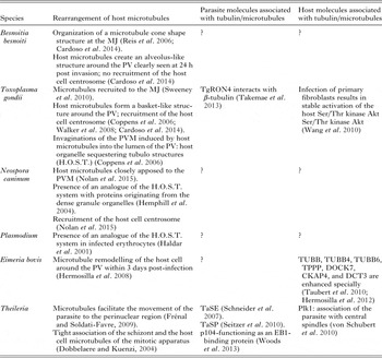
Several studies (see Table 1 for an overview) have described the remodelling of the host cell microtubule cytoskeleton during invasion by apicomplexan parasites (Coppens et al. Reference Coppens, Dunn, Romano, Pypaert, Zhang, Boothroyd and Joiner2006; Hermosilla et al. Reference Hermosilla, Schröpfer, Stowasser, Eckstein-Ludwig, Behrendt and Zahner2008; Walker et al. Reference Walker, Hjort, Smith, Tripathi, Hornick, Hinchcliffe, Archer and Hager2008; Romano et al. Reference Romano, Sonda, Bergbower, Smith and Coppens2013; Woods et al. Reference Woods, Theiler, Mühlemann, Segiser, Huber, Ansari, Pain and Dobbelaere2013; Cardoso et al. Reference Cardoso, Nolasco, Gonçalves, Cortes, Leitão and Soares2014; Nolan et al. Reference Nolan, Romano, Luechtefeld and Coppens2015). As such, modulation of the microtubule network by T. gondii and B. besnoiti occurs early during invasion and extends to the formation and establishment of the PV (Sehgal et al. Reference Sehgal, Bettiol, Pypaert, Wenk, Kaasch, Blader, Joiner and Coppens2005; Coppens et al. Reference Coppens, Dunn, Romano, Pypaert, Zhang, Boothroyd and Joiner2006; Walker et al. Reference Walker, Hjort, Smith, Tripathi, Hornick, Hinchcliffe, Archer and Hager2008; Romano et al. Reference Romano, Sonda, Bergbower, Smith and Coppens2013; Cardoso et al. Reference Cardoso, Nolasco, Gonçalves, Cortes, Leitão and Soares2014). Reis et al. (Reference Reis, Cortes, Viseu Melo, Fazendeiro, Leitão and Soares2006) demonstrated that upon B. besnoiti host cell entry, microtubules start to surround the parasite upon the first minutes of invasion forming a cone shape microtubule network; and a microtubule ring on the host cell is observed around the parasite entry site, probably corresponding to the moving junction (MJ) – a structure mediating a tight connection between the invading parasite and the host plasma membrane (reviewed in Tyler et al. Reference Tyler, Treeck and Boothroyd2011). In T. gondii, and most likely also in other Apicomplexa, the MJ is assembled by a macromolecular complex comprising several rhoptry neck proteins (RON2, RON4, RON5, RON8) and AMA1 (micronemal apical membrane antigen 1) (Alexander et al. Reference Alexander, Mital, Ward, Bradley and Boothroyd2005; Lebrun et al. Reference Lebrun, Michelin, El Hajj, Poncet, Bradley, Vial and Dubremetz2005; Besteiro et al. Reference Besteiro, Michelin, Poncet, Dubremetz and Lebrun2009; Straub et al. Reference Straub, Cheng, Sohn and Bradley2009). Other studies also showed that host cell microtubules surrounded the PV of T. gondii and B. besnoiti already after 15 min post invasion (Walker et al. Reference Walker, Hjort, Smith, Tripathi, Hornick, Hinchcliffe, Archer and Hager2008; Cardoso et al. Reference Cardoso, Nolasco, Gonçalves, Cortes, Leitão and Soares2014). Indeed, the presence of microtubule filaments at or near the MJ prior to invasion has been shown to increase the speed at which T. gondii starts to initiate the host cell invasion process after adhering to the host cell surface (Sweeney et al. Reference Sweeney, Morrissette, LaChapelle and Blader2010). On the other hand, host cell microtubule depolymerization effectively delayed parasite invasion, which could be explained by the fact that microtubules mediate the delivery of the RON complex to the plasma membrane at the site of the MJ (Sweeney et al. Reference Sweeney, Morrissette, LaChapelle and Blader2010). This hypothesis is supported by the observation that host cell β-tubulin binds to TgRON4, and that the C-terminal domain of β-tubulin interacts with TgRON4 during invasion (Takemae et al. Reference Takemae, Sugi, Kobayashi, Gong, Ishiwa, Recuenco, Murakoshi, Iwanaga, Inomata, Horimoto, Akashi and Kato2013). However, not all apicomplexans require the presence of a MJ during invasion: Theileria sporozoites enter their host cells through other mechanisms, which is morphologically characterized as a ‘self-zippering process’ between parasite and host cell surface membrane, also resulting in intracellular parasites surrounded by a parasitophorous vacuole membrane (PVM). However, in the case of Theileria, this membrane is rapidly degraded, and parasites reside free in the cytoplasm. In addition, no remodelling of the microtubule filament network has been described on the surface of host cells during Theileria invasion (reviewed in Shaw, Reference Shaw2003).
Rearrangement of host microtubules and parasite and host molecules that interact with the host microtubule cytoskeleton during invasion and replication

EB1, end-binding protein 1; MJ, moving junction; PV, parasitophorous vacuole; PVM, parasitophorous vacuole membrane; (?), not known.
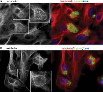
Upon the initiation of intracellular replication, T. gondii and B. besnoiti exhibit distinct differences. In cells infected with replicating B. besnoiti tachyzoites, the microtubules present a complex arrangement around the PV, creating a clearly visible alveolus-like structure (Fig. 1A). In contrast, microtubules in T. gondii infected host cells exhibit a basket-like structure (Fig. 1B), which is distinct from the alveolar arrangement found in B. besnoiti infected cells (Coppens et al. Reference Coppens, Dunn, Romano, Pypaert, Zhang, Boothroyd and Joiner2006; Reis et al. Reference Reis, Cortes, Viseu Melo, Fazendeiro, Leitão and Soares2006; Walker et al. Reference Walker, Hjort, Smith, Tripathi, Hornick, Hinchcliffe, Archer and Hager2008; Cardoso et al. Reference Cardoso, Nolasco, Gonçalves, Cortes, Leitão and Soares2014). In a recent study that compared microtubule interactions in N. caninum and T. gondii infected cells, microtubules were also closely apposed to N. caninum containing PVs, but at a statistically significant lower density compared with what was observed in T. gondii infected cells (Nolan et al. Reference Nolan, Romano, Luechtefeld and Coppens2015). Overall, this suggests that throughout invasion, the PVs of these three apicomplexan parasites interact with the host microtubule cytoskeleton, but they promote distinct arrangements of these polymers.
Host microtubule cytoskeleton rearrangement around the parasitophorous vacuole of Besnoitia besnoiti and Toxoplasma gondii. Indirect immunolocalization of host cell α-tubulin during infection by B. besnoiti and T. gondii. On the left side, in grey, α-tubulin shows the structure of host cell microtubules, and their organization surrounding the PVs. Zoomed areas correspond to detailed views of the close interaction between host cell microtubules and PVs, with microtubules delineated in white. On the right side α-tubulin is shown in red, parasites in green (polyclonal antibodies against B. besnoiti and T. gondii were used), and blue for DNA staining with DAPI. (A) Besnoitia besnoiti infecting Vero cells. (B) Toxoplasma gondii infecting Vero cells. Scale bar = 5 µm. Abbreviation: DAPI, 4′, 6-diamidino-2-phenylindole; PVs, parasitophorous vacuoles.

Walker et al. (Reference Walker, Hjort, Smith, Tripathi, Hornick, Hinchcliffe, Archer and Hager2008) observed that in macrophages actively invaded by T. gondii tachyzoites the host microtubule rearranged into ordered circular arrays, while this was not the case during phagocytosis of heat-killed tachyzoites. This suggested that these rearrangements require an active parasite intervention, and this raises the possibility that the delivery of parasite-secreted proteins into the host cell could be crucial for the microtubule rearrangement. Secretory proteins have been shown to mediate other intracellular interactions: for instance mitochondrial association factor 1 is a T. gondii protein that mediates the association of the PVM with host cell mitochondria (Pernas et al. Reference Pernas, Adomako-Ankomah, Shastri, Ewald, Treeck, Boyle and Boothroyd2014), and ER–PV interactions are mediated by T. gondii dense granule protein 3 (GRA3), which binds to calcium modulating ligand, an ER-anchoring host cell protein (Kim et al. Reference Kim, Ahn, Ryu and Nam2008). Thus, the importance of host cell microtubules during PV establishment could serve to position replicating parasites near to important host cell organelles such as the nucleus and centrosome (Coppens et al. Reference Coppens, Dunn, Romano, Pypaert, Zhang, Boothroyd and Joiner2006; Wang et al. Reference Wang, Weiss and Orlofsky2010; Cardoso et al. Reference Cardoso, Nolasco, Gonçalves, Cortes, Leitão and Soares2014), the host endoplasmic reticulum (Coppens et al. Reference Coppens, Dunn, Romano, Pypaert, Zhang, Boothroyd and Joiner2006), mitochondria (Sinai et al. Reference Sinai, Webster and Joiner1997; Pernas et al. Reference Pernas, Adomako-Ankomah, Shastri, Ewald, Treeck, Boyle and Boothroyd2014) and the Golgi complex (Coppens et al. Reference Coppens, Dunn, Romano, Pypaert, Zhang, Boothroyd and Joiner2006; Wang et al. Reference Wang, Weiss and Orlofsky2010; Cardoso et al. Reference Cardoso, Nolasco, Gonçalves, Cortes, Leitão and Soares2014). This could probably facilitate the acquisition of several important molecules and nutrients, but could also facilitate the delivery of parasite components into these organelles. This is supported by the observation that microtubules surrounding the PVM are short and stable microtubules, which maintain the PV structure and position near the host organelles (Coppens et al. Reference Coppens, Dunn, Romano, Pypaert, Zhang, Boothroyd and Joiner2006). Another hypothesis is that the recruitment of the host microtubules provides the mechanical support needed for the tachyzoites to divide and acquire a correct positioning inside the PV.
Toxoplasma gondii infected cells were shown to exhibit microtubule-induced double-membrane invaginations of the PVM into the lumen of the PV, which have been named Host Organelle Sequestering Tubulo structures (H.O.S.T.) (Coppens et al. Reference Coppens, Dunn, Romano, Pypaert, Zhang, Boothroyd and Joiner2006). H.O.S.T. are required for the scavenging of host's cholesterol and possibly other nutrients, serving as conduits for the delivery of host lysosomes and endocytic vesicles into the PV (reviewed in Laliberté and Carruthers, Reference Laliberté and Carruthers2008). The tubular conduits are coated by a dense collar of parasite proteins, including GRA7, which facilitates the sequestering of host organelles in the PV lumen, and consequently the scavenging of nutrients by T. gondii (Coppens et al. Reference Coppens, Dunn, Romano, Pypaert, Zhang, Boothroyd and Joiner2006).
Once inside the host cell, secreted GRA2, GRA4, GRA6 and GRA9 (Mercier et al. Reference Mercier, Dubremetz, Rauscher, Lecordier, Sibley and Cesbron-Delauw2002; Adjogble et al. Reference Adjogble, Mercier, Dubremetz, Hucke, Mackenzie, Cesbron-Delauw and Däubener2004) by T. gondii into the intravacuolar space, interact with a complex network of membrane-like material within the PV, being required for its reorganization. This network of thin tubules fills the lumen of the PV and is known as the intravacuolar network and probably it is responsible for the rosette arrangement of parasites inside the PV (Magno et al. Reference Magno, Lemgruber, Vommaro, Souza and Attias2005). A PV tubular network that forms the PV matrix has also been described in N. caninum PVs, and dense granule proteins are integral components of the PV matrix (Hemphill et al. Reference Hemphill, Vonlaufen, Naguleswaran, Keller, Riesen, Guetg, Srinivasan and Alaeddine2004; Guionaud et al. Reference Guionaud, Hemphill, Mevissen and Alaeddine2010).
In P. falciparum infected erythrocytes, the PV extends into the cytosol as a network of tubovesicular membranes to the periphery of the host cell [tubule vesicular network (TVN)]. The TVN might facilitate the parasite access to nutrients, and could be involved in lipid import from the erythrocyte surface (Lauer et al. Reference Lauer, Rathod, Ghori and Haldar1997; Haldar et al. Reference Haldar, Samuel, Mohandas, Harrison and Hiller2001; reviewed in Mbengue et al. Reference Mbengue, Yam and Braun-Breton2012).
Wang et al. (Reference Wang, Weiss and Orlofsky2010) were the first to provide evidence for the involvement of a host signalling pathway in the reorganization of the cytoskeleton of cells invaded by T. gondii. Infection of primary fibroblasts resulted in stable activation of the host Ser/Thr kinase Akt, a signalling regulatory kinase implicated in cell polarization during migratory responses in fibroblasts (Onishi et al. Reference Onishi, Higuchi, Asakura, Masuyama and Gotoh2007; Wang et al. Reference Wang, Weiss and Orlofsky2009), which is the upstream activator of Akt, phosphatidylinositol 3-kinase (PI3 K) and mTOR complex 2 (mTORC2) (reviewed in Franke, Reference Franke2008). mTORC2 is also able to directly regulate spatial cell growth through actin cytoskeleton remodelling by acting upstream Rho-GTPases (reviewed in Soares et al. Reference Soares, Marinho, Real and Antunes2014). Wang et al. (Reference Wang, Weiss and Orlofsky2010) showed that host mTORC2-Akt signalling is important for several aspects relating to cell reorganization, including the localization of the host centrosome, the organization of microtubules and the distribution of mitochondria and lysosomes surrounding the PV of T. gondii.
Microtubule remodelling of the host cell around the PV has also been observed in bovine endothelial cells infected with Eimeria bovis. This event begins within 3 days post infection (dpi), and becomes evident in the end stages of meront maturation. One part of these microtubules is post-translationally modified by acetylation, which is linked to the regulation of microtubule dynamics, mainly by increasing the binding of motor proteins, and thus promoting microtubule stability (reviewed in de Forges et al. Reference de Forges, Bouissou and Perez2012). This could serve the acquisition of organelles such as mitochondria and the endoplasm reticulum to the closer vicinity of the E. bovis PV during replication (Hermosilla et al. Reference Hermosilla, Schröpfer, Stowasser, Eckstein-Ludwig, Behrendt and Zahner2008). In E. bovis infected cells, the transcription levels of several tubulins (TUBB, TUBB4 and TUBB6) were increased. In the more advanced stage of infection, transcription was enhanced for proteins involved in microtubule cytoskeleton organization and regulation such as: (i) the polymerization-promoting protein (TPPP) that induces microtubule bundling; (ii) the cytoskeleton-associated protein 4 (CKAP4) involved in the anchoring of microtubules to membranes (e.g. endoplasmic reticulum); (iii) the dedicator of cytokinesis 7 (DOCK7) a guanine nucleotide exchange factor (Taubert et al. Reference Taubert, Wimmers, Ponsuksili, Jimenez, Zahner and Hermosilla2010; reviewed in Hermosilla et al. Reference Hermosilla, Ruiz and Taubert2012). Thus E. bovis infection impacts on molecular mechanisms that regulate the modulation of the host cell microtubule cytoskeleton.
Even in apicomplexan parasites that do not form a PV, the interaction with host microtubules during intracellular development seems to be a critical aspect. For example, in the case of Theileria schizonts, which are located freely in the host cell cytoplasm, the cross-talk between parasite and host microtubules facilitates the movement of the parasite to the perinuclear region of the host cell (reviewed in Shaw, Reference Shaw2003; Frénal and Soldati-Favre, Reference Frénal and Soldati-Favre2009). Moreover, Theileria proliferation is synchronized with host cell division, and the schizont is tightly associated with the host cell mitotic apparatus during host cell cytokinesis (reviewed in Dobbelaere and Kuenzi, Reference Dobbelaere and Kuenzi2004). The host cell mitotic kinase polo-like kinase 1 (Plk1) plays a fundamental role in the association of the parasite with central spindles (von Schubert et al. Reference von Schubert, Xue, Schmuckli-Maurer, Woods, Nigg and Dobbelaere2010). Nevertheless, the identity of parasite proteins involved in the interaction and association with host microtubules remains, for the most part, unknown. Surface-associated or secretory schizont proteins are most likely of major relevance. As an example, the Theileria annulata secreted protein TaSE was detected in infected cells in association with the parasite, and in the host cell cytoplasm co-localizing with α-tubulin (Schneider et al. Reference Schneider, Haller, Kullmann, Beyer, Ahmed and Seitzer2007). Taking in consideration this co-localization with α-tubulin, TaSE seems to play a role in the segregation of the schizont into the two daughter cells, which is supported by the fact that during mitosis and cytokinesis of the host cell, TaSE co-localizes with components of the mitotic spindle (reviewed in Frénal and Soldati-Favre, Reference Frénal and Soldati-Favre2009). In addition TaSE not only binds to α-tubulin, but indirectly also interacts with microtubules via scaffold proteins or with motor proteins (Schneider et al. Reference Schneider, Haller, Kullmann, Beyer, Ahmed and Seitzer2007).
Another T. annulata protein that stably localizes to the schizont surface, TaSP, could also be involved in mediating the interaction between the parasite surface and host cell microtubules. TaSP has an extracellular domain that could interact with the host cell microtubule network, contributing to the schizont separation during host cell division and to the regulation of microtubule assembly in the host cell. TaSP was found to co-localize with α-tubulin and γ-tubulin at the centrosome, and immunoprecipitated with γ-tubulin (Seitzer et al. Reference Seitzer, Gerber, Beyer, Dobschanski, Kullmann, Haller and Ahmed2010). Another schizont surface protein, p104, functions as an EB1-binding protein, which is an important regulator of microtubule dynamics and belongs to a vast family of +TIPs proteins that interact with the microtubule plus ends of the host cell. Thus, Theileria hijacks EB1 that docks to a consensus SxIP motif present in p104, thus gains access to the regulatory mechanisms that control the microtubule cytoskeleton dynamics. Other molecules could be involved in microtubule stabilization at the schizont surface, such as CLASP1 (CLIP-170 associating protein 1), another +TIP protein that stabilizes microtubules. This protein was found in great amounts at the schizont surface, and it also contains two SxIP motifs that could provide additional EB1 docking sites (Woods et al. Reference Woods, Theiler, Mühlemann, Segiser, Huber, Ansari, Pain and Dobbelaere2013). Therefore, the surface antigens p104 and TaSP [which are highly phosphorylated in a cell cycle dependent manner, during host cell S-phase (Wiens et al. Reference Wiens, Xia, von Schubert, Wastling, Dobbelaere, Heussler and Woods2014)] are involved in the interaction between Theileria and the host cell microtubule cytoskeleton, and could be of major importance for ensuring equal partitioning of the schizont to the two host cells during host cell division.
SOME, BUT NOT ALL, APICOMPLEXA RECRUIT THE HOST CELL CENTROSOME
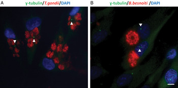
The centrosome has a major role in organizing the microtubule cytoskeleton in animal cells. Usually the centrosome is closely associated with the nucleus. However, during N. caninum (Nolan et al. Reference Nolan, Romano, Luechtefeld and Coppens2015) and T. gondii invasion (Coppens et al. Reference Coppens, Dunn, Romano, Pypaert, Zhang, Boothroyd and Joiner2006; Walker et al. Reference Walker, Hjort, Smith, Tripathi, Hornick, Hinchcliffe, Archer and Hager2008; Wang et al. Reference Wang, Weiss and Orlofsky2010; Romano et al. Reference Romano, Sonda, Bergbower, Smith and Coppens2013; Cardoso et al. Reference Cardoso, Nolasco, Gonçalves, Cortes, Leitão and Soares2014; Nolan et al. Reference Nolan, Romano, Luechtefeld and Coppens2015), the host cell centrosome is recruited away from the nuclear membrane and positioned near to the PVM (Fig. 2A). This process could be crucially involved in the mechanism by which these parasites effectively remodel host microtubules. In contrast, in B. besnoiti infected cells, the recruitment of the host cell centrosome towards the PV does not occur (Fig. 2B), and no additional foci of γ-tubulin that could act as non-centrosomal MTOCs were observed at the PV membrane. This must have implications regarding host cell microtubule dynamics in Besnoitia infected cells compared with cells infected with T. gondii or N. caninum. Thus, it seems that B. besnoiti remodelling of the host cell cytoskeleton depends neither on a centrosomal MTOC, nor on non-centrosomal MTOCs (Cardoso et al. Reference Cardoso, Nolasco, Gonçalves, Cortes, Leitão and Soares2014). Non-centrosomal MTOCs, similar to centrosomes, catalyze γ-tubulin-dependent microtubule nucleation and anchor microtubules (reviewed in Lüders and Stearns, Reference Lüders and Stearns2007). Interestingly, in T. gondii-infected cells, no additional clusters of γ-tubulin located in the vicinity of PVs could be detected, indicating that the rearrangement of host microtubules in T. gondii invasion/replication is probably dependent mostly on the interaction with the host centrosome that is recruited by the parasite (Cardoso et al. Reference Cardoso, Nolasco, Gonçalves, Cortes, Leitão and Soares2014).
Host centrosome–nucleus position in RPE-1 host cells infected with Toxoplasma gondii or Besnoitia besnoiti. Indirect immunolocalization of T. gondii and B. besnoiti infecting RPE-1 cells. Antibodies against γ-tubulin, B. besnoiti (polyclonal antibody) and T. gondii (polyclonal antibody) were used. DNA was stained with DAPI. The position of the centrosomes can be seen in green (γ-tubulin staining), in relation to the position of the parasites in each PV (red) and host cell nucleus (blue). Arrowheads indicate the host centrosome positioning. (A) Toxoplasma gondii 24 h post invasion – displacement of the centrosomes away from the nuclei and closer to the PVs. (B) Besnoitia besnoiti, 24 h post invasion – the position of the centrosome is maintained close to the host cell nucleus. Scale bar = 5 µm. Abbreviations: DAPI, 4′, 6-diamidino-2-phenylindole; PV, parasitophorous vacuole; RPE, retinal pigment epithelial.

Considering that the centrosome organizes microtubules that participate in intracellular trafficking, cell motility, cell adhesion, cell polarity and formation of the mitotic spindle (reviewed in Azimzadeh and Bornens, Reference Azimzadeh and Bornens2007), the repositioning of the centrosome in the vicinity of T. gondii PV could be important for the parasite to control the distribution of host organelles, modulation of the host cell cycle and cell migration. Meanwhile, the recruitment of the centrosome towards T. gondii PV is more evident in advanced stages of infection (24 h post invasion), without any significant difference in centrosome positioning in early infection (6 h post invasion) (Romano et al. Reference Romano, Sonda, Bergbower, Smith and Coppens2013; Cardoso et al. Reference Cardoso, Nolasco, Gonçalves, Cortes, Leitão and Soares2014). This again suggests that the remodelling of the host microtubules is not solely dependent on centrosome recruitment, especially in the first stages of PV establishment, since microtubules are observed at the PV after a few minutes following host cell entry, and the centrosome is detached from the nucleus only several hours later.
Since T. gondii infection induces the arrest of infected fibroblasts in the G2 phase of the cell cycle (Brunet et al. Reference Brunet, Pfaff, Abidi, Unoki, Nakamura, Guinard, Klein, Candolfi and Mousli2008), it is conceivable that the recruitment of the host cell centrosome facilitates the control over the host cell cycle. It is important to mention that in infected cells the recruited centrosome apparently maintains its integrity, as it consists of centrioles surrounded by a pericentriolar matrix [at least for NEDD1 and pericentrin (Romano et al. Reference Romano, Sonda, Bergbower, Smith and Coppens2013)].
Furthermore, overexpression of TBCCD1 [a centrosomal protein important for centrosome–nucleus connection in T. gondii infected cells (Gonçalves et al. Reference Gonçalves, Nolasco, Nascimento, Fanarraga, Zabala and Soares2010)], inhibits the recruitment of the centrosome at 18 h of invasion. Moreover, in these cells, T. gondii replication is significantly slower, suggesting that tachyzoite proliferation requires an efficient recruitment of the host cell centrosome, with the manipulation of the molecular mechanisms involved in the nucleus-centrosome connection. The observation that invasion of T. gondii into cells overexpressing TBCCD1 is not inhibited supports the hypothesis that the recruitment of the centrosome is important only for tachyzoites undergoing endodyogeny (Cardoso et al. Reference Cardoso, Nolasco, Gonçalves, Cortes, Leitão and Soares2014).
Thus, different apicomplexan parasites have evolved mechanisms to modulate the structure and functional activity of the microtubule cytoskeleton. However, some species such as those in the genus Toxoplasma, Neospora and Besnoitia remodel host cell microtubules already during host cell entry or at an early stage post invasion, while for others such as Eimeria and Theileria the current evidence suggests that they act on microtubule dynamics mainly at a later stage during intracellular proliferation.
Although several hypotheses have been proposed to explain these interactions, it is still not clear why parasites manipulate the host cell microtubule cytoskeleton to establish a successful infection, and the molecular mechanisms are only now starting to be uncovered.
ACTIN FILAMENT DYNAMICS IN CELLS INFECTED BY APICOMPLEXA
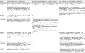
Several studies indicate that invasion by apicomplexan parasites triggers an important reorganization of the host cells actin microfilament system. The current data on host cell actin remodelling induced by apicomplexan parasites is summarized in Table 2. Even though the molecular mechanisms used by the parasites to trigger actin-filament rearrangements in infected cells are better understood compared with those responsible for the remodelling of the host cell microtubule cytoskeleton, they remain for the most part elusive.
Rearrangement of host actin filaments and parasite and host molecules that interact with the host actin cytoskeleton during invasion and replication

DC, dendritic cell; ERM, ezrin/radixin/moesin; MESA, mature-parasite-infected erythrocyte surface antigen; KAHRP, knob-associated histidine-rich protein; MJ, moving junction; PV, parasitophorous vacuole; PVM, parasitophorous vacuole membrane; PTA, podosome-type adhesions; RBC, red blood cells; (?) not known.
De novo polymerization of host actin is an important prerequisite for the entry of T. gondii tachyzoites into host cells. A host cell derived ring-shaped F-actin structure is formed at the MJ that disappears 10 min after tachyzoite entrance, which provides a solid anchor mechanism for pulling the parasite inside the host cell (reviewed in Frénal and Soldati-Favre, Reference Frénal and Soldati-Favre2009). Adding to this, the presence at the MJ of the host Arp2/3 complex involved in actin polymerization control, together with the nucleation-promoting factor, cortactin, supports de novo polymerization of the host actin (Gonzalez et al. Reference Gonzalez, Combe, David, Malmquist, Delorme, Leroy, Blazquez, Ménard and Tardieux2009). It is likely that the changes in the actin microfilaments during invasion include not only the induction of actin polymerization, but also a local disorganization of the cortical actin network to facilitate tachyzoite entrance. Considering this, and probably to overcome the resistance conferred by the host cortical actin, T. gondii expresses a protein named toxofilin (Bradley et al. Reference Bradley, Ward, Cheng, Alexander, Coller, Coombs, Dunn, Ferguson, Sanderson, Wastling and Boothroyd2005). This protein remodels the host cytoskeleton by upregulating actin filament turnover and retrograde flow, which loosens the local host cell actin meshwork and facilitates parasite internalization and invasion (Frénal and Soldati-Favre, Reference Frénal and Soldati-Favre2009). In addition, T. gondii tachyzoites lacking toxofilin had an increased difficulty in inducing cortical actin disassembly and exhibited delayed invasion kinetics (Delorme-Walker et al. Reference Delorme-Walker, Abrivard, Lagal, Anderson, Perazzi, Gonzalez, Page, Chauvet, Ochoa, Volkmann, Hanein and Tardieux2012). A recent study showed that about 20% of T. gondii toxofilin mutants fail to enter into the host cell and end up disengaging from the host cell membrane. This is in agreement with the view that both cortical disassembly and free actin availability is required for T. gondii complete invasion through the toxofilin activity (Bichet et al. Reference Bichet, Joly, Hadj Henni, Guilbert, Xémard, Tafani, Lagal, Charras and Tardieux2014).
Moreover, a functional relationship has also been established between host cell GTPases involved in cytoskeleton regulation and T. gondii invasion. For instance, host cell ADP-ribosylation factor-6 (ARF6) was shown to be recruited to the PVM (da Silva et al. Reference da Silva, da Silva, Cruz, Chavrier and Mortara2009). ARF6 is a small GTPase that is known to regulate membrane trafficking and actin cytoskeleton rearrangements at the plasma membrane. The RhoA and Rac1 GTPases, which are involved in the regulation of the organization and dynamics of the actin cytoskeleton (Hall, Reference Hall1998), were also observed to accumulate and be activated at the PVM upon T. gondii host entrance (Na et al. Reference Na, Zhu, Luo, Meng, Cui, Peng, Chen and Gomez-Cambronero2013). The recruitment and activation of these two small host GTPases could be implicated in the actin microfilament reorganization observed during PV formation.
Furthermore, by performing a high-throughput siRNA screen of a human siRNA library, a subset of host factors that appear to influence T. gondii invasion by regulating the host actin cytoskeleton were identified (Gaji et al. Reference Gaji, Huynh and Carruthers2013). Six of these identified factors (PTK9L – Twinfilin, actin binding protein homolog 2; PHPT1-Phosphohistidine phosphatase 1; MAPK7-Mitogen activated protein kinase 7; PTPRR – Protein tyrosine phosphatase, receptor type, R; MYLIP-Myosin regulatory light chain interacting protein; and PPIL2 – Peptidylprolyl isomerase (cyclophilin)-like 2) are antagonists of the actin cytoskeleton, as the downregulation of their expression causes increased development of cortical actin microfilaments with deposition of more F-actin near the PV periphery. Despite the fact that the mechanisms underlying how these proteins influence actin polymerization are unclear, the identification of these novel proteins can be of significance not only to understand T. gondii host cell invasion, but also to the understanding of other actin-dependent processes such as host cell migration (Gaji et al. Reference Gaji, Huynh and Carruthers2013).
Parasite–host cell F-actin interactions have also been described for other apicomplexans. In N. caninum infected cells, the actin microfilaments associate with the periphery of the PV, which might suggest a physical link between actin filaments and the pseudocyst membrane (Vonlaufen et al. Reference Vonlaufen, Gianinazzi, Müller, Simon, Björkman, Jungi, Leib and Hemphill2002). The molecular mechanisms and pathways underlying these actin rearrangements are far from being understood. Cryptosporidium parvum initiates structural changes in the host cell cortical actin upon attachment to the host cell surface, resulting in an accumulation of host cell actin filaments at the site of infection (Elliott et al. Reference Elliott, Coleman, Lane, May, Machesky and Clark2001; Feng et al. Reference Feng, Nie, Bonilla, Widmer, Sheoran and Tzipori2006). It is possible that this parasite employs signal cascades that are also involved in the formation of lamellipodia/filopodia. In this view the host phosphoinositide 3-kinase (PI3K) and Cdc42 (a small GTPase of the Rho-subfamily) are good candidates to induce actin remodelling during Cryptosporidium host cell invasion. In fact, the PI3K/cdc42/N-WASP pathway activates the Arp2/3 complex leading to the elongation and branching of actin filaments and formation of protrusions. Interestingly, inhibition of host Cdc42 reduced Cryptosporidium invasion up to 80% but never blocked host cell entry completely, which suggests that one or several alternative pathways exist. A possible, but not yet proven, alternative pathway for Cryptosporidium induced actin remodelling, involves integrins (like ITGA2/ITGB1, fundamental for the control of actin-based processes in lamellipodia and filopodia in cells), that could pass a signal via a tyrosin Src kinase, with the subsequent activation of cortactin and recruitment of the Arp2/3 complex (reviewed in Lendner and Daugschies, Reference Lendner and Daugschies2014).
Interestingly a similar strategy seems to be used by T. annulata that specifically promotes the formation of podosome-type adhesions (PTAs) and induces host cell polarization by triggering the organization of a single lamellipodium. Theileria annulata can manipulate the actin cytoskeleton by increasing the assembly of functional adhesion and signalling complexes, controlling proteins involved in cell–matrix interactions and cell signalling in lamellipodia and PTAs of infected macrophages (Baumgartner, Reference Baumgartner2011). Moreover, there seems to be de novo actin polymerization of F-actin in parasitized macrophages at host cell protrusions in the leading edge, allowing the forward extension of these cells necessary for their invasive characteristics (Ma and Baumgartner, Reference Ma and Baumgartner2013).
Thus, host cell actin cytoskeleton remodelling is a universal event during the apicomplexan infection process. Nevertheless parasites seem to have adapted their invading strategies to the specificities of the cytoskeleton of their target cells. For example, in the case of E. bovis entry into bovine endothelial cells, there is an accumulation of host cell actin at the PV that stabilizes the growing intracellular macromeront. Actin surrounds the PV within 3 dpi, and actin filament bundles form a thick layer in the final stages of meront maturation (15–20 dpi) (Hermosilla et al. Reference Hermosilla, Schröpfer, Stowasser, Eckstein-Ludwig, Behrendt and Zahner2008). Several actin-related genes are upregulated in cells containing developing macromeronts. This includes vinculin and ezrin (involved in actin cytoskeleton signalling) and others such as CAPG, CNN2, TAGLN and PALLD (Taubert et al. Reference Taubert, Wimmers, Ponsuksili, Jimenez, Zahner and Hermosilla2010). Meanwhile, in the final stages of macromeront development, when macromeronts rupture and merozoites are released, the expression of proteins that interact with actin, including α-actin-1, gelsolin, actin-like protein-2, gelsolin-like capping protein, tropomodulin-3 and transgelin are downregulated, and the same accounts for transcripts coding for β-tubulin 5 and 6. This suggests that the mechanisms regulating the microtubule and actin cytoskeleton are different, depending on the developmental stage of meront maturation. Thus, an upregulation is observed during meront formation (probably explained by the increasing need of structural support), and downregulation at the final phase of merogony, which could facilitate the release of merozoites (Lutz et al. Reference Lutz, Schmitt, Linder, Hermosilla, Zahner and Taubert2011; reviewed in Hermosilla et al. Reference Hermosilla, Ruiz and Taubert2012). In addition to the alterations in the actin filament system, E. bovis infected cells also exhibit alterations in the localization and overall organization of spectrin. Spectrin is a cytoskeletal protein that is closely linked to the cortical actin network lining the intracellular side of the plasma membrane. Spectrin forms tetramers that associate with short actin filaments, and provides a scaffold that ensures maintenance of plasma membrane integrity. Infection of endothelial cells with E. bovis results in accumulation of host cell spectrin around the PV 8 dpi, at the onset of parasite replication (Hermosilla et al. Reference Hermosilla, Schröpfer, Stowasser, Eckstein-Ludwig, Behrendt and Zahner2008).
Plasmodium falciparum infection of red blood cells (RBCs) induces dramatic host cell remodelling, which includes overall changes in shape and surface structures, and formation of several compartments in the host cell cytoplasm. Many of these alterations are also related to changes in the spectrin-actin cytoskeleton. During infection, the host spectrin elongates, the spectrin network becomes sparser and the spectrin tetramers appear more extended (Shi et al. Reference Shi, Liu, Li, Yin, Chong, Tan, Zhang and Lim2013). Maurer's clefts (MC) are the best-studied compartments formed in the RBC cytoplasm upon infection, but how these are formed is unclear and questions on their functional role have not been completely resolved so far. MC function as membrane-limited vacuolar structures important for trafficking of parasite-encoded proteins destined for the RBC membrane. Most of these proteins confer cytoadherence of infected RBCs to the endothelium of the microvasculature but it remains elusive how proteins are sorted and transported to and from these structures (reviewed in Mundwiler-Pachlatko and Beck, Reference Mundwiler-Pachlatko and Beck2013). The parasite adhesin P. falciparum erythrocyte membrane protein-1 (PfEMP1) is one of these proteins, and it mediates cytoadhesion by binding to host receptors at the surface of vascular endothelial cells. PfEMP1 localizes in specific regions at electron-dense protrusions of the erythrocyte membrane, referred to as knobs. Knob formation is a dynamic process. As the parasite matures from trophozoite to schizont, the density of knobs increases and in the end, knobs cover the entire RBC surface. The formation of these structures implicates several RBC cytoskeletal components, such as spectrin, ankyrin and actin, by probably altering the architecture of the sub-membrane skeleton and its interactions with membrane proteins. This results in increased rigidity and adhesiveness of the erythrocyte membrane (reviewed in Mbengue et al. Reference Mbengue, Yam and Braun-Breton2012). Recent electron microscopy studies revealed that each knob contains a spiral filament that coils into a shallow cone, and a layer of diffuse material that coats the upper surface of the conical frame. The outer coat of the knob skeleton connects with the erythrocyte spectrin cytoskeleton, mostly around the knob base. This interaction with the surrounding cytoskeleton could suggest a mechanical explanation for the enhanced cytoadherence of knobs (Watermeyer et al. Reference Watermeyer, Hale, Hackett, Clare, Cutts, Vakonakis, Fleck, Blackman and Saibil2016).
There are several proteins at MCs required for PfEMP1 trafficking (Maier et al. Reference Maier, Rug, O'Neill, Beeson, Marti, Reeder and Cowman2007; Spycher et al. Reference Spycher, Rug, Pachlatko, Hanssen, Ferguson, Cowman, Tilley and Beck2008), including PfEMP1 trafficking protein 1 (PfPTP1) that was recently shown to be required for transport of PfEMP1 to the surface of P. falciparum–infected erythrocytes. In the absence of PfPTP1, MC are reduced to globular structures, thus the protein is important for maintenance of the MC architecture. PfPTP1 has a role in linking MC to the host actin cytoskeleton, and is integrated in a large complex that includes skeleton-binding protein 1 (SBP1). It influences actin filament length or organization, suggesting that PfPTP1 has a possible function in host cell actin filament stabilization (Rug et al. Reference Rug, Cyrklaff, Mikkonen, Lemgruber, Kuelzer, Sanchez, Thompson, Hanssen, O'Neill, Langer, Lanzer, Frischknecht, Maier and Cowman2014). SBP1 is another MC protein that is essential for the placement of PfEMP1 at the erythrocyte surface, and it has a defined region that binds specifically to defined sub-domains of two major components of the RBC membrane skeleton, protein 4·1R and spectrin (Kats et al. Reference Kats, Proellocks, Buckingham, Blanc, Hale, Guo, Pei, Herrmann, Hanssen, Coppel, Mohandas, An and Cooke2015).
Several cytoskeleton binding proteins have been identified in P. falciparum, such as the Mature-parasite-infected erythrocyte surface antigen (MESA) that interacts with Band 4·1 in the erythrocyte cytoskeleton; or P. falciparum knob-associated histidine-rich protein (KAHRP), that binds the acidic terminal sequence (ATS) of PfEMP1 (termed VARC) and Spectrin-actin-4·1 complex, thus linking PfEMP1 to the cytoskeleton (Oh et al. Reference Oh, Voigt, Fisher, Yi, LeRoy and Derick2000). It was recently demonstrated that the full-length proteins KAHRP and PfEMP1 co-localized to membrane knobs in erythrocytes infected with the trophozoite stage P. falciparum. Four positively charged linear sequence motifs of high intrinsic mobility on KAHRP that interact electrostatically with VARC have been identified (Ganguly et al. Reference Ganguly, Ranjan, Kumar and Bhavesh2015). Another important protein is a PHISTb protein, ring-infected erythrocyte surface antigen (RESA) that contains a C-terminal spectrin-binding domain, which stabilizes the spectrin tetramer (Foley et al. Reference Foley, Tilley, Sawyer and Anders1991; Silva et al. Reference Silva, Cooke, Guillotte, Buckingham, Sauzet, Le Scanf, Contamin, David, Mercereau-Puijalon and Bonnefoy2005). Also, Tarr et al. (Reference Tarr, Moon, Hardege and Osborne2014) identified a cytoskeleton binding domain in RESA that again indicates that PHISTb proteins may act as cross-linkers of the erythrocyte cytoskeleton. Finally, another example of PHISTb proteins interaction with the erythrocyte cytoskeleton was recently demonstrated to be a member of the P. falciparum lysine-rich membrane-associated PHISTb (LyMP) protein family. It was shown that LyMP is exported into the RBC, where it interacts directly with the RBC membrane skeleton (Proellocks et al. Reference Proellocks, Herrmann, Buckingham, Hanssen, Hodges and Elsworth2014).
As it was already mentioned, after P. falciparum infection, the erythrocytes become more rigid, less deformable and more fragile (reviewed in Mbengue et al. Reference Mbengue, Yam and Braun-Breton2012). Parasite-exported proteins [like KAHRP and P. falciparum erythrocyte membrane protein 3 (PfEMP3)] and the extended spectrin tetramers may be responsible for the increased rigidity of the host cell cytoskeleton (Glenister et al. Reference Glenister, Coppel, Cowman, Mohandas and Cooke2002; Shi et al. Reference Shi, Liu, Li, Yin, Chong, Tan, Zhang and Lim2013).
APICOMPLEXAN PARASITES AND HOST CELL INTERMEDIATE FILAMENTS
Information on potential modulation of intermediate filaments by apicomplexan parasites is sparse. In fibroblasts, vimentin intermediate filament distribution markedly changed upon infection with T. gondii tachyzoites. While vimentin was largely localized around the nuclear periphery in non-invaded cells, filaments rapidly associated with the PVM periphery within 30 min post invasion. This association progressively became more pronounced with time, and at 18 h post invasion, an important network of intermediate filaments bundles encaged the PV (Halonen and Weidner, Reference Halonen and Weidner1994). In contrast, in endothelial cells infected with E. bovis, no marked changes in host cell vimentin intermediate filament distribution were evident during first merogony (Hermosilla et al. Reference Hermosilla, Schröpfer, Stowasser, Eckstein-Ludwig, Behrendt and Zahner2008).
During the development of T. gondii tissue cysts in murine astrocytes, immunostaining revealed the presence of glial fibrillary acidic protein (GFAP) intermediate filaments. The GFAP filaments were present on the cytoplasmic side of the cysts in close apposition to the cyst wall and arranged in a concentric layer measuring 5–10 µm in thickness. The GFAP filament wrapping seems to be important mainly in early cyst development since more mature cysts were surrounded by a GFAP layer that was diminished when compared with younger cysts (Halonen et al. Reference Halonen, Weiss and Chiu1998). The same intermediate filaments of GFAP were found in N. caninum infection, in close juxtaposition to the cytoplasmic side of the PVM in organotypic rat brain slice cultures (Vonlaufen et al. Reference Vonlaufen, Gianinazzi, Müller, Simon, Björkman, Jungi, Leib and Hemphill2002).
APICOMPLEXAN PARASITES AFFECT HOST CELL MIGRATION
Considering the pronounced effects of apicomplexan parasites on the host cell cytoskeleton, and the fact that some of these parasites affect the architecture of the sub-membrane cytoskeleton and the cell-matrix junctions, it seems likely that these changes could reflect on an altered migration of the host cell.
Most studies dealing with this aspect were carried out in T. gondii infected cells. For instance, T. gondii infection of dendritic cells (DCs) alters their motility and confers a hypermigratory phenotype (Lambert et al. Reference Lambert, Hitziger, Dellacasa, Svensson and Barragan2006, Reference Lambert, Dellacasa-Lindberg and Barragan2010). Thus, DCs could play an important role in the dissemination of T. gondii in an infected host. This is supported by the observation that parasitized DCs adoptively transferred to uninfected mice disseminate more quickly than non-infected DCs (reviewed in Blader and Saeij, Reference Blader and Saeij2009). Interestingly, the induction of increased cell migration through T. gondii infection is also exhibited by infected macrophages, but not by infected monocytes, T-cells, B-cells or NK-cells (Lambert et al. Reference Lambert, Dellacasa-Lindberg and Barragan2010).
The observed hypermigratory phenotype in DCs consists in rapid morphological transformation, altered adhesive properties, hypermotility, transmigration across endothelial monolayers in vitro, and responsiveness to chemokines, and was shown to account for T. gondii lineages types I, II and III (Weidner and Barragan, Reference Weidner and Barragan2014). Already 5–10 min following invasion of DCs, cytoskeletal actin redistribution occurred, leading to the disappearance of podosomes, which are known to limit rapid migration by strongly binding to the extracellular matrix. Other features included the appearance of a rounded cellular morphology with veils, ruffles, filopodia and lamellipodia linked to the protrusive activity of DCs. In addition, T. gondii invasion caused the redistribution of integrins (CD18 and CD11c), with a more prominent integrin staining of CD18 at the periphery of the cell membrane, and of CD11 in veil structures (Weidner et al. Reference Weidner, Kanatani, Hernández-Castañeda, Fuks, Rethi, Wallin and Barragan2013). Toxoplasma gondii infected DCs exhibited increase secretion of γ-aminobutyric acid (GABA), which is a neurotransmitter involved in cell migration, immunomodulation and metastasis. Interestingly, inhibition of GABA transporters or the GABA synthesizing enzyme, as well as blocking of the GABAA receptor, resulted in a reduction of hypermotility and transmigration, but did not alter the morphological changes in infected DCs described above (reviewed in Weidner and Barragan, Reference Weidner and Barragan2014). Finally, the hypermigratory phenotype in T. gondii infected DCs does not require de novo protein synthesis, since inhibition of parasite or host cell protein synthesis did not influence the morphological changes or hypermotility (Weidner et al. Reference Weidner, Kanatani, Hernández-Castañeda, Fuks, Rethi, Wallin and Barragan2013). Nearly identical features including a hypermigratory phenotype occur in DCs infected with N. caninum tachyzoites (Collantes-Fernandez et al. Reference Collantes-Fernandez, Arrighi, Álvarez-Garcıa, Weidner, Regidor-Cerrillo, Boothroyd, Ortega-Mora and Barragan2012).
The impact of T. gondii infection on host cell migration has also been addressed in other cell types, such as fibroblasts (Wang et al. Reference Wang, Weiss and Orlofsky2010) and epithelial cells (Cardoso et al. Reference Cardoso, Nolasco, Gonçalves, Cortes, Leitão and Soares2014). In these cases, infection leads to a loss of host cell motility. In an in vitro wound-healing assay, the migration of parasitized human foreskin fibroblasts (HFF) and retinal pigment epithelial cells (RPE-1) was delayed (Wang et al. Reference Wang, Weiss and Orlofsky2010; Cardoso et al. Reference Cardoso, Nolasco, Gonçalves, Cortes, Leitão and Soares2014). In contrast, RPE-1 cells infected with B. besnoiti tachyzoites did not show any motility impairment in wound-healing assays (Cardoso et al. Reference Cardoso, Nolasco, Gonçalves, Cortes, Leitão and Soares2014). It is possible that this difference in motile behaviour could be related to the above mentioned differences in recruitment of the centrosome (e.g. recruitment to the PV in T. gondii infected cells, but not in B. besnoiti infected cells) (Coppens et al. Reference Coppens, Dunn, Romano, Pypaert, Zhang, Boothroyd and Joiner2006; Walker et al. Reference Walker, Hjort, Smith, Tripathi, Hornick, Hinchcliffe, Archer and Hager2008; Wang et al. Reference Wang, Weiss and Orlofsky2010; Romano et al. Reference Romano, Sonda, Bergbower, Smith and Coppens2013; Cardoso et al. Reference Cardoso, Nolasco, Gonçalves, Cortes, Leitão and Soares2014). The participation of the centrosome in migration was previously documented by results showing that stimulation of cell motility was accompanied by centrosome reorientation towards the leading edge (Schmoranzer et al. Reference Schmoranzer, Fawcett, Segura, Tan, Vallee, Pawson and Gundersen2009; Vinogradova et al. Reference Vinogradova, Miller and Kaverina2009). This cell polarization could be compromised in T. gondii infected cells, but not in B. besnoiti infected cells. However, the mean angle of the centrosome towards the wound was quite similar in non-invaded RPE-1 cells, and in B. besnoiti and T. gondii infected cells (Cardoso et al. Reference Cardoso, Nolasco, Gonçalves, Cortes, Leitão and Soares2014). Overall, infection of RPE-1 cells by B. besnoiti and T. gondii did not have an obvious effect in directional cell polarization, but cell migration in T. gondii infected cells was impaired.
Transforming species of Theileria such as T. annulata trigger the motility and invasive behaviour of infected macrophages by controlling host cell membrane protrusions through F-actin dynamics, which assists parasite dissemination in the host (Baumgartner, Reference Baumgartner2011; Ma and Baumgartner, Reference Ma and Baumgartner2013, Reference Ma and Baumgartner2014). More recent studies determined that the parasite drives host cell motility and invasiveness by activating the host proto-oncogenic ser/thr kinase MAP4K4, which is induced by the inflammatory cytokine TNFα. TNFα induces the phosphorylation of the ERM (ezrin/radixin/moesin) family of cytoskeleton regulatory proteins in a MAP4K4-dependent manner, with this ERM phosphorylation likely occurring in lamellipodia, thus controlling parasite-dependent actin dynamics regulation. Supporting this, blocking TNFα secretion or MAP4K4 expression compromised the formation of polar F-actin-rich invasion structures, impairing host cell motility. Thus, Theileria has the capacity of inducing TNFα, activating the conserved kinase MAP4K4, in order to control actin cytoskeleton dynamics and thus cell motility (Ma and Baumgartner, Reference Ma and Baumgartner2014).
CONCLUDING REMARKS
Apicomplexan parasites exhibit a number of common morphological characteristics, which have been used to place them into the same phylum. However, they also share a number of features related to their interactions with the host cell, which is essential for intracellular survival, replication and further development. One aspect is their ability to modulate the host cell cytoskeletal filamentous networks composed of microtubules, actin microfilaments and intermediate filaments. In many instances, it still remains elusive to what extent this interaction and cross talk with the host cell cytoskeleton is beneficial for these parasites, but one can speculate that it enables them to (i) reposition crucial host cell organelles, (ii) hijack host cell nutrients, (iii) gain control of signalling pathways, (iv) interfere or promote host cell motility, and (v) control the host cell shape and adherence properties. Several recent studies have revealed effector parasite proteins and respective host cell targets that enable apicomplexans to modulate the host cell cytoskeleton.
Although being so similar in many ways, each apicomplexan species has its own peculiarities and requirements, and these are reflected in the differential influence that distinct apicomplexan species exert on the host cell cytoskeleton. As a word of caution, one should mention the possibility that some of the rearrangements induced upon the host cell cytoskeleton by invading parasites could just simply reflect host cell specific responses upon invasion, which are not actively induced by the parasites per se, and future approaches should address this point. In addition, it would be of great benefit to focus on three-dimensional cell culture models or organoid culture rather than on the two-dimensional in vitro cell culture that is currently mostly employed, simply to more accurately mimic what is going on in vivo. In any case, the elucidation of the molecular effector pathways that allow apicomplexan parasites to manipulate the host cell cytoskeleton in order to establish infection and propagate in their host is of major importance. These efforts could reveal new aspects of host–parasite interactions and parasite vulnerabilities that may be exploited for an efficient therapeutic and prophylactic research.
FINANCIAL SUPPORT
The authors thankfully acknowledge financial support by the Swiss National Science Foundation (grant No. 310030 146162 to A. H.), the Swiss Government Excellence Scholarship (reference No. 2015·0062 to R. C.) and Fundação para a Ciência e a Tecnologia (FCT), Portugal: project grants PTDC/CVT/71630/2006 to A. L. and H. S., PTDC/CVT/105470/2008 to H. S. and A. L. and fellowship SFRH/BD/38122/2007 to R. C.