




No CrossRef data available.
Article contents
Cross-stress tolerance and expression of stress-related proteins in osmotically desiccated entomopathogenic Steinernema feltiae IS-6
Published online by Cambridge University Press: 06 July 2005
Abstract
Infective juveniles (IJs) of the entomopathogenic nematode (EPN) Steinernema feltiae IS-6 can survive exposure to 24% glycerol solution by entering an osmotically desiccated state. Exposure of osmotically desiccated nematodes to extreme temperature assays (40 °C for 10 h and −20 °C for 360 h) resulted in gradual reduction in survival, whereas non-desiccated IJs died within a short exposure to the assay conditions. Through SDS-PAGE, a stress-related protein UNC-87 was found in osmotically desiccated IJs exposed to 40 °C for 3, 6, and 8 h, whose survival rates were 98·9±1·43, 78·5±5·87 and 20·9±4·93%, respectively. The protein was not found in IJs following exposure of osmotically desiccated individuals to 40 °C for 10 h, in which none of the IJs survived. After exposure to −20 °C for 360 h, the survival of osmotically desiccated EPNs with a weak band of UNC-87 was 13·0±3·32%. To identify other responsive proteins that are required for osmotic stress, we used 2-dimensional electrophoresis to analyse the proteins in osmotically desiccated EPNs. The results revealed that 10 novel protein spots and 10 up-regulated protein spots in osmotically desiccated IJs were detected by digital image analysis. Mass spectrometry analysis of 7 significant spots indicated that osmotic stress in desiccated IJs was associated with the induction of actin, Proteasome regulatory particle (ATPase-like), GroEL chaperonin, GroES co-chaperonin and transposase family member. It seems to show actin, UNC-87 and Proteasome regulatory particle may play distinct roles in specific aspects of organization of macromolecular structures under desiccation stress. GroEL and GroES are members of the Hsp60 family of chaperons.
Keywords
Information
- Type
- Research Article
- Information
- Copyright
- © 2005 Cambridge University Press
INTRODUCTION
The soil environment can be a highly suitable and favorable environment in which many organisms can thrive. However, unfavourable conditions in the soil threaten the persistence of the inhabiting life-forms, and especially those parasites that stay in the soil until a suitable host is found. Many multicellular organisms, including nematodes, have adopted unique survival mechanisms to resist environmental extremes. Once the environmental conditions deteriorate they enter a dormant state, which considerably prolongs their life-span and enables them to withstand the rigours of a fluctuating regime (Perry, 1998). One of the stimuli for entry into such a state is gradual loss of water from the nematode's body, which results in a reduction in metabolism, termed ‘quiescence’ (Perry, 1998; Glazer, 2002). In these dormant states metabolism is reduced to a level that cannot be detected (Womersley, Wharton and Higa, 1998). Induction of the dormant state by evaporative dehydration is called ‘anhydrobiosis’ and when water is removed by osmotic dehydration is called ‘osmobiosis’ (Perry, 1998).
Infective Juveniles (IJs) of entomopathogenic steinernematid and heterorhabditid nematodes have been shown to be efficient insect-killing parasites. The knowledge about the pathogenic process of these nematodes has been recently reviewed by Dowds and Peters (2002). The IJs live in the soil, search for a suitable host, and penetrate to the haemocoel by mechanical and enzymatic means (Abu-Hatab, Gaugler and Ehlers, 1998; Dowds and Peters, 2002). Shortly after entry, the nematodes release symbiotic bacteria. The bacteria associated with steinernematid and heterorhabditid nematodes belong to the genus Xenorhabdus and Photorhabdus, respectively (Frost et al. 1997). Once the nematode and its bacterial partner have overcome the immune system of a susceptible insect (Dunphy and Thurston, 1990) they kill it very quickly. The nematodes feed upon the rapidly multiplying bacteria and debris, and subsequently mature, mate, and produce two or more generations within the insect cadaver before emerging into the environment as IJs in search of new hosts.
These nematodes are currently used commercially as biological control agents of insect pests (Georgis and Manweiler, 1994; Gaugler and Han, 2002). The sensitivity of entomopathogenic nematodes (EPNs) to extreme environmental conditions and their poor stability in storage seems to hamper their full commercial exploitation (Kaya & Gaugler, 1993; Glazer, 2002; Grewal, 2002). It has been suggested that reduction of the metabolism of stored IJs by induction of desiccation will enable prolongation of their storage period (Glazer and Salame, 2000; Grewal, 2000, 2002; Grewal and Jagdale, 2002; Chen and Glazer, 2005). Furthermore, enhancement of EPN capabilities to withstand environmental extremes would increase their persistence and efficacy against insect pests (reviewed by Glazer, 2002).
IJs of EPNs can become desiccated under osmotic stress (Glazer and Salame, 2000; Chen and Glazer, 2004), under which the biochemical changes associated with desiccation indicated that the nematodes reduced their oxygen consumption (Grewal, 2000; Chen, Yang and Jiang, 2001). Osmotically desiccated nematodes exhibited reduced glycogen levels and increased trehalose levels (Chen et al. 2000; reviewed by Glazer, 2002). The free-living mycophagous nematode Aphelenchus avenae accumulated large amounts of trehalose in response to dehydration, and this correlated with its desiccation tolerance. Trehalose protects membranes and proteins from desiccation damage by replacing structural water, and contributes to the formation of an intracellular organic glass, which is thought to stabilize the cell's contents (Browne, Tunnacliffe and Burnell, 2002). Trehalose also plays an important role in enhanced freezing, cold and desiccation tolerance (reviewed by Jagdale and Grewal, 2003). The increased trehalose accumulation was also correlates well with cold pre-acclimation (Grewal and Jagdale, 2002).
However, several lines of evidence indicate that non-reducing sugars alone are not sufficient to confer a state of desiccation, and that further adaptations are required (reviewed by Browne et al. 2002). A common feature of the stress response is the induction of stress-related proteins, which were first discovered in cells exposed to slight hyperthermia (Ritossa, 1962, 1996; Tissieres, Mitchel and Tracy, 1974; Gabai and Sherman, 2002). A class of proteins known as LEA (late embryogenesis abundant) proteins occurs commonly in plants, accumulating during seed maturation and desiccation stress (Browne et al. 2002, 2004). The LEA proteins also accumulated in anhydrobiotic nematode A. avenae and S. feltiae (Solomon et al. 2000; Browne et al. 2002, 2004; Gal, Glazer and Koltai, 2003). It seems to indicate that some mechanisms of coping with water loss are conserved between plants and animals (Gal et al. 2003; Browne et al. 2004). Although previous studies have focused on the biochemical changes associated with water deficit in plant (Close, Kortt and Chandler, 1989; Hill et al. 1994; Thomashow, 1998; Dhaubhadel et al. 2002), and these adaptations may include changes in primary metabolism, alterations to cell membranes, osmotic adjustments via the accumulation of compatible solutes or hydrophilic proteins and the synthesis of stress-related proteins, such adaptations have not been defined for desiccated EPNs. In order to obtain a deeper understanding of the adaptations of water loss and the stress-related protein networks that are activated in S. feltiae during desiccation, in the present study we determined the differential proteins of S. feltiae IJs in response to osmotic desiccation by means of 2-dimensional polyacrylamide gel electrophoresis and mass spectrometry. We also demonstrated the cross-tolerance of EPNs to various environmental stresses.
MATERIALS AND METHODS
Nematode culture
Steinernema feltiae IS-6 was isolated from the soil of a citrus orchard in the Negev, a semiarid region in Israel (Solomon, Paperna and Glazer, 1999). The nematodes were reared at 25 °C in the last instar of the greater wax moth Galleria mellonella (L.), according to the method of Kaya & Stock (1997).
Osmotic desiccation
Water suspensions of S. feltiae IS-6 IJs were concentrated onto a 5 cm diameter filter paper (Whatman No. 1) in a vacuum filtration apparatus, and 0·2 g (approx. 450000 IJs) of IJs were transferred into a 200 ml flask that contained 30 ml of 24% (w/w) glycerol (24% w/w=3·529 Osmol/kg) (BIOLAB Ltd, Jerusalem, Israel). Immediately after transfer, the flask was stirred to ensure good mixing of the nematodes with the glycerol solution. The flask was incubated at 25 °C for 24 h, after which four 500 μl subsamples were withdrawn from the flask and transferred to 5 cm diameter plastic Petri dishes. The nematodes were considered to be osmotically desiccated when they had shrunk and did not respond to probing by the tip of a needle. However, in some cases shrunken nematodes were found to be dead after dehydration. Therefore, 7 ml of water was added to the Petri dish. The number of desiccated nematodes and returning to full motility were recorded. Each treatment included 4 replicates. Non-desiccated IJs served as control treatment.
Heat tolerance
The heat tolerance of osmotically desiccated IJs and non-desiccated nematodes, as control, were assessed by exposure to 40 °C for 10 h. The nematodes were transferred at a concentration of 5000 IJs/ml, in 500 μl aliquots, to 1·5 cm3 Eppendorf tubes and exposed to 40 °C in water bath. Each Eppendorf tube was closed by its cover, punctured by 3 small holes, during exposure to heat. After 0 h, 0·25 h, 0·5 h, 1 h, 2 h, 4 h, 6 h, 8 h and 10 h, the nematode suspensions were transferred from the Eppendorf tubes to 5 cm diameter plastic Petri dishes containing 7 ml of water. Each treatment consisted of 4 replicates. The Petri dishes were maintained at room temperature for 24 h and then the survival of nematodes in all treatments was examined with a stereoscopic microscope. The survival of the nematodes was recorded by counting the motile IJs, either without disturbance or after probing with the tip of a needle.
Freezing tolerance
Freezing tolerance of the osmotically desiccated IJs and non-desiccated nematodes, as controls, was assessed by exposing them to −20 °C for 360 h. The nematodes were transferred at a concentration of 5000 IJs/ml, in 500 μl aliquots, to 1·5 cm3 Eppendorf tubes, and exposed to −20 °C. After intervals of 0 h, 1 h, 2 h, 4 h, 6 h, 24 h, 72 h, 192 h, 288 h and 360 h the suspensions were transferred from the Eppendorf tubes to 5 cm diameter plastic Petri dishes containing 7 ml of distilled water. Each treatment consisted of 4 replicates. These Petri dishes were maintained at room temperature for 24 h and then the survival of the nematodes was determined with a stereoscopic microscope. The survival of the nematodes in all samples was recorded by counting the motile IJs, either without disturbance or after probing with the tip of a needle.
Protein extraction from osmotically desiccated IJs under extreme temperatures
The suspensions of osmotically desiccated IJs that had been exposed to a 40 °C water bath for 3 h, 6 h, 8 h and 10 h, or to −20 °C for 72 h, 192 h and 360 h were concentrated onto a 5 cm diameter filter paper in a vacuum filtration apparatus for protein extraction. Protein extraction from osmotically desiccated/temperature stressed nematodes was performed using methods described by Laemmli (1970) and Solomon et al. (2000).
Gel electrophoresis and analysis of protein
The proteins were analysed by 12% SDS-PAGE. Each lane was loaded with 20 μg protein and stained with Coomassie Brilliant Blue R-250. The relative contents of polypeptides and the apparent molecular weights were determined by applying the ChemiImager 4400 software (San Leandro, CA, USA) to the bands on the Coomassie-stained gels (Solomon et al. 2000).
Mg/NP-40 extraction and 2-dimensional gel electrophoresis
Proteins of osmotically desiccated nematode IJs were extracted with Mg/Np-40 extraction buffer. The extraction method was as described previously by Kim et al. (2001). Non-desiccated IJs served as control treatment. The protein was assayed with the Bicinchoninic Acid Protein Assay Kit, product codes BCA-1 and B9643 (Sigma, St Louis, MO, USA) according to the manufacturer's instruction manual.
Two-dimensional electrophoresis (2-DE) was performed according to the immobilized pH gradients (IPGs) principles and methods of Amersham Biosciences (Rehovot, Israel), with some modifications. For analytical and preparative gels, the 13 cm IPG strips (pH 3–10) (Amersham Pharmacia Biotech, Rehovot, Israel) were re-hydrated overnight with 250 μl of rehydration buffer (8 M urea, 2% CHAPS, IPG buffer 2% (v/v), 0·3% DTT and 0·002% bromophenol blue), containing 200 μg protein, at room temperature. Isoelectric focusing (IEF) was conducted at 18 °C with a Multiphor II (Pharmacia, Rehovot, Israel). The running conditions were as follows: 300 V for 15 min, followed by 500 V for 15 min, 1000 V for 15 min, 1500 V for 15 min, 2000 V for 15 min, 2500 V for 15 min, 3000 V for 15 min, and finally 3500 V for 5 h. The focused strips were equilibrated twice for 15 min in 10 ml of equilibration solution. The first equilibration was performed in a solution containing 50 mM Tris-HCl, pH 8·8, 6 M urea, glycerol 30% (v/v), SDS 2% (w/v), bromophenol blue 0·002%, and 2 mM tributylphosphine (TBP); the second equilibration was the same except that TBP was replaced with 2·5% (w/v) iodoacetamide. The second-dimension electrophoresis was performed by SDS-PAGE in a vertical slab of 12·5% acrylamide using a SE 600 Series Vertical Slab Gel Unit (Hoefer Scientific Instruments, Minnesota St, San Francisco, US). The protein spots in analytical gels were visualized by staining with silver nitrate as described by Blum, Beier and Gross (1987).
Gel matching for protein quantification was performed with the Z3 software (Compugen Inc., Israel) and spot pairs were confirmed visually.
Analysis of proteins of osmotically desiccated IJs
Mass-spectrometry electrospray-ion-trap analysis of proteins was performed at the Smoler Proteomics Center of the Department of Biology, Technion – Israel Institute of Technology. Silver-stained spots were cut and in-gel proteolysed with trypsin. The resulting peptides were resolved by reverse-phase HPLC and microsprayed directly into the electrospray-ion-trap mass spectrometer (DecaXP, ThermoFinnigan. San Jose, CA). The collected data were compared with the results of simulated proteolysis and fragmentation of known proteins in the NCBI-nr database by means of the Pep-Miner software (Beer et al. 2004).
Statistical analysis
Nematode survival at the end of each experiment was expressed as percentages. The General Linear Model Procedure of SAS (1988) was used for analysis of variance. The significance of differences between values in Fig. 1 and Fig. 2 was determined by using Scheffe's test at P<0·05.

Fig. 1. The effect of exposure to 40±0·1 °C for 10 h on survival of osmotically desiccated Steinernema feltiae IS-6 infective juveniles (IJs). Values are expressed as percentages of viable nematodes in samples obtained from each treatment. At least 200 individuals were counted in each sample. Bars represent standard error of the means. Bars with same letter(s) do not differ significantly according to Scheffe's test (P<0·05). F=999·999; D.F.=19, 60; P<0·05.

Fig. 2. Effect of exposure to −20±0·3 °C for 15 days on survival of osmotically desiccated Steinernema feltiae IS-6 IJs. Values are expressed as percentages of viable nematodes in samples obtained from each treatment. At least 200 individuals were counted in each sample. Bars represent standard error of the means. Bars with same letter(s) do not differ significantly according to Scheffe's test (P<0·05). F=999·999; D.F.=21, 66; P<0·05.
RESULTS
Osmotic desiccation and heat tolerance
The effects of high temperature on the osmotically desiccated IJs are shown in Fig. 1. Exposure of osmotically desiccated IJs to 40 °C for 10 h resulted in gradual reduction in survival rate. Significant reduction in viability was observed after 6 h when survival declined to less than 80% (P<0·05), whereas non-desiccated IJs died within a short exposure to 40 °C. Exposure to heat for 1 h resulted in over 65% reduction in viability. Within 2 h complete mortality was recorded.
Osmotic desiccation and freezing tolerance
Osmotically desiccated IJs showed a high level of freezing tolerance following exposure to −20 °C (Fig. 2). Significant reduction in viability (P<0·05) was observed after 192 h when survival declined to 68·6%±1·8. Exposure of non-desiccated IJs to freezing conditions resulted in a drastic reduction in their survival within 1 h. Within 24 h complete mortality was recorded.
Analysis of stress-related protein responsiveness under extreme temperatures
The question we addressed was whether exposure of osmotically desiccated IJs to extreme temperatures elicited responsiveness of the stress-related proteins. Analysis by SDS-PAGE revealed a significant protein band that was accumulated in the osmotically desiccated IJs, but not in non-desiccated IJs (Fig. 3A). During the heat- and freezing-tolerance experiments, this band was found in the osomotically desiccated IJs exposed to 40 °C for 3, 6 and 8 h, but not in those exposed to 40 °C for 10 h (Fig. 3B), none of which survived. The significant band was also found in the osmotically desiccated IJs exposed to −20 °C for 72, 192 and 360 h, but for 360 h it was weaker than that for both 72 and 192 h (Fig. 3C).

Fig. 3. SDS-PAGE (12% acrylamide gel) profile of stress-related proteins. Molecular markers are shown on the right. Each lane was loaded with 20 μg protein. (The samples were taken from osmotically desiccated IJs under extreme temperatures.) The arrow indicates the significant protein band. (A) NDIJs, non-desiccated IJs. DIJs, desiccated IJs. (B) 0 h, 3 h, 6 h, 8 h and 10 h, nematodes were exposed in glycerol concentration of 24% for 24 h, then exposed to 40 °C for 0 h, 3 h, 6 h, 8 h, and 10 h. (C) 0 h, 72 h, 192 h, and 360 h – nematodes were exposed in glycerol concentration of 24% for 24 h, then exposed to −20 °C for 0 h, 72 h, 192 h, and 360 h.
Protein responsiveness of EPN IJs under osmotic stress
To identify other responsive proteins that are required for osmotic stress, the 2-DE technique was used to analyse the proteins in osmotically desiccated IJs. After 2-D gel separation and silver staining, more than 1000 proteins were detected by digital image analysis, and at least 400 proteins gave reproducible staining patterns for all samples estimated by visual observation and by spot intensity using Z3 software. Twenty proteins showed significant and reproducible differences in abundance between osmotically desiccated and non-desiccated IJs. Ten novel protein spots in osmotically desiccated IJs were detected by digital image analysis, and 10 other protein spots were up-regulated, expressing at least twice as much density as the spots of non-desiccated nematodes (Fig. 4 and Table 1).

Fig. 4. 2-D gel analysis of non-desiccated and osmotically desiccated Steinernema feltiae IS-6 IJ proteins extracted with Mg/Np-40 extraction buffer. In the first dimension, isoelectric focusing, 200 μg of protein was separated on a 13-cm IPG strip with a linear gradient of 3–10. In the second dimension, 12·5% SDS-PAGE (12·5% acrylamide gel) was used, with a well for Mr (kDa) standards. Proteins were visualized by silver staining. Molecular markers are shown on the left. The circle indicated that proteins changed significantly in osmotically desiccated IJs compared with non-desiccated IJs.

Amino acid sequence analysis of proteins
In 2-D gels (Fig. 4) 7 protein spots (spots 2, 5, 6, 9, 15, 16 and 18) were analysed by LC-MS/MS and a software tool, Pep-Miner (http://www.haifa.il.ibm.com/projects/verification/bioinformatics). This suggested that individual spots comprised between 1 and 2 proteins such that a total of 5 putative identifications could be made (Table 2). Spot 5 was a member of the transposase family (5D107) (Caenorhabditis elegans), a protein of unknown function. There were 2 proteins – actin (41·8 kDa) (C. elegans) and proteasome regulatory particle, ATPase-like (C. elegans), found at spot 9 in the osmotically desiccated IJs. Spot 16 was GroES co-chaperonin (Lactobacillus plantarum WCFS1). Spot 18 was GroEL chaperonin (L. plantarum WCFS1). Spots 2, 6 and 15 had no significant match in the NCBI database (Table 2). The protein in the significant band (Fig. 3) was UNCoordinated locomotion UNC-87, calponin (41·6 kDa).

DISCUSSION
The present results indicate that exposure of S. feltiae IS-6 to 24% glycerol solution increased their heat and freezing tolerance, as evidenced by their increased survival rates under exposure to 40 and −20 °C. This is in agreement with previous findings, which dormancy induced by one factor (osmotic stress, in the present study) provided cross-protection against other stresses (Demeure and Freckman, 1981; Crowe and Crowe, 1992; Glazer and Salame, 2000).
It is important to realize that in the complexity of a real soil system these factors interact. For example, solute concentrations increase as the soil dries; therefore, desiccation stress of nematodes is commonly preceded by exposure to osmotic stress. It is likely that the physiological and biochemical mechanisms involved in tolerance of different stresses also interact.
In the present study we showed that exposure of osmotically desiccated IJs to extreme temperatures elicited responsiveness of the stress-related protein, UNC-87. It is an actin-bundling protein and highlights the calponin-like repeats as a novel actin-binding module (Kranewitter, Ylanne and Gimona, 2001). The findings in the present study emphasize that such a protein may have an important role in the mechanisms that protect EPNs from environmental hazards. UNC-87 may act as a structural component of the nematode muscle by cross-linking actin filaments into stable bundles (Kranewitter et al. 2001). A structural role has also been postulated for calponin (Gimona and Mital, 1998; Leinweber et al. 1999), which bundles filaments at low ionic strength (Tang et al. 1997).
The present study indicated that 20 proteins in the 2-DE image of IJs exhibited significant and reproducible changes in abundance under osmotic desiccation. Spot 9 whose amino acid sequence was analysed indicated that osmotic desiccation of S. feltiae IJs was associated with the induction of the following proteins: actin, and proteasome regulatory particle. Actin is a ubiquitous protein that is involved in the formation of the filaments, which constitute a major component of the cytoskeleton. Interaction with myosin provides the basis of muscular contraction and many aspects of cell motility. Each actin protomer binds 1 molecule of ATP and an Mg2+ ion complexed with ATP or ADP. Actin exists as a monomer at low salt concentrations, but filaments form rapidly as the salt concentration rises, leading to the hydrolysis of ATP. Polymerization is regulated by so-called capping proteins. The ATPase domain of actin is similar to the ATPase domains of hexokinase and hsp70 proteins (Lodish et al. 2000). This may suggest that actin plays an important role in maintenance of the cell skeleton and the rapid formation of filaments in our nematodes, as they shrink because of osmotic stress.
The proteasome regulatory particle necessitates the provision of ATP for transcription during changes in the cell skeleton under osmotic stress (Pena and Garesse, 1993; Gonczy et al. 2000; Lodish et al. 2000; Davy et al. 2001). There was 34% identity in amino acids and 51% positivity between AAA-type ATPase (C. elegans) and proteasome regulatory particle (Fig. 5).

Fig. 5. Homology between predicted CDS, AAA-type ATPase (XA897) (C. elegans) (A) and proteasome regulatory particle, ATPase-like (C. elegans) (B). Identical amino acids are highlighted in the black box and similar amino acids are in the grey box. Identities=43/124 (34%), positives=65/124 (51%), gaps=7/124 (5%) (http://www.ncbi.nlm.nih.gov/blast/).
In this study we have shown that GroEL is a novel accumulated protein, and that GroES was up-regulated in the osmotically desiccated IJs compared with non-desiccated IJs under osmotic stress. These proteins may act to repair damaged proteins or in the maintenance of homeostasis under osmotic stress. This hypothesis is consistent with the previous observations that GroEL and GroES are members of the Hsp60 family of chaperons (Hennequin et al. 2001). Heat shock proteins were the evolutionarily highly conserved stress-inducible or constitutive proteins that maintain homeostasis in eukaryotic and prokaryotic cells (Zugel and Kaufmann, 1999). The groESL operon of Clostridium difficile contains groES and groEL, the latter of which could be one of the adhesions mediating adherence to target cells after stress (Hennequin et al. 2001). The GroELS operon seems to be both positively and negatively regulated, as it possesses both a CIRCE and a σ32 consensus binding site (Hennequin et al. 2001). The proteomic analysis of logarithmic growth of an adaptive response in several lactic acid bacteria revealed that heat-shock chaperones were always up-regulated presumably to repair acid-induced damage of proteins and/or to facilitate the folding of neosynthesized proteins (Guchte et al. 2002). However, the subset of acid-induced heat-shock proteins varied between species although DnaK and GroEL were often identified (Guchte et al. 2002). Kilstrup et al. (1997) used 2-DE to identify proteins induced upon osmotic upshift in Lactococcus lactis. They observed that largely the same proteins were induced after salt stress and heat-shock, although the induction factors varied. Among the proteins identified were the general stress proteins GroES, GroEL, and DnaK. Commonly, GroEL and GroES occur in bacteria, accumulating during acid, salt and heat-shock stresses (Guchte et al. 2002). The presence of the GroEL and GroES in osmotically desiccated EPN IJs suggests that some mechanisms of coping with osmotic stress are conserved between bacteria and nematodes.
Gal et al. (2003) identified over 90 expressed sequence tags that are differentially expressed during exposure to desiccation stress in S. feltiae IS-6 IJs. In the present study we did not find any proteins which corresponded to those found by Gal et al. (2003). This is probably due to the fact that in the present study we observed many proteins which were up-regulated but focused only on few. Thus, other spots may have included proteins of which their genes were identified (for example LEA protein).
In conclusion, we elucidated parts of the molecular mechanisms that drive the response of S. feltiae IS-6 IJs to osmotic desiccation stress, by examining some of the molecular events that take place in nematodes during dehydration. Our findings imply that exposure of S. feltiae IS-6 IJs to glycerol solution at a particular concentration induced a physiological state that enhanced their resistance to environmental stresses such as extreme temperatures. This special state of the nematodes was associated with the accumulation of significant stress-related proteins. This procedure should be considered for long-term storage of EPNs and as a means for preserving them during transportation. The results showed that all changes in protein abundance depended on their synthesis and breakdown, or on the up-regulation of the osmotic process for EPNs. Greater understanding of the qualitative and quantitative variations that affect proteins during this process will provide the basis for improvement of the induction process. Using genomic and proteomic tools will enable us to understand such complex responses.
We are indebted to Dr Micheal Willson for his review and modification of the paper. We also thank Mrs L Salame for technical assistance. The work was supported by Research Grant 116/3-1 from the German Ministry of Science, and by MASHAV, the Israeli Ministry of Foreign Affairs.
References
REFERENCES
Abu-Hatab, M.,
Gaugler, R. and
Ehlers, R. U. (
1998).
Influence of culture method on Steinernema glaseri lipids.
Journal of Parasitology
84,
215–221.CrossRefGoogle Scholar
Beer, I.,
Barnea, E.,
Ziv, T. and
Admon, A. (
2004).
Improving large-scale proteomics by clustering of mass spectrometry data.
Proteomics
4,
950–960.CrossRefGoogle Scholar
Blum, H.,
Beier, H. and
Gross, H. J. (
1987).
Improved silver staining of plant proteins, RNA and DNA in polyacrylamide gels.
Electrophoresis
8,
93–99.CrossRefGoogle Scholar
Browne, J. A.,
Dolan, K. M.,
Tyson, T.,
Goyal, K.,
Tunnacliffe, A. and
Burnell, A. M. (
2004).
Dehydration-Specific Induction of Hydrophilic Protein Genes in the Anhydrobiotic Nematode Aphelenchus avenae.
Eukaryotic Cell
3,
966–975.CrossRefGoogle Scholar
Browne, J.,
Tunnacliffe, A. and
Burnell, A. (
2002).
Anhydrobiosis: plant desiccation gene found in a nematode.
Nature
416,
38.CrossRefGoogle Scholar
Chen, S. and
Glazer I. (
2004).
Effect of rapid and gradual Increase of osmotic stress on survival of entomopathogenic nematodes.
Phytoparasitica
32,
486–497.CrossRefGoogle Scholar
Chen, S. and
Glazer, I. (
2005).
A novel method for long-term storage of the entomopathogenic nematode Steinernema feltiae at room temperature.
Biological Control
32,
104–110.CrossRefGoogle Scholar
Chen, S.,
Yang, H. and
Jiang, S. (
2001).
Morphology and oxygen consumption of entomopathogenic nematode Steinernema carpocapsae BJ in anhydrobiosis.
Acta Entomologica Sinica
44,
62–66.Google Scholar
Chen, S.,
Yang, H. and
Jiang, S. (
2000).
Studies on the biochemical characters of Steinernema carpocapsae BJ in anhydrobiosis.
Acta Parasitologica et Medica Entomologica Sinica
7,
30–34.Google Scholar
Close, T. J.,
Kortt, A. A. and
Chandler, P. M. (
1989).
A c-DNA based comparison of dehydration induced proteins (dehydrins) in barley and corn.
Plant Molecular Biology
13,
95–108.CrossRefGoogle Scholar
Crowe, J. H. and
Crowe, L. M. (
1992).
Preservation of liposomes by freeze drying. In
Liposome Technology (
ed. Gregoriadis, G.), pp.
229–252.
CRC Press,
Boca Raton, FL,
USA.
Davy, A.,
Bello, P.,
Thierry-Mieg, N.,
Vaglio, P.,
Hitti, J.,
Doucette-Stamm, L.,
Thierry-Mieg, D.,
Reboul, J.,
Boulton, S.,
Walhout, A. J. M.,
Coux, O. and
Vidal, M. (
2001).
A protein-protein interaction map of the Caenorhabditis elegans 26S proteasome.
EMBO Reports
2,
821–828.CrossRefGoogle Scholar
Demeure, Y. and
Freckman, D. W. (
1981).
Recent advances in the study of anhydrobiotic nematodes. In
Plant Parasitic Nematodes (
ed. Zuckerman, B. M. and Rohde, R. A.), pp.
205–226.
Academic Press,
New York.CrossRef
Dhaubhadel, S.,
Browning, K. S.,
Gallie, D. R. and
Krishna, P. (
2002).
Brassinosteroid functions to protect the translational machinery and heat-shock protein synthesis following thermal stress.
The Plant Journal
29,
681–691.CrossRefGoogle Scholar
Dowds, B. C. A. and
Peters, A. (
2002).
Virulence mechanisms. In
Entomopathogenic Nematology (
ed. Gaugler. R.), pp.
79–98.
CABI Publishing,
Wallingford,
UK.CrossRef
Dunphy, G. B. and
Thurston, G. S. (
1990).
Insect inmunity. In
Entomopathogenic Nematodes in Biological Control (
ed. Gaugler, R. and Kaya, H. K.), pp.
301–323.
CRC Press,
Boca Raton, FL,
USA.
Frost, S.,
Dowds, B.,
Boemare, N. and
Stackebrandt, E. (
1997).
Xenorhabdus and Photorhabdus spp.: Bugs that kill bugs.
Annual Review of Microbiology
51,
47–72.Google Scholar
Gabai, V. L. and
Sherman, M. Y. (
2002).
Invited review: interplay between molecular chaperones and signaling pathways in survival of heat shock.
Journal of Applied Physiology
92,
1743–1748.CrossRefGoogle Scholar
Gal, T. Z.,
Glazer, I. and
Koltai, H. (
2003).
Differential gene expression during desiccation stress in Insect-killing nematode Steinernema feltiae IS-6.
Journal of Parasitology
89,
761–766.CrossRefGoogle Scholar
Gaugler, R. and
Han, R. (
2002).
Production Technology. In
Entomopathogenic Nematology (
ed. Gaugler. R.), pp.
289–310.
CABI Publishing,
Wallingford,
UK.CrossRef
Georgis, R. and
Manweiler, S. A. (
1994).
Entomopathogenic nematodes: a developing biological control technology.
Agricultural Zoology Reviews
6,
63–94.Google Scholar
Gimona, M. and
Mital, R. (
1998).
The single CH domain of calponin is neither sufficient nor necessary for F-actin binding.
Journal Cell Science
111,
1813–1821.Google Scholar
Glazer, I. and
Salame, L. (
2000).
Osmotic survival of the entomopathogenic nematode Steinernema carpocapsae.
Biological Control
18,
251–257.CrossRefGoogle Scholar
Glazer, I. (
2002).
Survival biology. In
Entomopathogenic Nematology (
ed. Gaugler. R.), pp.
169–187.
CABI Publishing,
Wallingford,
UK.CrossRef
Gonczy, P.,
Echeverri, C.,
Oegema, K.,
Coulson, A.,
Jones, S. J. M.,
Copley, R. R.,
Duperon, J.,
Oegema, J.,
Brehm, M.,
Cassin, E.,
Hannak, E.,
Kirkham, M.,
Pichler, S.,
Flohrs, K.,
Goessen, A.,
Leidel, S.,
Alleaume, A. M.,
Martin, C.,
Özlü, N.,
Bork, P. and
Hyman, A. A. (
2000).
Functional genomic analysis of cell division in C. elegans using RNAi of genes on chromosome III.
Nature, London
408,
331–336.Google Scholar
Grewal, P. S. (
2002).
Formulation and application technology. In
Entomopathogenic Nematology (
ed. Gaugler, R.), pp.
265–287.
CABI Publishing,
Wallingford,
UK.CrossRef
Grewal, P. S. and
Jagdale, G. B. (
2002).
Enhanced trehalose accumulation and desiccation survival of entomopathogenic nematodes through cold preacclimation.
Biocontrol Science and Technology
12,
533–545.CrossRefGoogle Scholar
Grewal, P. S. (
2000).
Anhydrobiotic potential and long-term storage of entomopathogenic nematodes (Rhabditida: Steinernematidae).
International Journal for Parasitology
30,
995–1000.CrossRefGoogle Scholar
Guchte, M. V. D.,
Serror, P.,
Chervaux, C.,
Smokvina, T.,
Ehrlich, S. D. and
Maguin, E. (
2002).
Stress responses in lactic acid bacteria.
Antonie van Leeuwenhoek
82,
187–216.CrossRefGoogle Scholar
Hennequin, C.,
Collignon, A. and
Karjalainen, T. (
2001).
Analysis of expression of GroEL (Hsp60) of Clostridium difficile in response to stress.
Microbial Pathogenesis
31,
255–260.CrossRefGoogle Scholar
Hennequin, C.,
Porcheray, F.,
Waligora-Dupriet, A. J.,
Collignon, A.,
Barc, M. C.,
Bourlioux, P. and
Karjalainen, T. (
2001).
GroEL (Hsp60) of Clostridium difficile is involved in cell adherence.
Microbiology
147,
87–96.CrossRefGoogle Scholar
Hill, R. D.,
Hladun, L. S.,
Scherer, S. and
Potts, M. (
1994).
Water stress proteins of Nostoc commune (Cyanobacteria) are secreted with UV-A/B-absorbing pigments and associated with 1,4-β-D-Xylanxylanohydrolase activity.
Journal of Biological Chemistry
269,
7726–7734.Google Scholar
Jagdale, G. B. and
Grewal, P. S. (
2003).
Acclimation of entomopathogenic nematodes to novel temperatures: trehalose accumulation and the acquisition of thermotolerance.
International Journal for Parasitology
33,
145–152.CrossRefGoogle Scholar
Kaya, H. K. and
Gaugler, R. (
1993).
Entomopathogenic nematodes.
Annual Review of Entomology
38,
181–206.CrossRefGoogle Scholar
Kaya, H. K. and
Stock, S. P. (
1997).
Techniques in insect nematology. In
Manual of Techniques in Insect Pathology (
ed. Lacey, L.), pp.
281–324.
Academic Press,
San Diego, CA,
USA.CrossRef
Kilstrup, M.,
Jacobsen, S.,
Hammer, K. and
Vogensen, F. K. (
1997).
Induction of heat shock proteins Dnak, GroEL, and GroES by salt stress in Lactococcus lactis.
Applied and Environmental Microbiology
63,
1826–1837.Google Scholar
Kim, S. T.,
Cho, K. S.,
Jang, Y. S. and
Kang, K. Y. (
2001).
Two-dimensional electrophoretic analysis of rice proteins by polyethylene glycol fractionation for protein arrays.
Electrophoresis
22,
2103–2109.3.0.CO;2-W>CrossRefGoogle Scholar
Kranewitter, W. J.,
Ylanne, J. and
Gimona M. (
2001).
UNC-87 Is an Actin-bundling Protein.
The Journal of Biological Chemistry
276,
6306–6312.CrossRefGoogle Scholar
Laemmli, U. K. (
1970).
Cleavage of structural proteins during the assembly of the head of bacteriophage T4.
Nature, London
227,
680–685.CrossRefGoogle Scholar
Leinweber, B.,
Tang, J. X.,
Stafford, W. F., III and
Chalovich, J. M. (
1999).
Calponin interaction with (alpha-actinin-actin): Evidence for a structural role for calponin.
Biophysical Journal
77,
3208–3217.CrossRefGoogle Scholar
Lodish, H.,
Berk, A.,
Zipursky, S. L.,
Matsudaira, P.,
Baltimore, D. and
Darnell, J. (
2000).
Cell motility and shape II: microtubules and intermediate filaments. In
Molecular Cell Biology, pp.
751–794.
New York.
Pena, P. and
Garesse, R. (
1993).
The beta subunit of the Drosophila melanogaster ATP synthase: cDNA cloning, amino acid analysis and identification of the protein in adult flies.
Biochemical and Biophysical Research Communications
195,
785–791.CrossRefGoogle Scholar
Perry, R. N. (
1998).
Survival of terrestrial organisms. In Survival of Entomopathogenic Nematodes (ed. Glazer, I., Richardson, P., Boemare, N. and Coudert, F.), pp. 7–13. European Commission Press, UK.
Ritossa, F. (
1962).
A new puffing pattern induced by temperature shock and DNP in Drosophila.
Experientia
18,
571–573.CrossRefGoogle Scholar
Ritossa, F. (
1996).
Discovery of the heat shock response.
Cell Stress Chaperones
1,
97–98.2.3.CO;2>CrossRefGoogle Scholar
SAS (
1988).
SAS/STAT User's Guide, release 6.12.
SAS Institute Inc.,
Cary, NC,
USA.
Solomon, A.,
Salomon, R.,
Paperna, I. and
Glazer, I. (
2000).
Desiccation stress of entomopathogenic nematodes induces the accumulation of a novel heat-stable protein.
Parasitology
121,
409–416.CrossRefGoogle Scholar
Solomon, A.,
Paperna, I. and
Glazer, I. (
1999).
Desiccation survival of the entomopathogenic nematode Steinernema feltiae: Induction of anhydrobiosis.
Nematology
1,
61–68.CrossRefGoogle Scholar
Tang, J. X.,
Szymanki, P. T.,
Janmey, P. A. and
Tao, T. (
1997).
Electrostatic effects of smooth muscle calponin on actin assembly.
European Journal of Biochemistry
247,
432–440.CrossRefGoogle Scholar
Thomashow, M. F. (
1998).
Role of cold-responsive genes in plant freezing tolerance.
Plant Physiology
118,
1–7.CrossRefGoogle Scholar
Tissieres, A.,
Mitchell, H. K. and
Tracy, U. M. (
1974).
Protein synthesis in salivary glands of Drosophila melanogaster: relation to chromosome puffs.
Journal of Molecular Biology
85,
389–398.CrossRefGoogle Scholar
Womersley, C. Z.,
Wharton, D. A. and
Higa, L. M. (
1998).
Survival biology. In
The Physiology and Biochemistry of Free-Living and Plant-Parasitic Nematodes (
ed. Perry, R. N. and Wright, D. J.), pp.
271–302.
CABI Publishing,
London.
Zugel, U. and
Kaufmann, S. H. (
1999).
Role of heat shock proteins in protection from and pathogenesis of infectious diseases.
Clinical Microbiology Reviews
12,
19–39.Google Scholar

Fig. 1. The effect of exposure to 40±0·1 °C for 10 h on survival of osmotically desiccated Steinernema feltiae IS-6 infective juveniles (IJs). Values are expressed as percentages of viable nematodes in samples obtained from each treatment. At least 200 individuals were counted in each sample. Bars represent standard error of the means. Bars with same letter(s) do not differ significantly according to Scheffe's test (P<0·05). F=999·999; D.F.=19, 60; P<0·05.
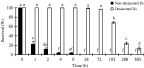
Fig. 2. Effect of exposure to −20±0·3 °C for 15 days on survival of osmotically desiccated Steinernema feltiae IS-6 IJs. Values are expressed as percentages of viable nematodes in samples obtained from each treatment. At least 200 individuals were counted in each sample. Bars represent standard error of the means. Bars with same letter(s) do not differ significantly according to Scheffe's test (P<0·05). F=999·999; D.F.=21, 66; P<0·05.
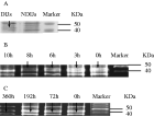
Fig. 3. SDS-PAGE (12% acrylamide gel) profile of stress-related proteins. Molecular markers are shown on the right. Each lane was loaded with 20 μg protein. (The samples were taken from osmotically desiccated IJs under extreme temperatures.) The arrow indicates the significant protein band. (A) NDIJs, non-desiccated IJs. DIJs, desiccated IJs. (B) 0 h, 3 h, 6 h, 8 h and 10 h, nematodes were exposed in glycerol concentration of 24% for 24 h, then exposed to 40 °C for 0 h, 3 h, 6 h, 8 h, and 10 h. (C) 0 h, 72 h, 192 h, and 360 h – nematodes were exposed in glycerol concentration of 24% for 24 h, then exposed to −20 °C for 0 h, 72 h, 192 h, and 360 h.
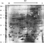
Fig. 4. 2-D gel analysis of non-desiccated and osmotically desiccated Steinernema feltiae IS-6 IJ proteins extracted with Mg/Np-40 extraction buffer. In the first dimension, isoelectric focusing, 200 μg of protein was separated on a 13-cm IPG strip with a linear gradient of 3–10. In the second dimension, 12·5% SDS-PAGE (12·5% acrylamide gel) was used, with a well for Mr (kDa) standards. Proteins were visualized by silver staining. Molecular markers are shown on the left. The circle indicated that proteins changed significantly in osmotically desiccated IJs compared with non-desiccated IJs.
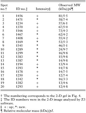
Table 1. Abundance ratio of proteins during exposure of osmotically desiccated EPN IJs compared with non-desiccated IJs
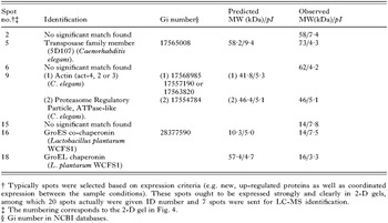
Table 2. List of proteins identified by LC-MS/MS isolated from 2-D electrophoresis gel
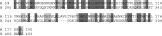
Fig. 5. Homology between predicted CDS, AAA-type ATPase (XA897) (C. elegans) (A) and proteasome regulatory particle, ATPase-like (C. elegans) (B). Identical amino acids are highlighted in the black box and similar amino acids are in the grey box. Identities=43/124 (34%), positives=65/124 (51%), gaps=7/124 (5%) (http://www.ncbi.nlm.nih.gov/blast/).
 You have
Access
- 19
- Cited by