Introduction
Magnetic resonance imaging (MRI) is one of the most significant developments in medical imaging in the 20th century. Since its inception in the early 1970s, MRI has evolved into an indispensable modality for routine clinical diagnosis. In clinical medicine, MRI is attractive because it is completely non-invasive and yet provides images with exquisite soft tissue contrast. In its early days, MRI was used primarily for anatomic imaging; however, its role in biomedical research has expanded significantly in the past 20 years largely owing to its ability to provide physiological and functional measures. Throughout the history of MRI, its development has been related to the increase of magnet strength. In fact, the past three decades have seen an increase in the strength of magnets from 0.015 to 9.4 T. Despite early doubts about the feasibility and utility of high-field MR systems in human subjects, it is now clear that high fields have benefits for a variety of applications.
Functional MRI
Functional MRI (fMRI) is a revolutionary development of the past decade that makes it possible to use MRI to non-invasively map areas of increased neuronal activity in the human brain without the use of an exogenous contrast agent Reference Lagopoulos(1). It is generally accepted that the signal-to-noise ratio (SNR) in MR images scales linearly with the field strength. Furthermore, as a susceptibility phenomenon, blood oxygenation level dependent (BOLD) contrast is expected to increase with field strength. Another interesting aspect of high-field fMRI is the detection of an initial dip in the signal, which is believed to arise from an initial increase in deoxyhaemoglobin concentration before the haemodynamic response takes place, and this is thought to be more specific to the site of neuronal activation than the hyperaemic BOLD response. In addition to increases in the SNR and BOLD contrast with increasing magnetic field strength, the dependence on the magnetic field is also a function of the size of the vessels contributing to the BOLD signal. Large vessel contributions scale linearly with the field strength, whereas small vessel contributions scale quadratically. This improves the spatial specificity of BOLD-based fMRI.
MR spectroscopy
Besides increased SNR, in vivo MR spectroscopy benefits from high magnetic fields because of increased spectral resolution. In MRI, the magnetic field experienced by a nucleus is not only determined by the applied magnetic field but also by its chemical environment Reference Lagopoulos(2). In particular, the electron distribution of molecules may be perturbed by the external magnetic field, leading to a small but significant modification to the magnetic field at a nucleus surrounded by the electronic distribution. This causes a shift in its resonance frequency. This shift is commonly known as chemical shift and is an important signature used in MR spectroscopy for the identification of a specific nucleus. Because it is induced by the external magnetic field, it is proportional to the external field and scales up with magnetic field increments, leading to an increase in spectral separation between chemically different nuclei and hence better spectral resolution. In addition to the proton, there are other nuclei of biological interest, including 31P, 13C, 23Na and 19F. These nuclei have a much lower in vivo sensitivity because of their low biological concentration and/or low natural abundance. The increase in SNR has proven to be tremendously beneficial for the in vivo study of these nuclei.
Diffusion tensor imaging
The increase in SNR at high fields has also been important for newly emerging applications where primarily the SNR has been a limiting factor. One such application is diffusion tensor imaging (DTI), which acquires diffusion-weighted images along six or more directions in space to map the directional dependence of water diffusion Reference Saether, Das and Lagopoulos(3). DTI has generated a great deal of interest in both basic research and clinical medicine because it can be used to visualise microstructural tissue organisation in the human brain. SNR gain at high fields can significantly improve the quality of DTI.
MR angiography
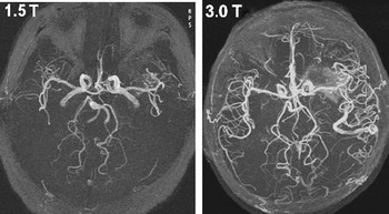
In MRI, blood vessels can be imaged with MR angiography (MRA). One well-established approach is the time-of-flight (TOF) technique, which derives the vessel contrast based on the shortening of the effective T1 of moving blood. In these types of images, vessels appear brighter than the background. High magnetic field provides an SNR advantage as well as a contrast advantage because it lengthens the T1 of the stationary tissue Reference Al-Kwifi, Emery and Wilman(4). A remarkable example of improved MRA is shown in Fig. 1, where three-dimensional TOF images obtained at 1.5 and 3T are compared. The 3T images exhibit not only a substantial increase in the SNR but also an increase in the vessel contrast, allowing enhanced visualisation of small vessels.
MR angiograms obtained at 1.5 and 3 T from a patient with an arteriovenous malformation. Both images were acquired with no contrast agent. It is evident that 3T offers better background tissue suppression and improved resolvability of smaller vessels. (Image courtesy of K. Thulborn and X.J. Zhou, University of Illinois at Chicago.)

The byte
High-field MR neuroimaging is changing the field of in vivo neuro-MRI in existing and clinically significant ways. Although there are inherent pitfalls associated with high-field imaging, the benefits that have emerged across many applications will no doubt outweigh these in time.
Researchers have enjoyed the benefits of high-field imaging for years, and now its advantages are spilling into the clinical arena and are beginning to be realised. With the current growth of the number of 3T clinical systems, clinical applications of high-field imaging will become routine, and hopefully, this will further improve both the throughput and the quality of diagnostic imaging and ultimately provide enhanced patient care.