INTRODUCTION
Since the introduction of combination antiretroviral therapy (cART), life expectancies among people living with HIV (PLWH) have increased, while rates of medical morbidity and mortality have decreased. However, HIV-associated neurocognitive disorders (HAND), especially milder forms of neurocognitive impairment (NCI), have persisted in the cART era (Heaton et al., Reference Heaton, Franklin, Ellis, McCutchan, Letendre, LeBlanc, Corkran, Duarte, Clifford, Woods, Collier, Marra, Morgello, Mindt, Taylor, Marcotte, Atkinson, Wolfson, Gelman, McArthur, Simpson, Abramson, Gamst, Fennema-Notestine, Jernigan, Wong and Grant2011). Frascati criteria, the current international research nosology for HAND, were developed to meet the need for updated, operationalized classification methods, especially as literature emerged showing that even mild NCI in HIV was associated with a range of unfavorable functional outcomes (Antinori et al., Reference Antinori, Arendt, Becker, Brew, Byrd, Cherner, Clifford, Cinque, Epstein, Goodkin, Gisslen, Grant, Heaton, Joseph, Marder, Marra, McArthur, Nunn, Price, Pulliam, Robertson, Sacktor, Valcour and Wojna2007). Using Frascati criteria, HAND prevalence ranges from 30 to 50%, depending on comorbidities and HIV disease treatment histories (Heaton et al., Reference Heaton, Clifford, Franklin, Woods, Ake, Vaida, Ellis, Letendre, Marcotte, Atkinson, Rivera-Mindt, Vigil, Taylor, Collier, Marra, Gelman, McArthur, Morgello, Simpson, McCutchan, Abramson, Gamst, Fennema-Notestine, Jernigan, Wong and Grant2010; Saloner & Cysique, Reference Saloner and Cysique2017). The majority of those with HAND are classified with mild-to-moderate impairment (>1 SD below the mean in two cognitive domains), which is further divided into asymptomatic neurocognitive impairment (ANI; meeting criteria for cognitive, but not functional decline) and mild neurocognitive disorder (MND; meeting criteria for both cognitive and functional decline; Antinori et al., Reference Antinori, Arendt, Becker, Brew, Byrd, Cherner, Clifford, Cinque, Epstein, Goodkin, Gisslen, Grant, Heaton, Joseph, Marder, Marra, McArthur, Nunn, Price, Pulliam, Robertson, Sacktor, Valcour and Wojna2007).
There remains controversy regarding Frascati criteria’s clinical applicability. Some investigators have proposed that the Frascati criteria for milder HAND are too liberal, resulting in high false positive classifications, overestimation of HAND, and undue distress for patients (Gisslén, Price, & Nilsson, Reference Gisslén, Price and Nilsson2011; Meyer, Boscardin, Kwasa, & Price, Reference Meyer, Boscardin, Kwasa and Price2013; Torti, Focà, Cesana, & Lescure, Reference Torti, Focà, Cesana and Lescure2011). Consequently, Meyer et al. (Reference Meyer, Boscardin, Kwasa and Price2013) advocate for stricter cognitive impairment cutoffs (1.5 SD below the mean) and a smaller test battery (i.e., 3–5 domains versus 7 domains). Although false positive misclassifications may result in adverse consequences, detecting HIV-associated neurological dysfunction at its earliest point may be important for early intervention. For example, detection of early CNS dysfunction may allow for cognitive neurorehabilitation interventions and mitigate risk for NCI-related adverse functional outcomes (Iudicello, Hussain, Watson, Morgan, & Heaton, Reference Iudicello, Hussain, Watson, Morgan, Heaton, Stern and Alosco2019). In addition, constraining cutoffs to 1.5 SD below the mean may result in a loss of sensitivity for detecting brain disorders (Heaton, Miller, Taylor, & Grant, Reference Heaton, Miller, Taylor and Grant2004; Heaton et al., Reference Heaton, Marcotte, Mindt, Sadek, Moore, Bentley, McCutchan, Reicks and Grant2004) and the probability that PLWH with clinically significant neurocognitive difficulties may be overlooked.
An alternate, slightly more conservative approach relative to Frascati for detecting NCI in PLWH is the global deficit score (GDS; Carey et al., Reference Carey, Woods, Gonzalez, Conover, Marcotte, Grant and Heaton2004) method. The GDS, an objective, algorithmic approach to classifying NCI, is often used in research settings and has demonstrated reliability in detecting NCI in HIV (Cysique et al., Reference Cysique, Letendre, Ake, Jin, Franklin, Gupta, Shi, Yu, Wu, Abramson, Grant and Heaton2010; Heaton et al., Reference Heaton, Grant, Butters, White, Kirson, Atkinson, McCutchan, Taylor, Kelly, Ellis, Wolfson, Velin, Marcotte, Hesselink, Jernigan, Chandler, Wallace and Abramson1995; Kabuba, Anitha Menon, Franklin, Heaton, & Hestad, Reference Kabuba, Anitha Menon, Franklin, Heaton and Hestad2017; Kanmogne et al., Reference Kanmogne, Kuate, Cysique, Fonsah, Eta, Doh, Njamnshi, Nchindap, Franklin, Ellis, McCutchan, Binam, Mbanya, Heaton and Njamnshi2010) and is related to functional- and health-related outcomes (Andrade et al., Reference Andrade, Deutsch, Celano, Duarte, Marcotte, Umlauf, Atkinson, McCutchan, Franklin, Alexander, McArthur, Marra, Grant and Collier2013; Hinkin et al., Reference Hinkin, Hardy, Mason, Castellon, Durvasula, Lam and Stefaniak2004). The GDS is a weighted average of neuropsychological test deficit scores ranging from 0 (no impairment) to 5 (severe impairment); therefore, it considers both number and severity of deficits. Blackstone et al. (Reference Blackstone, Moore, Franklin, Clifford, Collier, Marra, Gelman, McArthur, Morgello, Simpson, Ellis, Atkinson, Grant and Heaton2012) illustrated that GDS-determined NCI nearly guarantees meeting Frascati criteria for mild NCI, though the opposite is not necessarily true. Additionally, those impaired using only Frascati criteria had less neurocognitive and functional deficits than those impaired using the GDS methodology, further suggesting the GDS approach is more conservative than Frascati criteria.
In addition to neurocognitive assessment, non-invasive magnetic resonance imaging (MRI) techniques have enhanced understanding of HIV-related brain abnormalities and may have potential in diagnosing HAND, as there is no reliable biomarker correlate of HAND. Structural MRI studies report cortical and subcortical gray matter atrophy and white matter atrophy as well as increased abnormal white matter volume, and these findings have persisted into the cART era (Becker et al., Reference Becker, Maruca, Kingsley, Sanders, Alger, Barker, Goodkin, Martin, Miller, Ragin, Sacktor and Selnes2012; Kallianpur et al., Reference Kallianpur, Shikuma, Kirk, Shiramizu, Valcour, Chow, Souza, Nakamoto and Sailasuta2013; Nichols et al., Reference Nichols, Gates, Soares, Moffat, Rae, Brew and Cysique2019). With respect to cognitive functioning, greater brain atrophy correlates with widespread neurocognitive deficits (Alakkas et al., Reference Alakkas, Ellis, Watson, Umlauf, Heaton, Letendre, Collier, Marra, Clifford, Gelman, Sacktor, Morgello, Simpson, McCutchan, Kallianpur, Gianella, Marcotte, Grant and Fennema-Notestine2018; Bonnet et al., Reference Bonnet, Amieva, Marquant, Bernard, Bruyand, Dauchy, Mercié, Greib, Richert, Neau, Catheline, Dehail, Dabis, Morlat, Dartigues and Chene2013; Cohen et al., Reference Cohen, Harezlak, Schifitto, Hana, Clark, Gongvatana, Paul, Taylor, Thompson, Alger, Brown, Zhong, Campbell, Singer, Daar, McMahon, Tso, Yiannoutsos and Navia2010; Patel et al., Reference Patel, Kolson, Glosser, Matozzo, Ge, Babb, Mannon and Grossman2002; Thames et al., Reference Thames, Foley, Wright, Panos, Ettenhofer, Ramezani, Streiff, El-Saden, Goodwin, Bookheimer and Hinkin2012). Magnetic resonance spectroscopy (MRS) studies demonstrate cerebral metabolic abnormalities indicative of neuroinflammation (e.g., elevated choline and myo-inositol) and neuronal loss (e.g., reduced N-acetyl-aspartate and glutamate) across cortical and subcortical tissues (Chang et al., Reference Chang, Lee, Yiannoutsos, Ernst, Marra, Richards, Kolson, Schifitto, Jarvik, Miller, Lenkinski, Gonzalez and Navia2004; Cysique et al., Reference Cysique, Moffat, Moore, Lane, Davies, Carr, Brew and Rae2013; Ernst, Jiang, Nakama, Buchthal, & Chang, Reference Ernst, Jiang, Nakama, Buchthal and Chang2010; Harezlak et al., Reference Harezlak, Buchthal, Taylor, Schifitto, Zhong, Daar, Alger, Singer, Campbell, Yiannoutsos, Cohen and Navia2011; Paul et al., Reference Paul, Ernst, Brickman, Yiannoutsos, Tate, Cohen and Navia2008). PLWH with the greatest evidence of neuroinflammation and neuronal loss are at higher risk for HAND, with decrements in executive function, working memory, learning, recall, and complex motor skills (Ernst et al., Reference Ernst, Jiang, Nakama, Buchthal and Chang2010; Mohamed, Barker et al., Reference Mohamed, Barker, Skolasky, Selnes, Moxley, Pomper and Sacktor2010; Mohamed, Barker, Skolasky, & Sacktor, Reference Mohamed, Barker, Skolasky and Sacktor2018; Paul et al., Reference Paul, Ernst, Brickman, Yiannoutsos, Tate, Cohen and Navia2008). Given these findings, some authors have advocated for the inclusion of neuroimaging biomarkers in HAND diagnostic decision-making (Ances & Hammoud, Reference Ances and Hammoud2014; Masters & Ances, Reference Masters and Ances2014; Saloner & Cysique, Reference Saloner and Cysique2017).
Those who have criticized current HAND nosology suggest Frascati criteria over-diagnose HAND, potentially resulting in a decrease in power to detect associations between HAND and imaging markers (Meyer et al., Reference Meyer, Boscardin, Kwasa and Price2013; Torti et al., Reference Torti, Focà, Cesana and Lescure2011). However, analyses of neuroimaging data have not been reported. Therefore, the current study aims to examine structural and metabolite brain imaging markers in PLWH who are impaired via Frascati criteria but unimpaired via Meyer criteria compared to those that are concordantly unimpaired and concordantly impaired. Additionally, Meyer et al. (Reference Meyer, Boscardin, Kwasa and Price2013) recommended exploring other commonly used classifications, including the GDS; therefore, we also examined those that are impaired via GDS but unimpaired via Meyer criteria compared to those that are concordanly unimpaired or concordantly impaired. These findings may help determine if discrepantly classified PLWH exhibit neuroanatomical profiles that more closely resemble the concordantly impaired or the concordanly unimpared groups.
METHODS
Participants
The present cross-sectional study included 241 PLWH in the NIH-funded CNS HIV Anti-Retroviral Therapy Effects Research (CHARTER) study who completed comprehensive neuropsychological testing, neuromedical assessment, and high-resolution multichannel structural MRI and MRS between 2004 and 2007 (Jernigan et al., Reference Jernigan, Archibald, Fennema-Notestine, Taylor, Theilmann, Julaton, Notestine, Wolfson, Letendre, Ellis, Heaton, Gamst, Franklin, Clifford, Collier, Gelman, Marra, McArthur, McCutchan, Morgello, Simpson and Grant2011). Participants were excluded from analysis if there was evidence of severe confounding comorbid conditions as described in Antinori et al. (Reference Antinori, Arendt, Becker, Brew, Byrd, Cherner, Clifford, Cinque, Epstein, Goodkin, Gisslen, Grant, Heaton, Joseph, Marder, Marra, McArthur, Nunn, Price, Pulliam, Robertson, Sacktor, Valcour and Wojna2007) and Heaton et al. (Reference Heaton, Clifford, Franklin, Woods, Ake, Vaida, Ellis, Letendre, Marcotte, Atkinson, Rivera-Mindt, Vigil, Taylor, Collier, Marra, Gelman, McArthur, Morgello, Simpson, McCutchan, Abramson, Gamst, Fennema-Notestine, Jernigan, Wong and Grant2010), current substance use disorder, or insufficient neuropsychological data. Additionally, one participant was excluded for meeting criteria for Meyer impairment but being classified as unimpaired via Frascati criteria (see Supplemental Figure 1).
Participants were recruited and examined at one of five sites: Johns Hopkins University (Baltimore, MD, n = 38); Icahn School of Medicine at Mount Sinai (New York, NY, n = 56); University of California, San Diego (San Diego, CA, n = 68); University of Texas Medical Branch (Galveston, TX, n = 53); and University of Washington (Seattle, WA, n = 26). The procedures were approved by the Institutional Review Board at each university, and all participants provided written informed consent.
Neuropsychological Assessment
Participants completed comprehensive neuropsychological testing, which assesses seven cognitive domains commonly affected by HIV (verbal fluency, working memory, speed of information processing, executive functioning, learning, recall, and motor). Specific tests are described in detail in Heaton et al., Reference Heaton, Clifford, Franklin, Woods, Ake, Vaida, Ellis, Letendre, Marcotte, Atkinson, Rivera-Mindt, Vigil, Taylor, Collier, Marra, Gelman, McArthur, Morgello, Simpson, McCutchan, Abramson, Gamst, Fennema-Notestine, Jernigan, Wong and Grant2010. Raw test scores were converted to T-scores corrected for the effects of age, education, sex, and race (Heaton et al., Reference Heaton, Marcotte, Mindt, Sadek, Moore, Bentley, McCutchan, Reicks and Grant2004; Heaton, Taylor, & Manly, Reference Heaton, Taylor and Manly2003; Norman et al., Reference Norman, Moore, Taylor, Franklin, Cysique, Ake, Lazarretto, Vaida and Heaton2011).
Clinical ratings (Frascati criteria)
As referenced in the Antinori et al. (Reference Antinori, Arendt, Becker, Brew, Byrd, Cherner, Clifford, Cinque, Epstein, Goodkin, Gisslen, Grant, Heaton, Joseph, Marder, Marra, McArthur, Nunn, Price, Pulliam, Robertson, Sacktor, Valcour and Wojna2007) presentation of Frascati criteria, clinical ratings (CR) are described in detail by Woods et al. (Reference Woods, Rippeth, Frol, Levy, Ryan, Soukup, Hinkin, Lazzaretto, Cherner, Marcotte, Gelman, Morgello, Singer, Grant and Heaton2004). Demographically corrected T-scores were not averaged per domain to obtain a domain T-score, which would allow a good score on one test to obscure or offset a very poor score on another test. Instead, more weight is given to the most impaired scores per domain: if there are two domain scores at that level, this defines the level of the domain, whereas if there is only one score at that level the domain rating is one rating score less (Woods et al., Reference Woods, Rippeth, Frol, Levy, Ryan, Soukup, Hinkin, Lazzaretto, Cherner, Marcotte, Gelman, Morgello, Singer, Grant and Heaton2004). Tests and domains were then assigned a CR, using a 9-point scale ranging from 1 (i.e., above average functioning) to 9 (i.e., severe impairment). Performance in a domain is considered to be impaired if the domain CR is greater than or equal to 5 (CR of 5 is defined as “mild impairment” and is equivalent to a T-score of 35–39). A participant was classified with HAND if at least two domains of the seven domains assessed were in the impaired range (i.e., CR≥5). However, if learning and memory were the only impaired domains, the participant had to demonstrate impaired recall retention (i.e., forgetting or impaired retrieval) as well as impaired learning, in order to avoid double-penalization for learning deficits. The CR methodology operationalizes and is consistent with current Frascati criteria for classifying HAND (Antinori et al., Reference Antinori, Arendt, Becker, Brew, Byrd, Cherner, Clifford, Cinque, Epstein, Goodkin, Gisslen, Grant, Heaton, Joseph, Marder, Marra, McArthur, Nunn, Price, Pulliam, Robertson, Sacktor, Valcour and Wojna2007).
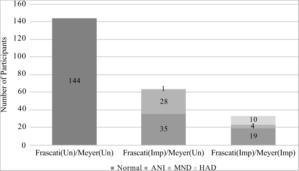
The 13-item Lawton-Brody ADL questionnaire (Lawton & Brody, Reference Lawton and Brody1969) was used to assess functional impairment (vs. best level of prior functioning) in performing instrumental activities of daily living. The 33-item Patient’s Assessment of Own Functioning (PAOFI; Chelune, Heaton, & Lehman, Reference Chelune, Heaton, Lehman, Goldstein and Tarter1986) was used to assess current cognitive difficulties in everyday life. Functional decline was defined as at least two types of evidence of decreased everyday functioning. Those who were classified as impaired using Frascati CR guidelines were classified with ANI, MND, or HIV-associated dementia (HAD). In line with Frascati criteria described in Antinori et al. (Reference Antinori, Arendt, Becker, Brew, Byrd, Cherner, Clifford, Cinque, Epstein, Goodkin, Gisslen, Grant, Heaton, Joseph, Marder, Marra, McArthur, Nunn, Price, Pulliam, Robertson, Sacktor, Valcour and Wojna2007), those demonstrating mild NCI (i.e., at least 1 SD below the mean in at least two cognitive domains) but not endorsing functional impairment were classified as ANI, those demonstrating mild NCI and also endorsing decline in functional abilities were classified as MND, and those with major NCI (i.e., at least 2 SD below the mean in at least two cognitive domains) and more severe functional decline were classified as HAD (Figure 1).
Distribution of subjects per group using Frascati Clinical Rating versus Meyer Criteria. Note. Frascati(Un)/Meyer(Un) = Unimpaired by both criteria; Frascati(Imp)/Meyer(Un) = Impaired by Frascati criteria and Unimpaired by Meyer; Frascati(Imp)/Meyer(Imp) = Impaired by both criteria. HAND subtypes defined using Frascati criteria: ANI = asymptomatic neurocognitive impairment; MND = mild neurocognitive disorder; HAD = HIV-associated dementia.

Meyer criteria
Meyer et al. (Reference Meyer, Boscardin, Kwasa and Price2013) recommend analyses be limited to three to five cognitive domains, the threshold for impairment be lowered to less than or equal to −1.5 standard deviations below the demographically corrected mean, and the average domain score be used to define an “abnormal” domain. Meyer et al. (Reference Meyer, Boscardin, Kwasa and Price2013) do not recommend which domains be removed from consideration; we selected verbal fluency and speed of information processing as these domains show the lowest prevalence of impairment in HIV (Heaton et al., Reference Heaton, Franklin, Ellis, McCutchan, Letendre, LeBlanc, Corkran, Duarte, Clifford, Woods, Collier, Marra, Morgello, Mindt, Taylor, Marcotte, Atkinson, Wolfson, Gelman, McArthur, Simpson, Abramson, Gamst, Fennema-Notestine, Jernigan, Wong and Grant2011), leaving five cognitive domains (learning, delayed recall, working memory, complex motor, and executive function). Individual test T-scores were averaged in each domain to create domain T-scores, and a participant was classified as impaired via Meyer criteria if at least two domains had an average T-score of less than or equal to 35 (equivalent to Meyer’s recommendations of ≤ −1.5 standard deviations below the average domain score). Participants were categorized into three groups for Frascati CR versus Meyer analyses: (1) a dually unimpaired group [Frascati(Un)/Meyer(Un)]: unimpaired by Frascati CR and Meyer criteria; (2) Frascati CR-only impairment [Frascati(Imp)/Meyer(Un)]: impaired by Frascati CR but unimpaired by Meyer criteria; and (3) a dually impaired group [Frascati(Imp)/Meyer(Imp)]: impaired by Frascati CR and Meyer criteria.
Global Deficit Score (GDS)
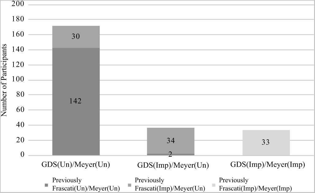
To generate the GDS, individual test T-scores were first converted to deficit scores, which range from 0 (T-score ≥ 40; unimpaired) to 5 (T < 20; severely impaired). Deficit scores were averaged across all tests in the seven domains assessed to obtain a GDS and global impairment was defined by a GDS ≥ 0.5. To compare patterns of impairment by domain, tests in each domain were averaged to obtain a domain deficit score (DDS); an impaired domain was defined as a DDS > 0.5. These cutoffs are commonly utilized and have previously been shown to yield the best sensitivity to specificity ratio (Carey et al., Reference Carey, Woods, Gonzalez, Conover, Marcotte, Grant and Heaton2004; Heaton et al., Reference Heaton, Marcotte, Mindt, Sadek, Moore, Bentley, McCutchan, Reicks and Grant2004). Participants were categorized into three groups for GDS versus Meyer analyses: (1) a dually unimpaired group [GDS(Un)/Meyer(Un)]: unimpaired by GDS and Meyer criteria; (2) GDS-only impairment [GDS(Imp)/Meyer(Un)]: impaired by GDS but unimpaired by Meyer criteria; and (3) a dually impaired group [GDS(Imp)/Meyer(Imp)]: impaired using both Meyer and GDS criteria (Figure 2).
Distribution of subjects per group using GDS versus Meyer Criteria. Note. Depicts number of participants that were previously Frascati(Imp)/Meyer(Un) via Frascati versus Meyer Criteria and are GDS(Un)/Meyer(Un) via GDS versus Meyer Criteria (n = 30) and those that were Frascati(Un)/Meyer(Un) via Frascati versus Meyer Criteria and GDS(Un)/Meyer(Un) via GDS versus Meyer criteria (n = 2).

Cognitive reserve variables
Participants also were administered the Reading Subtest from the Wide Range Achievement Test-3 (WRAT; Wilkinson, Reference Wilkinson1993). The WRAT is a reliable estimator of premorbid IQ, despite cognitive decline associated with HIV-infection (Casaletto et al., Reference Casaletto, Cattie, Franklin, Moore, Woods, Grant, Heaton and Group2014; Olsen et al., Reference Olsen, Fellows, Rivera-Mindt, Morgello, Byrd and Bank2015). Additionally, the Hollingshead Index of Social Status (Hollingshead, Reference Hollingshead1975) was calculated using a weighted average of educational and occupational attainment.
Neuromedical and Psychiatric Assessment
Medical history and HIV disease history were gathered via standardized neuromedical evaluation. Common medical comorbidities listed in Tables 1 and 2 were determined by self-report or taking medication for the condition. Psychiatric comorbidities were assessed using the Composite International Diagnostic Interview (World Health Organization, 1997), which is a computer-based, fully structured interview to assess lifetime and current affective and substance use disorders in line with the Diagnostic and Statistical Manual of Mental Disorders-IV (DSM-IV). A senior clinician (R.K.H.) reviewed all medical, educational, and psychiatric histories to classify each participant into comorbidity subgroups as defined by Antinori et al. (Reference Antinori, Arendt, Becker, Brew, Byrd, Cherner, Clifford, Cinque, Epstein, Goodkin, Gisslen, Grant, Heaton, Joseph, Marder, Marra, McArthur, Nunn, Price, Pulliam, Robertson, Sacktor, Valcour and Wojna2007) and Heaton et al. (Reference Heaton, Clifford, Franklin, Woods, Ake, Vaida, Ellis, Letendre, Marcotte, Atkinson, Rivera-Mindt, Vigil, Taylor, Collier, Marra, Gelman, McArthur, Morgello, Simpson, McCutchan, Abramson, Gamst, Fennema-Notestine, Jernigan, Wong and Grant2010). Participants with severe comorbidities were classified as “confounded” and were excluded from this study. Participants with mild-to-moderate comorbidities were classified as having “contributing” comorbidities and were included in this study. These mild-to-moderate comorbidities could have contributed to NCI, but the timing or severity of the NCI suggests an HIV component. The remainder had no more than minimal comorbidities that were considered “incidental” to a HAND diagnosis.
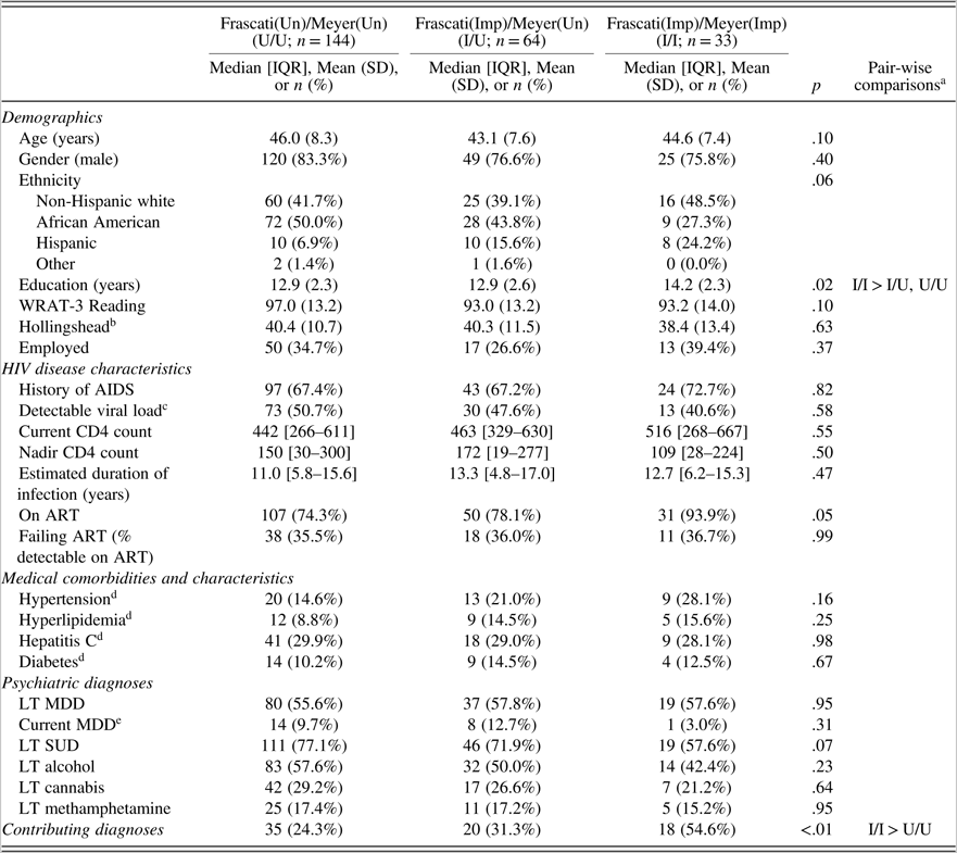
Participant characteristics by clinical rating (Frascati) versus Meyer impairment grouping

Note. Frascati(Un)/Meyer(Un) = Unimpaired by both criteria; Frascati(Imp)/Meyer(Un) = Impaired by Frascati CR criteria and Unimpaired by Meyer; Frascati(Imp)/Meyer(Imp) = Impaired by both criteria. WRAT = Wide Range Achievement Test, third edition; ART = antiretroviral therapy; BMI = body mass index; LT = lifetime; MDD = major depressive disorder; SUD = substance use disorder.
a Pairwise comparisons were examined using Tukey’s H.S.D. (α = 0.05) for continuous outcomes and Bonferroni-adjustments (α = 0.05/3 = 0.0167) for dichotomous outcomes.
b n = 236.
c n = 239.
d n = 231.
e n = 240.
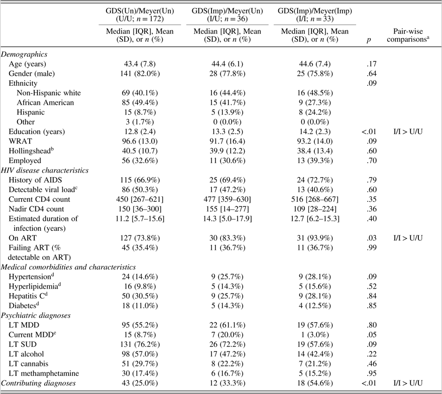
Participant characteristics by GDS versus Meyer grouping

Note. GDS(Un)/Meyer(Un) = Unimpaired by both Meyer and GDS criteria; GDS(Imp)/Meyer(Un) = Impaired by GDS criteria and Unimpaired by Meyer; GDS(Imp)/Meyer(Imp) = impaired by both criteria. WRAT = Wide Range Achievement Test, third edition; ART = antiretroviral therapy; BMI = body mass index; LT = lifetime; MDD = major depressive disorder; SUD = substance use disorder.
a Pairwise comparisons were examined using Tukey’s H.S.D. (α = 0.05) for continuous outcomes and Bonferroni-adjustments (α = 0.05/3 = 0.0167) for dichotomous outcomes.
b n = 236.
c n = 239.
d n = 231.
e n = 240.
Nadir CD4+ T-cell count was estimated via a combination of self-report and medical records. Current CD4+ T-cell count was measured with flow cytometry, and HIV RNA level was measured by ultra-sensitive PCR (Amplicor, Roche Diagnostic System, Indianapolis IN; lower limit of detection <50 copies/ml) in a CLIA-certified clinical laboratory.
Neuroimaging Assessments
MRI data were acquired on six General Electric 1.5-Tesla scanners across five sites, annually reviewed for quality; because scanner differences (e.g., hardware upgrades) can influence neuroimaging measurements, we included a “scanner” variable in statistical analyses to account for these effects between sites (Fennema-Notestine et al., Reference Fennema-Notestine, Gamst, Quinn, Pacheco, Jernigan, Thal, Buckner, Killiany, Blacker, Dale, Fischl, Dickerson and Gollub2007) as in our prior work (Fennema-Notestine et al., Reference Fennema-Notestine, Ellis, Archibald, Jernigan, Letendre, Notestine, Taylor, Theilmann, Julaton, Croteau, Wolfson, Heaton, Gamst, Franklin, Clifford, Collier, Gelman, Marra, McArthur, McCutchan, Morgello, Simpson and Grant2013; Jernigan et al., Reference Jernigan, Archibald, Fennema-Notestine, Taylor, Theilmann, Julaton, Notestine, Wolfson, Letendre, Ellis, Heaton, Gamst, Franklin, Clifford, Collier, Gelman, Marra, McArthur, McCutchan, Morgello, Simpson and Grant2011). Four series were acquired for structural morphometric analysis: coronal two-dimensional T2- and proton-density (PD)-weighted fast spin echo sequences (section thickness = 2.0 mm), and three-dimensional sagittal T1- and PD-weighted spoiled gradient recalled acquisitions (section thickness = 1.3 mm; as in Fennema-Notestine et al., Reference Fennema-Notestine, Ellis, Archibald, Jernigan, Letendre, Notestine, Taylor, Theilmann, Julaton, Croteau, Wolfson, Heaton, Gamst, Franklin, Clifford, Collier, Gelman, Marra, McArthur, McCutchan, Morgello, Simpson and Grant2013; Jernigan et al., Reference Jernigan, Archibald, Fennema-Notestine, Taylor, Theilmann, Julaton, Notestine, Wolfson, Letendre, Ellis, Heaton, Gamst, Franklin, Clifford, Collier, Gelman, Marra, McArthur, McCutchan, Morgello, Simpson and Grant2011). MRS was performed using a standardized point-resolved spectroscopy protocol (echo time = 35 ms, repetition time = 3000 ms; as in Anderson et al., Reference Anderson, Fennema-Notestine, Umlauf, Taylor, Clifford, Marra, Collier, Gelman, McArthur, McCutchan, Simpson, Morgello, Grant and Letendre2015).
Multi-channel structural MRI
As described previously (Fennema-Notestine et al., Reference Fennema-Notestine, Ellis, Archibald, Jernigan, Letendre, Notestine, Taylor, Theilmann, Julaton, Croteau, Wolfson, Heaton, Gamst, Franklin, Clifford, Collier, Gelman, Marra, McArthur, McCutchan, Morgello, Simpson and Grant2013; Jernigan et al., Reference Jernigan, Archibald, Fennema-Notestine, Taylor, Theilmann, Julaton, Notestine, Wolfson, Letendre, Ellis, Heaton, Gamst, Franklin, Clifford, Collier, Gelman, Marra, McArthur, McCutchan, Morgello, Simpson and Grant2011), we used the multi-channel structural images in a semi-automated workflow to measure cortical and subcortical gray matter; total cerebral and abnormal (e.g., hyperintense regions on T2-weighted images) white matter; and ventricular and cerebral sulcal CSF, as well as supra-tentorial cranial vault volume to account for individual differences in head size. The workflow includes inspection for motion and other artifacts, re-slicing to a standard space, intrasubject mutual information registration, bias-correction with nonparametric non-uniformity normalization, removal of non-brain tissue, three-tissue segmentation (gray matter, white matter, and CSF), abnormal white matter designation, and anatomical labeling performed by trained anatomists. This approach includes the identification of cerebral white matter regions with abnormal MRI signal characteristics that are segmented as gray matter but anatomically located within the white matter.
Single-voxel MRS
As described previously (Anderson et al., Reference Anderson, Fennema-Notestine, Umlauf, Taylor, Clifford, Marra, Collier, Gelman, McArthur, McCutchan, Simpson, Morgello, Grant and Letendre2015), three voxels were acquired: frontal gray matter (20 × 20 × 20 mm and 64 acquisitions), frontal white matter (20 × 20 × 20 mm and 64 acquisitions), and basal ganglia (20 × 20 × 15mm and 96 acquisitions). MRS concentrations of N-acetyl-aspartate, choline, myo-inositol, and creatine were quantified using LCModel with water suppression (Provencher, Reference Provencher2001). Water suppression allows for examination of absolute metabolite levels; although ratios to creatine have been commonly reported in the past for standardization across sites and studies, that approach is limited by evidence that HIV infection independently affects creatine levels directly, confounding the interpretation of ratio values and existing findings (as in Anderson et al., Reference Anderson, Fennema-Notestine, Umlauf, Taylor, Clifford, Marra, Collier, Gelman, McArthur, McCutchan, Simpson, Morgello, Grant and Letendre2015; Jansen, Backes, Nicolay, & Kooi, Reference Jansen, Backes, Nicolay and Kooi2006). Only metabolite estimates for adequate spectra (standard deviation < 21) were used; therefore, sample size varied by MRS metabolite. Structural segmentation was used to estimate the proportion of relevant tissue volume within each MRS voxel (e.g., amount of gray matter in frontal gray matter voxel) to control for variability in individual sampling.
Statistical Analyses
First, ANOVAs or Kruskal-Wallis tests for continuous variables and Chi-square tests for categorical variables were used in order to compare both Frascati CR versus Meyer and GDS versus Meyer group differences on demographics, HIV disease characteristics, medical comorbidities, and psychiatric diagnoses. To control for multiple comparisons, Tukey’s Honest Significant Difference test was applied to ANOVA tests, and Bonferroni correction was applied for Kruskal-Wallis and Chi-square tests.
Multivariable linear regression analyses were performed to examine the associations between group and log-transformed MRI structural volumes and MRS metabolites. Both age and scanner were included as covariates in every model. Additionally, supratentorial cranial vault volume was included in models predicting MRI structural volume analyses to control for differences in head size, and proportion of relevant tissue in each voxel was included in MRS analyses. Standardized beta coefficients are reported in text and tables. For significant findings, an effect size that is analogous to Cohen’s d was estimated by dividing the regression coefficient by the residual standard deviation. These analyses were run twice: once to compare groups using Frascati CR and Meyer criteria, and again to compare GDS and Meyer criteria. In all multivariable linear regression analyses, the impaired/unimpaired group was specified as the reference group to compare differences between the concordant impaired and concordant unimpaired groups. JMP version 13.0.0 was used for all analyses.
RESULTS
Sample Characteristics
Most participants were non-Hispanic white men with an average age of 43.9 years and 13.1 years of education. About 78% of participants were being prescribed antiretroviral therapy (ART), and 51% were virally suppressed (i.e., <50 copies/ml plasma).
Frascati CR versus Meyer criteria
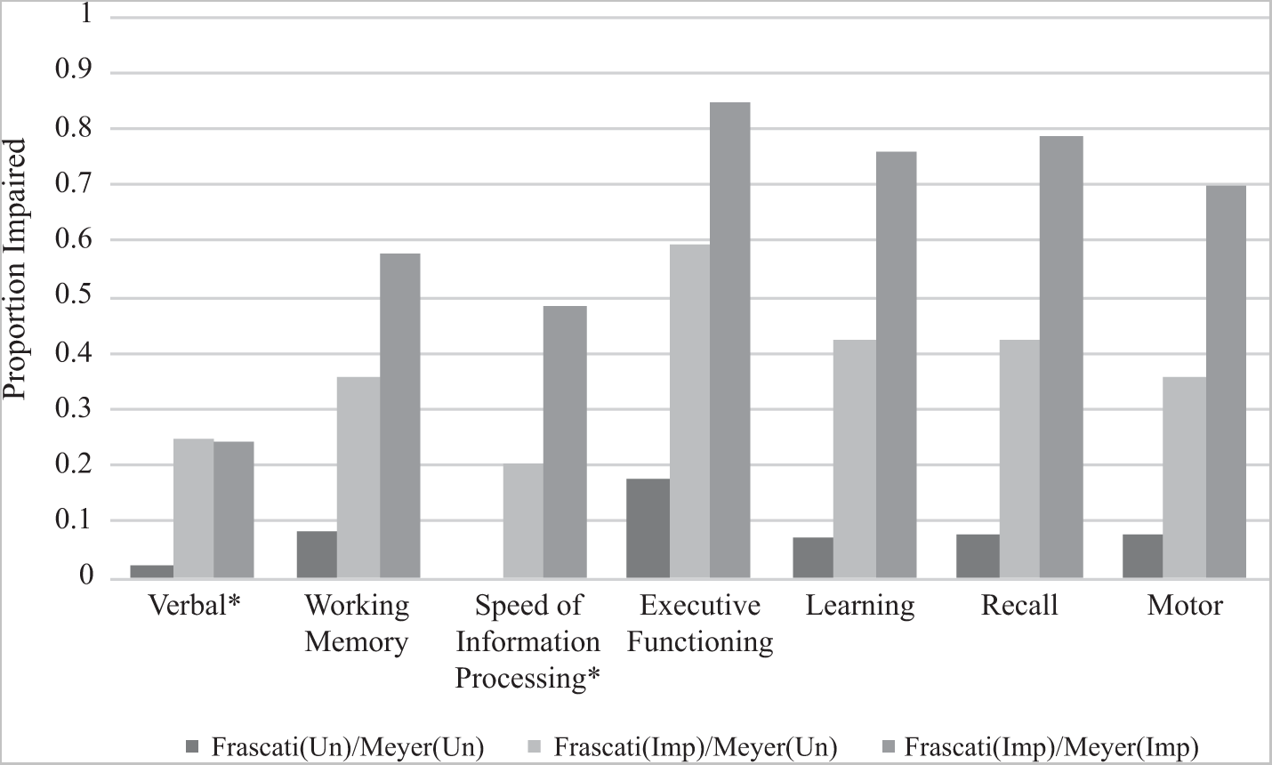
Participants’ characteristics by Frascati CR versus Meyer Impairment grouping are displayed in Table 1. The groups had similar demographics, HIV disease characteristics, medical comorbidities, and psychiatric diagnoses. However, after correcting for multiple comparisons, the Frascati(Imp)/Meyer(Imp) group had higher average education than both the Frascati(Un)/Meyer(Un) and Frascati(Imp)/Meyer(Un) groups, and the Frascati(Imp)/Meyer(Imp) group had a greater proportion classified with contributing comorbidities than the Frascati(Un)/Meyer(Un) group. Of note, the proportion of those demonstrating evidence of functional impairment and classified as MND or HAD (Figure 1) was similar in the Frascati(Imp)/Meyer(Un) group (45.3%) and Frascati(Imp)/Meyer(Imp) group (42.4%); put another way, 29.9% of the functionally impaired participants in the Frascati(Imp) group were classified as normal by the Meyer criteria. Proportion impaired, as defined by a CR of ≥5, in each cognitive domain by group is displayed in Figure 3. A stair-step pattern (Frascati(Un)/Meyer(Un)< Frascati(Imp)/Meyer(Un) < Frascati(Imp)/Meyer(Imp) was observed for all domains except for verbal fluency in which the Frascati(Imp)/Meyer(Un) and Frascati(Imp)/Meyer(Imp) groups had similar rates of impairment.
Neuropsychological impairment rates by clinical rating (CR; Frascati) versus Meyer group. Note. Frascati(Un)/Meyer(Un) = Unimpaired by both criteria; Frascati(Imp)/Meyer(Un) = Impaired by Frascati CR criteria and Unimpaired by Meyer; Frascati(Imp)/Meyer(Imp) = Impaired by both criteria. Impairment defined as a CR ≥ 5. *Domain not included in Meyer criteria

GDS versus Meyer criteria
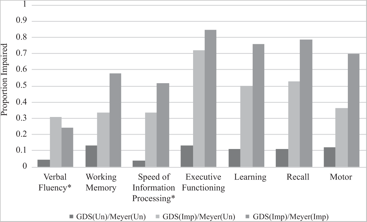
Participants’ characteristics by GDS versus Meyer Impairment grouping are displayed in Table 2. The groups were similar on most characteristics; however, the GDS(Imp)/Meyer(Imp) group had more education, a greater proportion prescribed antiretroviral therapy, and a greater proportion of those classified as having contributing comorbidities than the GDS(Un)/Meyer(Un) group. Thirty of 64 participants (46.9%) who were categorized as Frascati(Imp)/Meyer(Un) using Frascati CR criteria were classified as GDS(Un)/Meyer(Un) using GDS methodology, and 2 of 144 (1.4%) participants who were previously classified as Frascati(Un)/Meyer(Un) using Frascati CR criteria were classified as GDS(Imp)/Meyer(Un) using GDS methodology (Figure 2). Proportion impaired, as defined by DDS > 0.5, in each cognitive domain by group is displayed in Figure 4. A similar stair-step pattern of proportion impaired GDS(Un)/Meyer(Un) < GDS(Imp)/Meyer(Un) < GDS(Imp)/Meyer(Imp) was observed for all cognitive domains except for verbal fluency.
Neuropsychological impairment rates by GDS versus Meyer group. GDS(Un)/Meyer(Un) = Unimpaired by both Meyer and GDS criteria; GDS(Imp)/Meyer(Un) = Impaired by GDS criteria and Unimpaired by Meyer; GDS(Imp)/Meyer(Imp) = impaired by both criteria. Note. Impairment defined as a domain deficit score (DDS) > 0.5. *Domain not included in Meyer criteria.

Frascati CR versus Meyer Criteria Imaging Measures
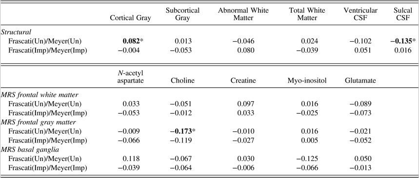
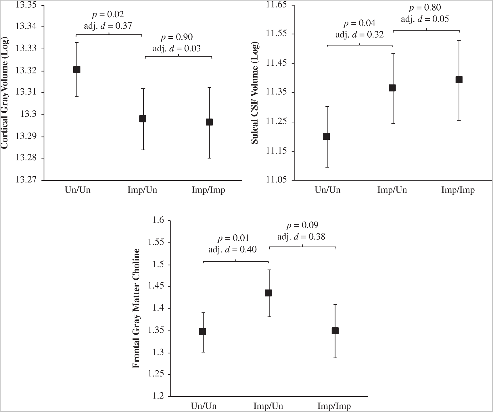
Standardized beta coefficients (β) from the linear regression models predicting structural MRI and MRS variables that compare Frascati CR versus Meyer Frascati(Un)/Meyer(Un) and Frascati(Imp)/Meyer(Imp) groups to the Frascati(Un)/Meyer(Imp) group are displayed in Table 3, and predicted means and effect sizes from the significant finding are displayed in Figure 5.
Standardized beta coefficients for structural MRI and MRS measures comparing Frascati clinical ratings versus Meyer criteria

Note. Frascati(Un)/Meyer(Un) = Unimpaired by both criteria; Frascati(Imp)/Meyer(Un) = Impaired by Frascati CR criteria and Unimpaired by Meyer; Frascati(Imp)/Meyer(Imp) = Impaired by both criteria. CSF = cerebrospinal fluid.
All analyses were in comparison to the Frascati(Imp)/Meyer(Un) group derived from Frascati CR versus Meyer criteria. Predicted means for significant relationships are presented in Figure 5.
* p < 0.05.
Predicted means with error bars denoting standard error for cortical gray matter volume (log), sulcal CSF volume (log), and frontal gray matter choline for the Frascati CR versus Meyer groups. Note. Un/Un = Unimpaired by both criteria; Imp/Un = Impaired by Frascati CR criteria and Unimpaired by Meyer; Imp/Imp = Impaired by both criteria; adj. d = adjusted Cohen’s d. The Frascati(Imp)/Meyer(Un) group had less cortical gray matter, greater sulcal CSF volume, and greater FGM choline than the Frascati(Un)/Meyer(Un) group. The Frascati(Imp)/Meyer(Un) group did not significantly differ from the Frascati(Imp)/Meyer(Imp) group on these measures.

Structural MRI
After adjusting for scanner, age, and cerebral vault volume, the Frascati(Imp)/Meyer(Un) group had less cortical gray matter volume (β = 0.08, p = .02) and more sulcal CSF volume (β = −0.14, p = .04) than the Frascati(Un)/Meyer(Un) group. The Frascati(Imp)/Meyer(Un) group did not significantly differ from the Frascati(Imp)/Meyer(Imp) on any MRI structural variables.
MRS
After accounting for relevant covariates, choline in the frontal gray matter was significantly greater in the Frascati(Imp)/Meyer(Un) group compared to the Frascati(Un)/Meyer(Un) group (β = −0.17, p = .01). The Frascati(Imp)/Meyer(Un) group also tended to have higher choline in frontal gray matter than the Frascati(Imp)/Meyer(Imp) group, but this was not significant (β = −0.12, p = .08) nor did any other Frascati(Imp)/Meyer(Un) versus Frascati(Imp)/Meyer(Imp) MRS metabolite comparisons differ significantly.
GDS versus Meyer Criteria Imaging Measures
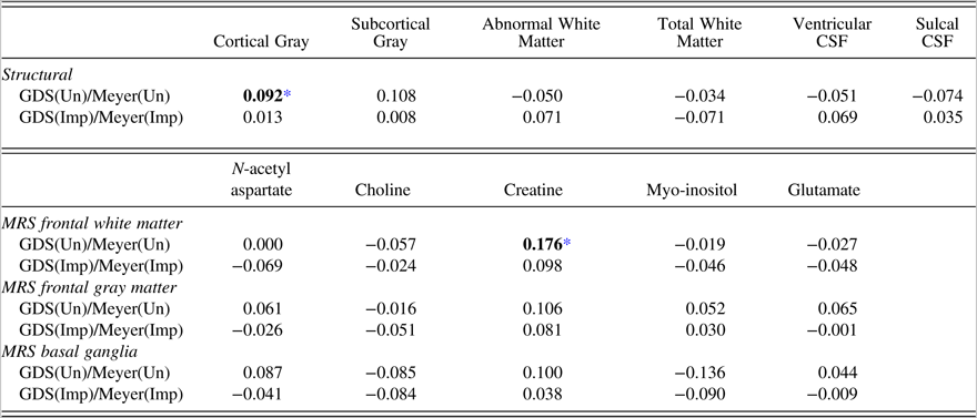
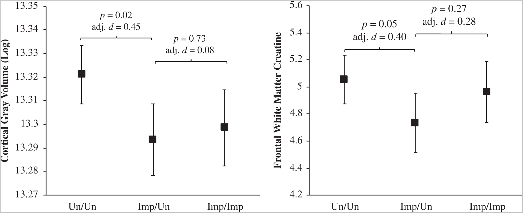
Standardized beta coefficients (β) from the linear regression models predicting structural MRI and MRS variables that compare GDS versus Meyer GDS(Un)/Meyer(Un) and GDS(Imp)/Meyer(Imp) groups to the GDS(Un)/Meyer(Imp) group are displayed in Table 4, and predicted means and effect sizes from the significant finding are displayed in Figure 6.
Standardized beta coefficients for structural MRI and MRS measures comparing GDS versus Meyer criteria

Note. GDS(Un)/Meyer(Un) = Unimpaired by both Meyer and GDS criteria; GDS(Imp)/Meyer(Un) = Impaired by GDS criteria and Unimpaired by Meyer; GDS(Imp)/Meyer(Imp) = impaired by both criteria. CSF = cerebrospinal fluid.
All analyses were in comparison to the GDS(Imp)/Meyer(Un) group derived from GDS versus Meyer criteria. Predicted means for significant relationships are presented in Figure 6.
* p < 0.05.
Predicted means with error bars denoting standard error for cortical gray matter volume (log) and frontal white matter creatine for the GDS versus Meyer groups. Note. Un/Un = Unimpaired by both Meyer and GDS criteria; Imp/Un = Impaired by GDS criteria and Unimpaired by Meyer; Imp/Imp = impaired by both criteria; adj. d = adjusted Cohen’s d. The GDS(Imp)/Meyer(Un) group had less cortical gray matter and less FGM choline than the GDS(Un)/Meyer(Un) group. The GDS(Imp)/Meyer(Un) group did not significantly differ from the GDS(Imp)/Meyer(Imp) group on these measures.

Structural MRI
When analyzing the structural MRI variables using the GDS versus Meyer criteria, the GDS(Imp)/Meyer(Un) group had significantly less cortical gray matter volume than the GDS(Un)/Meyer(Un) group (β = 0.9, p = .02), after adjusting for covariates. As seen in the Frascati CR versus Meyer criteria analyses, the GDS(Imp)/Meyer(Un) group did not statistically differ from the GDS(Imp)/Meyer(Imp) group on any MRI structural variables.
MRS
When comparing the groups derived from the GDS versus Meyer criteria, the GDS(Imp)/Meyer(Un) group had significantly less creatine in the frontal white matter than the GDS(Un)/Meyer(Un) group (β = 0.18, p = .05). The GDS(Imp)/Meyer(Un) and the GDS(Imp)/Meyer(Imp) groups did not differ significantly on any MRS metabolites.
DISCUSSION
Optimal classification of NCI within the context of HIV enables evaluation of potential mechanisms of CNS injury and prediction of functional outcomes. It is also clinically relevant, as false positive and false negative errors are both important: false positive errors that are communicated to patients could cause undue stress, and false negative diagnoses can result in missed opportunities to more closely monitor or intervene. This study utilized neuroimaging to examine different NCI criteria and help determine the optimal criteria for detecting NCI within the context of HIV with regards to brain integrity. Our findings indicate that those classified as impaired using Frascati criteria or GDS criteria, but unimpaired by the Meyer criteria, differ neuroanatomically from those with unimpaired cognition. Based on these findings, the Meyer criteria fail to capture a sizable group of PLWH with cortical atrophy. The group that was not captured by the Meyer criteria is not small; over a quarter (26%) of the sample was in the discordant Frascati(Imp)/Meyer(Un) group when classifying using Frascati CR, and 15% of the sample was in the discordant GDS(Imp)/Meyer(Un) group when classifying using GDS. While Meyer et al. (Reference Meyer, Boscardin, Kwasa and Price2013) are correct that false positives will negatively affect power to detect associations between cognitive impairment and brain integrity, it is important to also note that false negatives reduce power as well, which should be considered when examining optimal NCI criteria within the context of HIV.
Consistent across the Frascati CR and GDS criteria, the discordant group displayed similarly small cortical gray matter volumes compared to concordant impaired group, and significantly smaller volumes of cortical gray matter compared to the concordant unimpaired group. While it is difficult to compare this study, which splits HAND into two groups (i.e., Impaired/Unimpaired and Impaired/Impaired) to other imaging studies examining HAND as one group, our findings were somewhat consistent with other studies. Recent cART era studies comparing neuroanatomical profiles of PLWH to seronegative controls have identified cortical thinning, particularly in frontal and temporal structures, as a prevalent neuroimaging correlate within the context of HIV (Bonnet et al., Reference Bonnet, Amieva, Marquant, Bernard, Bruyand, Dauchy, Mercié, Greib, Richert, Neau, Catheline, Dehail, Dabis, Morlat, Dartigues and Chene2013; Cohen, Seider, & Navia, Reference Cohen, Seider and Navia2015; Thompson et al., Reference Thompson, Dutton, Hayashi, Toga, Lopez, Aizenstein and Becker2005). Additionally, these findings are consistent with a recent study by the CHARTER group in an overlapping population that found those classified as MND and HAD had smaller cortical gray matter volumes relative to those without NCI (Alakkas et al., Reference Alakkas, Ellis, Watson, Umlauf, Heaton, Letendre, Collier, Marra, Clifford, Gelman, Sacktor, Morgello, Simpson, McCutchan, Kallianpur, Gianella, Marcotte, Grant and Fennema-Notestine2018). Furthermore, reductions in frontal gray matter have been detected in neurocognitively intact PLWH in the absence of subcortical and white matter changes (Towgood et al., Reference Towgood, Pitkanen, Kulasegaram, Fradera, Kumar, Soni, Sibtain, Reed, Bradbeer, Barker and Kopelman2012). Thus, while the Meyer criteria classify individuals in the discordant Impaired/Unimpaired group as neurocognitively unimpaired, the subtler neurocognitive deficits defined by Frascati CR and GDS criteria may include early HIV-related CNS injury. Although discordant Impaired/Unimpaired individuals also displayed some evidence of subcortical gray and white matter tissue injury compared to concordant unimpaired individuals, these differences were not statistically significant and are therefore consistent with observations that PLWH in the cART era may be less vulnerable to subcortical and white matter damage than the pre-cART era (O’Connor, Zeffiro, & Zeffiro, Reference O’Connor, Zeffiro and Zeffiro2018). Given that our sample was characterized by high rates of impairment in executive functioning, learning, and recall, it is not surprising that discordant Impaired/Unimpaired and concordant unimpaired individuals were best differentiated by our estimate of cortical gray matter volume, which encompasses frontal and temporal structures that support memory and higher-order thinking.
Discordant Impaired/Unimpaired individuals also displayed evidence of neurochemical abnormalities in cortical gray and white matter tissues. For the Frascati CR versus Meyer comparisons, the Frascati(Imp)/Meyer(Un) had higher levels of choline, a marker of cellular membrane turnover that is elevated with glial proliferation and neuroinflammatory responses, in frontal gray matter than the concordant unimpaired group. Previous research has shown that PLWH display elevated levels of choline in the context of both chronic-infection (Harezlak et al., Reference Harezlak, Buchthal, Taylor, Schifitto, Zhong, Daar, Alger, Singer, Campbell, Yiannoutsos, Cohen and Navia2011; Mohamed, Lentz et al., Reference Mohamed, Barker, Skolasky, Selnes, Moxley, Pomper and Sacktor2010) and early infection (Lentz et al., Reference Lentz, Kim, Kim, Soulas, Lee, Venna, Halpern, Rosenberg, Williams and González2011; Valcour et al., Reference Valcour, Chalermchai, Sailasuta, Marovich, Lerdlum, Suttichom, Suwanwela, Jagodzinski, Michael, Spudich, van Griensven, de Souza, Kim and Ananworanich2012) compared to seronegative controls. Research has also shown higher choline is associated with worse neurocognitive functioning (Alakkas et al., Reference Alakkas, Ellis, Watson, Umlauf, Heaton, Letendre, Collier, Marra, Clifford, Gelman, Sacktor, Morgello, Simpson, McCutchan, Kallianpur, Gianella, Marcotte, Grant and Fennema-Notestine2018; Chang et al., Reference Chang, Lee, Yiannoutsos, Ernst, Marra, Richards, Kolson, Schifitto, Jarvik, Miller, Lenkinski, Gonzalez and Navia2004; Paul et al., Reference Paul, Ernst, Brickman, Yiannoutsos, Tate, Cohen and Navia2008). The neurocognitive ramifications associated with choline may in part be mediated by monocyte activation, a known driver of neural injury that has been linked to choline in PLWH (Anderson et al., Reference Anderson, Fennema-Notestine, Umlauf, Taylor, Clifford, Marra, Collier, Gelman, McArthur, McCutchan, Simpson, Morgello, Grant and Letendre2015; Kamat et al., Reference Kamat, Lyons, Misra, Uno, Morgello, Singer and Gabuzda2012; Lentz et al., Reference Lentz, Kim, Kim, Soulas, Lee, Venna, Halpern, Rosenberg, Williams and González2011). However, while myo-inositol, another marker of neuroinflammation, was greatest in the Frascati(Imp)/Meyer(Un) group, this difference was not statistically significant. Myo-inositol is more variable than choline when measured using a 1.5 Tesla scanner, so this variability may be affecting our power to detect a significant difference, or the neuroinflammatory differences between groups may be more specific to choline. For the GDS versus Meyer comparisons, the GDS(Imp)/Meyer(Un) group had lower levels of creatine, a marker of brain energy metabolism, in frontal white matter than the GDS(Un)/Meyer(Un) group. Although creatine has been employed as a reference marker in MRS studies to generate metabolite/creatine ratios (Jansen et al., Reference Jansen, Backes, Nicolay and Kooi2006), we have reported correlations between frontal white matter creatine levels and HIV RNA in plasma and CSF, nadir CD4 count, and CSF biomarkers of monocyte and macrophage activation (Anderson et al., Reference Anderson, Fennema-Notestine, Umlauf, Taylor, Clifford, Marra, Collier, Gelman, McArthur, McCutchan, Simpson, Morgello, Grant and Letendre2015). Thus, the altered frontal white matter brain energy metabolism in GDS(Imp)/Meyer(Un), compared to GDS(Un)/Meyer(Un) individuals, may reflect neural dysfunction related to HIV-associated neurotoxicity and neuroinflammation.
Analyses comparing impairment groupings’ demographics suggest that these three groups were fairly comparable with the exception of formal educational attainment. The concordant impaired group had the highest level of education for both analyses. This is surprising given the ample evidence showing that greater education is protective against cognitive impairment in PLWH (Foley et al., Reference Foley, Ettenhofer, Kim, Behdin, Castellon and Hinkin2012; Maki et al., Reference Maki, Rubin, Valcour, Martin, Crystal, Young, Weber, Manly, Richardson, Alden and Anastos2015; Tozzi et al., Reference Tozzi, Balestra, Bellagamba, Corpolongo, Salvatori, Visco-Comandini, Vlassi, Giulianelli, Galgani, Antinori and Narciso2007). Education, which is often used as a proxy variable for cognitive reserve, is a protective factor against HAND (Morgan et al., Reference Morgan, Woods, Smith, Weber, Scott and Grant2012; Stern, Silva, Chaisson, & Evans, Reference Stern, Silva, Chaisson and Evans1996). However, cognitive reserve as estimated from pre-morbid IQ (i.e., WRAT) and the Hollingshead Index did not differ among the groups. In fact, the concordant unimpaired group had the highest pre-morbid IQ but also had the lowest educational attainment. The concordant impaired group had the most years (quantity) of education, but the quality of their education and/or occupational attainment may have been lower. All neuropsychological tests were adjusted for the effects of education; therefore, the educational normative adjustments with T-scores (in the potential case of lower education quality) may be negatively impacting the concordant impaired group resulting in greater impairment rates.
The three groups were fairly similar on most HIV disease characteristics. The concordant impaired group was more likely to be on ART, but this was only significant in the GDS versus Meyer comparisons and may result from earlier policies that ART not be initiated until severe immunosuppression was detected. Of note, the proportion of those on ART that were not virally suppressed did not differ across groups. While the specific medical and psychiatric comorbidities did not differ between the three groups, the concordant impaired group had a greater proportion with mild-to-moderate (i.e., “contributing” vs. “incidental”) comorbidities compared to both the concordant unimpaired and discordant groups. However, this comparison was only significantly different with regard to the concordant unimpaired group. We chose to exclude those with severe (i.e., “confounded”) comorbidities, as confounding conditions preclude diagnosis of HAND whereas contributing conditions were included because they may exacerbate HIV-associated NCI or brain abnormalities and NC impairment, but are unlikely to fully account for them. This decision is further supported by a recent neuroimaging study by the CHARTER group, which found that those with “contributing” comorbidities were fairly comparable to those with “incidental” comorbidities (Saloner et al., Reference Saloner, Heaton, Campbell, Chen, Franklin, Ellis, Collier, Marra, Clifford, Gelman, Sacktor, Morgello, McCutchan, Letendre, Grant and Fennema-Notestine2019). However, greater comorbidity burden has been shown to increase risk for impairment (Heaton et al., Reference Heaton, Clifford, Franklin, Woods, Ake, Vaida, Ellis, Letendre, Marcotte, Atkinson, Rivera-Mindt, Vigil, Taylor, Collier, Marra, Gelman, McArthur, Morgello, Simpson, McCutchan, Abramson, Gamst, Fennema-Notestine, Jernigan, Wong and Grant2010; Patel et al., Reference Patel, Thames, Arbid, Panos, Castellon and Hinkin2013), and some groups have found comorbid conditions and sociodemographic risk factors are associated with a greater risk of NCI than HIV status (Maki et al., Reference Maki, Rubin, Valcour, Martin, Crystal, Young, Weber, Manly, Richardson, Alden and Anastos2015). Additionally, comorbidity burden may also contribute to volumetric abnormalities in PLWH (Masters & Ances, Reference Masters and Ances2014; Lake et al., Reference Lake, Popov, Post, Palella, Sacktor, Miller, Brown and Becker2017). Therefore, the concordant impaired group would be expected to have more widespread cognitive impairments and a higher prevalence of comorbidities and brain atrophy. The aim of this study was to use neuroimaging to help assess neurocognitive criteria for identifying CNS differences. This study and the Meyer criteria do not address the relative contribution of HIV versus comorbidities to the CNS difference. Therefore, future studies that include an HIV-uninfected comparison groups are needed to parse apart the effect of HIV infection compared to comorbidities with respect to imaging measures and cognition.
Several limitations of our study should be considered. First, this study was cross-sectional in nature; therefore, we cannot make inferences with regard to causality or timing of NCI in relation to structural or metabolic brain abnormalities. Longitudinal studies examining long-term neuropsychological outcomes of these groups in conjunction with imaging measures are warranted. Next, because we did not study HIV-uninfected controls, we cannot assess if the observed associations are totally or primarily due to HIV infection; therefore, these results should be interpreted within the context of HIV rather than fully due to HIV. Additionally, while the overall sample was fairly large, some groups, particularly the concordant impaired group, had a smaller sample size. Standardized betas were utilized in order to compare magnitude of the associations; however, this study may be underpowered to observe some differences between the discordant group and the concordant impaired group. Also, generalizability of the current findings to other populations (e.g., to women) should be considered. For example, this sample was predominantly men. Additionally, while the rate of viral suppression was similar to Center for Disease Control and Prevention (CDC) estimates for viral suppression in the United States, viral suppression was somewhat lower than CDC estimates for viral suppression of those engaged in medical care (80% at <200 copies/ml; Center for Disease Control and Prevention, 2016). Lastly, this study analyzed global measures of brain structure acquired on 1.5 Tesla systems. Future studies should utilize scanners with greater field strength (e.g., 3 Tesla) as well as examine specific brain structures and other imaging modalities such as functional MRI and diffusion tensor imaging that have been shown to be sensitive to cognitive functioning in HIV (Ances et al., Reference Ances, Sisti, Vaida, Liang, Leontiev, Perthen, Buxton, Benson, Smith, Little, Richman, Moore and Ellis2009; Cysique et al., Reference Cysique, Soares, Geng, Scarpetta, Moffat, Green, Brew, Henry and Rae2017; du Plessis et al., Reference du Plessis, Paul, Hoare, Stein, Taylor, Meintjes and Joska2017; Masters & Ances, Reference Masters and Ances2014).
In a large group of demographically and geographically diverse PLWH, we found that both Frascati and GDS criteria captured a group of PLWH with worse brain integrity as compared to those without NCI. While Frascati and GDS criteria may be improved by further research, these findings suggest that the cutoff for mild impairment in HIV research studies should remain at 1 SD below the mean (i.e., the cutoff used in Frascati and GDS criteria). Future neuroimaging studies that track and attempt to predict longitudinal progression of HIV-related NCI are needed in order to assess the possible clinical utility of neuroimaging in helping to identify those at risk for HIV-related NCI.
ACKNOWLEDGEMENTS
The CNS HIV Anti-Retroviral Therapy Effects Research (CHARTER) N01 MH2205 and HHSN271201000036C, to I. Grant; R01 MH107345, to R.K. Heaton and S.L. Letendre; and P30 MH062512, to R.K. Heaton.
The CNS HIV Anti-Retroviral Therapy Effects Research (CHARTER) group is affiliated with the Johns Hopkins University, Mount Sinai School of Medicine, University of California, San Diego, University of Texas, Galveston, University of Washington, Seattle, Washington University, St. Louis, and is headquartered at the University of California, San Diego and includes: Director: Igor Grant, M.D.; Co-Directors: Scott L. Letendre, M.D., Ronald J. Ellis, M.D., Ph.D., Thomas D. Marcotte, Ph.D.; Center Manager: Donald Franklin, Jr.; Neuromedical Component: Ronald J. Ellis, M.D., Ph.D. (P.I.), J. Allen McCutchan, M.D., Terry Alexander, R.N.; Laboratory, Pharmacology and Immunology Component: Scott Letendre, M.D. (P.I.), Edmund Capparelli, Pharm.D.; Neurobehavioral Component: Robert K. Heaton, Ph.D. (P.I.), J. Hampton Atkinson, M.D., Steven Paul Woods, Psy.D., Matthew Dawson; Virology Component: Davey M. Smith, M.D. (P.I.); Imaging Component: Christine Fennema-Notestine, Ph.D. (P.I.), Michael J. Taylor, Ph.D., Rebecca Theilmann, Ph.D.; Data Management Component: Anthony C. Gamst, Ph.D. (P.I.), Clint Cushman,; Statistics Component: Ian Abramson, Ph.D. (P.I.), Florin Vaida, Ph.D.; Protocol Coordinating Component: Thomas D. Marcotte, Ph.D. (P.I.), Jennifer Marquie-Beck, M.P.H.; Johns Hopkins University Site: Justin McArthur (P.I.), Vincent Rogalski; Mount Sinai School of Medicine Site: Susan Morgello, M.D. (Co-P.I.) and David Simpson, M.D. (Co-P.I.), Letty Mintz, N.P.; University of California, San Diego Site: J. Allen McCutchan, M.D. (P.I.), Will Toperoff, N.P.; University of Washington, Seattle Site: Ann Collier, M.D. (Co-P.I.) and Christina Marra, M.D. (Co-P.I.), Trudy Jones, M.N., A.R.N.P., Sheryl Storey, PA-C; University of Texas, Galveston Site: Benjamin Gelman, M.D., Ph.D. (P.I.), Eleanor Head, R.N., B.S.N.; and Washington University, St. Louis Site: David Clifford, M.D. (P.I.), Muhammad Al-Lozi, M.D., Mengesha Teshome, M.D.
The views expressed in this article are those of the authors and do not reflect the official policy or position of the United States Government.
CONFLICT OF INTEREST
The authors declare no conflict of interest.
FUNDING
This research was supported by National Institute of Health awards N01 MH22005, HHSN271201000036C, and HHSN271201000030C. Ms. Campbell and Ms. Hussain were supported by T32 DA031098 from the National Institute on Drug Abuse. Mr. Saloner was supported by T32AA013525 from the National Institute on Alcohol Abuse and Alcoholism. The study was more broadly supported by the HIV Neurobehavioral Research Center (HNRC) Award P30 MH062512.
SUPPLEMENTARY MATERIAL
To view supplementary material for this article, please visit https://doi.org/10.1017/S1355617719000985