Introduction
Bipolar disorder (BD) is characterized by episodes of hypo/mania and depression. Episodes of depression are more frequent, last longer and cause more morbidity than episodes of mania (Hirschfeld et al., Reference Hirschfeld, Lewis and Vornik2003; Judd and Akiskal, Reference Judd and Akiskal2003). Depressive mood states are characterized by reduced expectation of positive and increased expectation of negative outcomes (Andersen et al., Reference Andersen, Spielman and Bargh1992; Strunk et al., Reference Strunk, Lopez and DeRubeis2006), while hypo/manic mood states are characterized by increased expectation of positive outcomes (Johnson et al., Reference Johnson, Eisner and Carver2009). Anticipation of future positive or negative outcomes may evoke a variety of emotional and motivational states prior to task performance (Robinson et al., Reference Robinson, Yager, Cogan and Saunders2014) thus affecting future behavior. The evoked emotional and motivational states may also potentially trigger symptoms of depression and hypo/mania in affected individuals. Understanding neural underpinnings of anticipatory processing in individuals with BD v. healthy controls (HC) can enhance our understanding of emotion and cognitive deficits in BD and yield neural targets to guide interventions for individuals with BD.
Studies of anticipation-related brain functioning revealed that anticipatory processing in individuals with BD was associated with aberrant patterns of activity (Nusslock et al., Reference Nusslock, Almeida, Forbes, Versace, Frank, Labarbara, Klein and Phillips2012; Caseras et al., Reference Caseras, Lawrence, Murphy, Wise and Phillips2013; Chase et al., Reference Chase, Nusslock, Almeida, Forbes, LaBarbara and Phillips2013; Yip et al., Reference Yip, Worhunsky, Rogers and Goodwin2015; Schreiter et al., Reference Schreiter, Spengler, Willert, Mohnke, Herold, Erk, Romanczuk-Seiferth, Quinlivan, Hindi-Attar, Banzhaf, Wackerhagen, Romund, Garbusow, Stamm, Heinz, Walter and Bermpohl2016; Kollmann et al., Reference Kollmann, Scholz, Linke, Kirsch and Wessa2017) and functional connectivity (Manelis et al., Reference Manelis, Almeida, Stiffler, Lockovich, Aslam and Phillips2016; Schreiter et al., Reference Schreiter, Spengler, Willert, Mohnke, Herold, Erk, Romanczuk-Seiferth, Quinlivan, Hindi-Attar, Banzhaf, Wackerhagen, Romund, Garbusow, Stamm, Heinz, Walter and Bermpohl2016) in striatal, prefrontal (PFC) and parietal cortices. For example, loss anticipation was characterized by lower ventral striatum (VS) activation (Yip et al., Reference Yip, Worhunsky, Rogers and Goodwin2015) and sparser frontal-striatal-parietal functional connectivity in individuals with BD v. HC (Manelis et al., Reference Manelis, Almeida, Stiffler, Lockovich, Aslam and Phillips2016). Reward anticipation in BD was characterized by increased VS (Nusslock et al., Reference Nusslock, Almeida, Forbes, Versace, Frank, Labarbara, Klein and Phillips2012), left ventrolateral PFC (Chase et al., Reference Chase, Nusslock, Almeida, Forbes, LaBarbara and Phillips2013) and anterior cingulate cortical (Kollmann et al., Reference Kollmann, Scholz, Linke, Kirsch and Wessa2017) activation in some studies, but decreased VS activation (Schreiter et al., Reference Schreiter, Spengler, Willert, Mohnke, Herold, Erk, Romanczuk-Seiferth, Quinlivan, Hindi-Attar, Banzhaf, Wackerhagen, Romund, Garbusow, Stamm, Heinz, Walter and Bermpohl2016) in other studies. Greater depressive symptom severity was related to lower VS activation during reward anticipation across psychiatric diagnoses including BD (Hägele et al., Reference Hägele, Schlagenhauf, Rapp, Sterzer, Beck, Bermpohl, Stoy, Ströhle, Wittchen, Dolan and Heinz2015).
It remains unknown how anticipation-related brain activation changes over time and how BD is associated with these longitudinal changes. No longitudinal neuroimaging study focused on anticipatory processing thus far. Longitudinal neuroimaging studies of BD that focused on other types of processing showed that while activation in subcortical regions did not change over 1 year despite improvement in depression symptoms in individuals with BD (Marchand et al., Reference Marchand, Lee, Thatcher, Thatcher, Jensen and Starr2007), longitudinal changes in mood state corresponded to changes in functional connectivity between amygdala and PFC regions during performance of an emotional attentional task (Cerullo et al., Reference Cerullo, Fleck, Eliassen, Smith, DelBello, Adler and Strakowski2012). A recent study of youth recruited across BD and non-BD diagnoses revealed that longitudinal improvement in depression severity was associated with longitudinal decreases in right amygdala and the left ventrolateral PFC activity during performance of emotional working memory task (Bertocci et al., Reference Bertocci, Bebko, Dwojak, Iyengar, Ladouceur, Fournier, Versace, Perlman, Almeida, Travis, Gill, Bonar, Schirda, Diwadkar, Sunshine, Holland, Kowatch, Birmaher, Axelson, Horwitz, Frazier, Arnold, Fristad, Youngstrom, Findling and Phillips2017).
In the current study, we aim to examine longitudinal changes in brain activation during anticipation of monetary gains and losses. Given that cross-sectional studies indicated that neural activity and connectivity during reward and loss anticipation differed between individuals with BD and HC (e.g. Nusslock et al., Reference Nusslock, Almeida, Forbes, Versace, Frank, Labarbara, Klein and Phillips2012; Caseras et al., Reference Caseras, Lawrence, Murphy, Wise and Phillips2013; Chase et al., Reference Chase, Nusslock, Almeida, Forbes, LaBarbara and Phillips2013; Yip et al., Reference Yip, Worhunsky, Rogers and Goodwin2015; Manelis et al., Reference Manelis, Almeida, Stiffler, Lockovich, Aslam and Phillips2016), we hypothesized that these groups would also differ in patterns of longitudinal changes in brain activation during these conditions. The specific brain regions in which longitudinal changes might occur as well as the direction of those changes were difficult to predict as no previous study examined those questions. While it was possible that individuals with BD and HC had different longitudinal patterns of brain activation in the regions identified in the cross-sectional studies (e.g. VS, PFC), it was also possible that the longitudinal effects would be observed in different brain regions.
Materials and methods
Participants
The study was approved by the University of Pittsburgh Institutional Review Board. All participants provided written informed consent before participation in the study. Participants were recruited from local and university communities, counseling and medical centers, outpatient clinics and from other research studies through advertisements and referrals. Individuals with BD (Type-I; n = 35) and HC (n = 19) were right-handed, native English speakers matched on age, gender and intelligence quotient (IQ). All were scanned twice: at baseline and 6 months.
All diagnoses were made by a trained clinician and confirmed by a psychiatrist(s) according to Diagnostic and Statistical Manual of Mental Disorders (DSM)-IV criteria and the Structured Clinical Interview for DSM-IV, Research Version (SCID-P; First et al., Reference First, Spitzer, Gibbon and Williams1995). Current depression severity was measured using the Hamilton Rating Scale for Depression (HDRS-25; Hamilton, Reference Hamilton1960) on the day of scan. Current mania was measured using the Young Mania Rating Scale (YMRS; Young et al., Reference Young, Biggs, Ziegler and Meyer1978) on the day of scan. The 6-month changes in depression/mania were measured as the difference in the HDRS-25/YMRS scores collected at baseline and at 6 months. Lifetime depression and mania were measured using the MOODS-SR questionnaire (Dell'Osso et al., Reference Dell'Osso, Armani, Rucci, Frank, Fagiolini, Corretti, Shear, Grochocinski, Maser, Endicott and Cassano2002). HC had no personal and family history of psychiatric disorders. All participants had a YMRS score ⩽10 on the day of the scan. No individual with BD was hypo/manic at either baseline or 6-month scans.
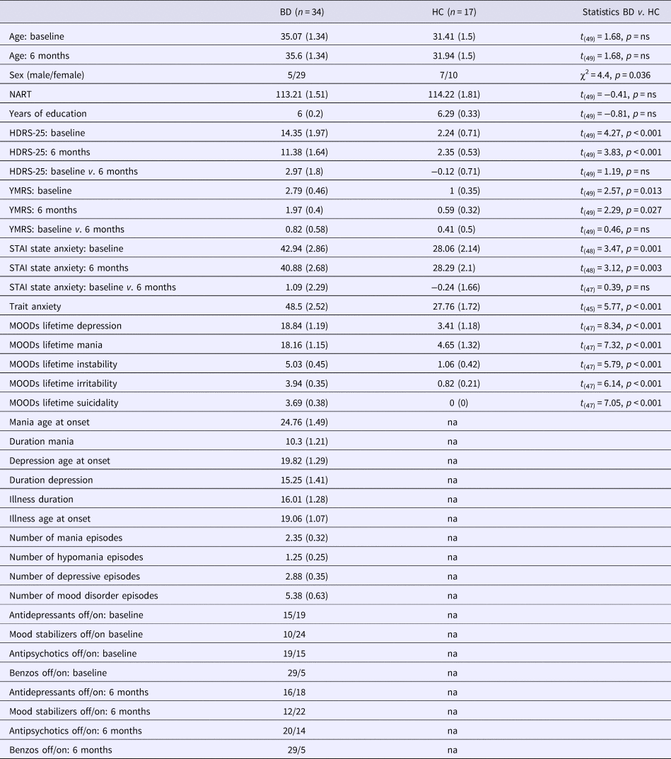
We recruited both euthymic and depressed individuals with BD to be able to examine trait, rather than state, characteristics of BD. Tables 1 and 2 report participants’ demographic and clinical characteristics at both visits. Exclusion criteria included history of head injury, neurodevelopmental disorders, systemic medical illness, cognitive impairment (score <24 in the Mini-Mental State Examination; Folstein et al., Reference Folstein, Folstein and McHugh1975), premorbid IQ < 85 measured by the National Adult Reading Test (Blair and Spreen, Reference Blair and Spreen1989), current alcohol/drug abuse, metal in the body, pregnancy, left-handedness and claustrophobia. Data from three participants were excluded from analyses due to excessive motion in the scanner (>2 mm), leaving 34 BD (16 depressed and 18 euthymic individuals) and 17 HC in the longitudinal dataset.
Demographic and clinical information for individuals with BD and HCs

The table reports means and standard deviations (in parentheses).
HDRS-25, Hamilton Rating Scale for Depression; na, not applicable; NART IQ, National Adult Reading Test intelligence quotient; YMRS, Young Mania Rating Scale. Years of Education, a level of education with range from 1 (less than 7th grade) to 8 (graduate or professional degree).
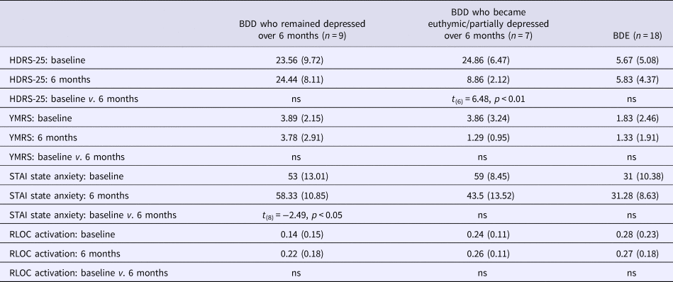
Depression, mania and anxiety scores and percent signal changes in the RLOC for individuals with bipolar disordered who were depressed at baseline and 6 months, those who improved their depression over 6 months and those who were euthymic at baseline and 6 months

The table reports means and standard deviations (in parentheses). All euthymic individuals with BD who were euthymic at baseline, remained euthymic at 6 months. BDD, individuals with bipolar disorder who were depressed at baseline; BDE, individuals with bipolar disorder who were euthymic at baseline.
Task
Participants performed a card number guessing task (Forbes et al., Reference Forbes, Hariri, Martin, Silk, Moyles, Fisher, Brown, Ryan, Birmaher, Axelson and Dahl2009) at baseline and at 6-month scans. The task examines reward/loss anticipation and processing. During the task, participants were presented with a 4-s question mark and had to guess whether the number was greater than ‘5’ by pressing a corresponding button. After that, a 6-s screen presented participants with an anticipatory cue suggesting the possibility of either winning $1 (indicated with upward arrow) or losing 50 cents (indicated with downward arrow). There were 12 trials of each type. Each trial was followed by a 1-s feedback screen showing the trial outcome. In 50% of trials, the outcome was the one predicted by the cue (money win or loss), and in other 50% of trials, it was a ‘no-change’ outcome (no money would be gained or lost). The feedback screens were followed by a 9-s inter-trial interval.
Neuroimaging data acquisition and analyses
Data acquisition
Neuroimaging data were acquired at the University of Pittsburgh using a Siemens MAGNETOM TrioTim 3T MR system. Functional data (240 volumes) were collected using a gradient-echo, echo-planar sequence (voxel size: 3.2 × 3.2 × 3.1 mm, TR = 2000 ms, TE = 28 ms, FOV = 205, FA = 90°, 39 slices), and structural data were collected using MPRAGE (voxel size: 1 × 1 × 1 mm, TR = 2200 ms, TE = 3.29 ms, FOV = 256, FA = 9, 192 slices). The two neuroimaging visits were completed 6 months apart.
Preprocessing
Images were preprocessed and analyzed using FSL5.0.9 (http://www.fmrib.ox.ac.uk/fsl). Preprocessing included nonlinear noise reduction using SUSAN (http://fsl.fmrib.ox.ac.uk/fsl/fslwiki/SUSAN); motion correction with MCFLIRT (Jenkinson et al., Reference Jenkinson, Bannister, Brady and Smith2002), non-brain removal using BET (Smith, Reference Smith2002), spatial smoothing with a Gaussian kernel of full width at half maximum = 6 mm; multiplicative mean intensity normalization of the volume at each time point; high-pass temporal filtering (Gaussian-weighted least-squares straight line fitting, with σ = 30.0 s). We used ICA-AROMA (Pruim et al., Reference Pruim, Menne, van Rooij, Llera, Buitelaa and Beckmann2015) to remove motion artifacts and, potentially, scanner-related noise. A hemodynamic response function was modeled using a Gamma function.
Neuroimaging data analyses
After preprocessing, functional magnetic resonance imaging (fMRI) data were submitted to a first-level analysis with number guessing, win anticipation, loss anticipation and feedback periods as regressors. The first-level analyses were conducted separately for baseline and 6-month follow-up scans. White matter and cerebrospinal fluid time courses were extracted from the preprocessed functional data and used as confound variables of no interest in the first-level analyses. BOLD images were registered to the high-resolution structural (MPRAGE) images using FLIRT (Jenkinson et al., Reference Jenkinson, Bannister, Brady and Smith2002), the high-resolution images were registered with the MNI152_T1_2 mm template using FNIRT (Andersson et al., Reference Andersson, Jenkinson and Smith2007), and the two resulting transformations were concatenated and applied to the original BOLD image to transform it to MNI space. In the second-level analysis, the longitudinal changes in brain activation were calculated by subtracting brain activation at 6-month scan from brain activation at baseline. This analysis was performed for each participant separately using a fixed effects analysis in FEAT.
Group-level analyses examined a Time (baseline/6-month) × Group (HC/BD) interaction separately for Win and Loss anticipation, as well as for Win v. Loss anticipation using FLAME1 (FMRIBs Local Analysis of Mixed Effects). Age, gender and IQ were used as covariates in this analysis to factor out the effects of these variables on the results. Multiple comparisons correction (p-corrected <0.05) was performed using Gaussian random field theory. Significant clusters of activation were determined by thresholding Z-statistic images in the whole brain at z > 3.09 (uncorrected voxel-wise p < 0.001) and a family-wise error corrected cluster significance threshold of p < 0.05 (Worsley, Reference Worsley, Jezzard, Matthews and Smith2001).
Percent signal changes were extracted from the brain regions showing a significant Time × Group interaction using Featquery. The longitudinal changes in brain activation were calculated as the differences in activation from baseline to 6-month follow-up scan. A stepwise regression analysis was used to understand the effect of depression and mania symptoms at baseline (HDRS-25 and YMRS scores), the changes in depression and mania symptoms over 6 months and lifetime burden of depression and mania symptoms (MOODs scores) on the longitudinal changes in brain activation in the BD group. Two individuals with BD had missing MOODs scores for lifetime depression and mania. These scores were imputed using predictive mean matching in the ‘mice’ package in R (https://cran.r-project.org/web/packages/mice/mice.pdf).
The effect of medications on brain activation was examined by looking at the relationship between the number of psychotropics used and the longitudinal changes in brain activation, and by comparing the longitudinal changes in brain activation for individuals with BD who were ON v. OFF antidepressant medications, ON v. OFF antipsychotic medications, and ON v. OFF mood stabilizers.
Results
The longitudinal changes in anticipation-related brain activation for individuals with BD v. HC
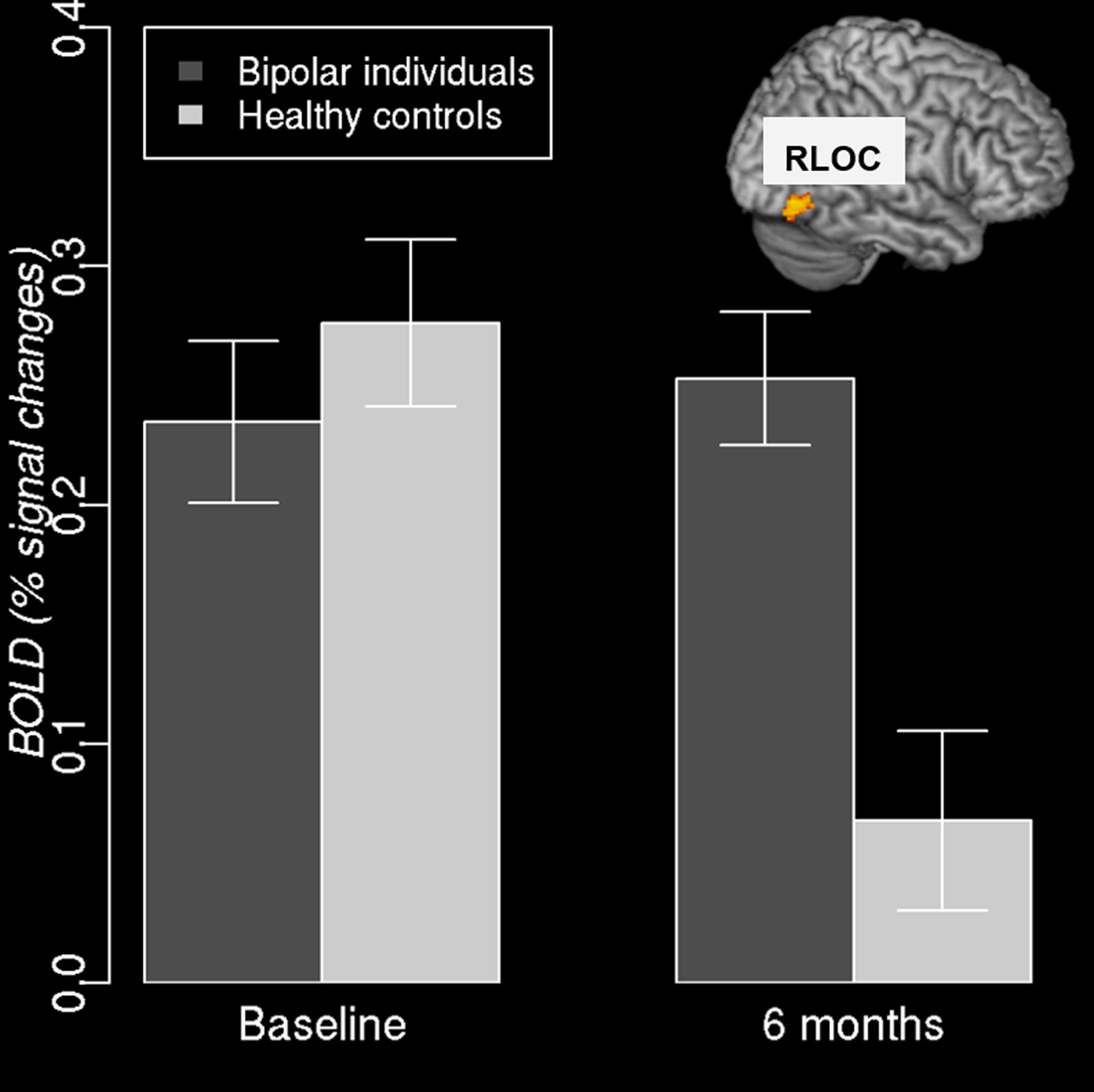
The whole brain analysis revealed that individuals with BD significantly differed from HC in the magnitude of longitudinal changes in right inferior lateral occipital cortex [RLOC, nvox = 104, zmax = 4.11, (48, −72, −8)] activation during anticipation of monetary loss. The further analysis of percent signal changes in the RLOC during loss anticipation conducted in R showed that the result was due to significant differences in RLOC activation at 6 months with HC having significantly lower activation in this region than individuals with BD (t (49) = 3.9, p < 0.001, Fig. 1). HC and individuals with BD did not differ in RLOC activation at baseline (p > 0.1). The magnitude of the longitudinal changes in RLOC activation did not depend on whether BD were depressed or euthymic at baseline or at 6-month scan (p > 0.1 for both baseline and 6-month follow-up; Table 2).
RLOC activation at baseline and 6-month scans for individuals with BD and HC.

There were no significant between-group differences in the longitudinal changes during reward anticipation, and no interaction between Group (BD/HC) and anticipation condition (Win/Loss anticipation), at least at the threshold we have chosen.
The effect of clinical symptoms on the longitudinal changes in the RLOC
The longitudinal changes in the RLOC were calculated as the differences in percent signal changes between RLOC activation at baseline and RLOC activation at the 6-month follow-up scan. The first analysis used a one-way analysis of variance to compare the differences in RLOC activation over 6 months in depressed individuals with BD (BDD), euthymic individuals with BD (BDE) and HC. It revealed a significant effect of group (F 2,48 = 8.53, p < 0.01). A multiple comparisons Tukey HSD post-hoc test showed that BDD and BDE did not differ from each other but had significantly smaller RLOC changes than HC (p < 0.01).
Based on the SCID-IV assessment, 7 of 16 depressed individuals became euthymic (n = 5) or partially depressed (n = 2), but no initially euthymic individual became depressed at 6 months. Of all individuals with BD, one depressed individuals experienced one manic and one depressive episodes, while two euthymic individuals experienced one depressive episode over the 6-month period. The comparison of BDD that remained depressed over 6 months (n = 9), BDD that improved their mood state over 6 months (n = 7), BDE (n = 18) and HC showed the main effect of group (F 3,47 = 5.8, p < 0.01) with patient groups not different from each other, but having smaller RLOC changes than HC.
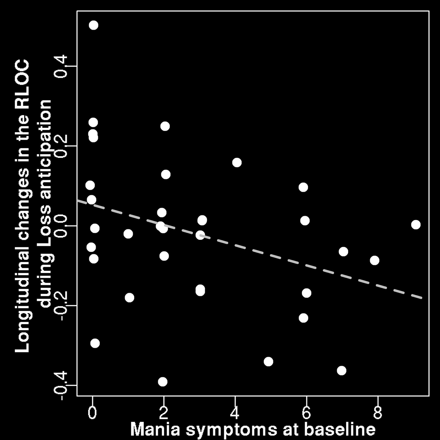
We further examined the effect of depression and hypo/mania symptoms at baseline (HDRS-25 and YMRS scores), the changes in depression and mania symptoms over 6 months and lifetime burden of depression and mania symptoms (MOODs scores) on the longitudinal changes in the RLOC during anticipation of monetary loss in individuals with BD. A stepwise regression analysis was implemented using the stepAIC algorithm in R that performed stepwise model selection based on the akaike information criterion (AIC) statistics. The best model selected by the algorithm included hypo/mania symptoms at baseline as the only predictor of the changes in RLOC activation over time. This analysis showed that lower hypo/mania symptom severity at baseline predicted greater longitudinal changes in the RLOC during Loss anticipation (F 1,32 = 4.56, p < 0.05, R 2 = 0.125, Fig. 2) in individuals with BD.
The relationship between subthreshold hypo/mania symptoms at baseline (the YMRS scores) and longitudinal changes between baseline and 6-month scans in RLOC activation during anticipation of monetary loss in individuals with BD.

The effect of psychotropic medications
The longitudinal changes in the RLOC activation during loss anticipation did not depend on whether individuals with BD were ON or OFF antidepressants, antipsychotics or mood stabilizers. There was no significant correlation between RLOC activation changes over 6 months and the number of psychotropic medications that the individuals with BD used at baseline or 6-month scan, or the changes in the number of medications over 6 months (p > 0.1). The exploratory analysis compared the RLOC activation changes during loss anticipation over 6 months in individuals with BD who remained on the same combination and dosage of medications over 6 months (two BDD and six BDE) and those who changed their medication therapy. No significant group differences were found (p > 0.1). The online Supplementary Materials include Fig. S1 that plots anticipation-related RLOC activation across baseline/6 months scans, and various combinations of mood state at baseline, mood improvement and changes in medications over 6 months. While the sample size did not allow us to conduct statistical comparisons among these groups, one can see that the patterns of the RLOC activation changes over 6 months were similar across different groups of patients and were dissimilar to the activation patterns observed in HC.
Discussion
Anticipatory processing is impaired in individuals with mood disorders (Andersen et al., Reference Andersen, Spielman and Bargh1992; Strunk et al., Reference Strunk, Lopez and DeRubeis2006; Hägele et al., Reference Hägele, Schlagenhauf, Rapp, Sterzer, Beck, Bermpohl, Stoy, Ströhle, Wittchen, Dolan and Heinz2015). Our study is the first to examine the extent to which BD is associated with abnormal longitudinal patterns of brain activation during anticipation of monetary reward and loss. The study resulted in two main findings. First, HC, but not individuals with BD, showed reduction over 6 months in RLOC activation during anticipation of monetary losses. Second, in individuals with BD, longitudinal changes in RLOC activation during anticipation of monetary losses were independent of participants’ mood state (depressed or euthymic), psychotropic medications, depression symptoms severity at scan, lifetime burden of depression and mania symptoms or changes in depression and mania symptoms between the scans. Instead, more aberrant brain activation patterns (i.e. smaller longitudinal reductions in the RLOC over 6 months) during anticipation of monetary loss were predicted by more severe subthreshold mania symptoms at baseline (no participant was in hypo/manic state at any scan though).
Previous cross-sectional studies of mood disordered individuals implicated striatal, medial temporal, ventrolateral PFC and anterior cingulate cortices in reward anticipation (Nusslock et al., Reference Nusslock, Almeida, Forbes, Versace, Frank, Labarbara, Klein and Phillips2012; Caseras et al., Reference Caseras, Lawrence, Murphy, Wise and Phillips2013; Chase et al., Reference Chase, Nusslock, Almeida, Forbes, LaBarbara and Phillips2013; Yip et al., Reference Yip, Worhunsky, Rogers and Goodwin2015; Schreiter et al., Reference Schreiter, Spengler, Willert, Mohnke, Herold, Erk, Romanczuk-Seiferth, Quinlivan, Hindi-Attar, Banzhaf, Wackerhagen, Romund, Garbusow, Stamm, Heinz, Walter and Bermpohl2016; Kollmann et al., Reference Kollmann, Scholz, Linke, Kirsch and Wessa2017). The current study highlighted the role of the occipital cortex during loss anticipation in the longitudinal context. While the occipital cortex has not been the main focus of neuroimaging studies of BD, there is evidence suggesting abnormal structure and function of this region in BD. Here, individuals with BD showed increased occipital asymmetry (i.e. occipital bending) and enlarged lateral ventricles that together could increase pressure on subcortical structures (Maller et al., Reference Maller, Anderson, Thomson, Rosenfeld, Daskalakis and Fitzgerald2015). In that study, occipital bending was four times more prevalent in BD than in HC. A recent voxel-based meta-analysis showed that individuals with BD had increased gray matter volume of the right occipital cortex (among other regions) compared with HC (Wise et al., Reference Wise, Radua, Via, Cardoner, Abe, Adams, Amico, Cheng, Cole, de Azevedo Marques Périco, Dickstein, Farrow, Frodl, Wagner, Gotlib, Gruber, Ham, Job, Kempton, Kim, Koolschijn, Malhi, Mataix-Cols, McIntosh, Nugent, O'Brien, Pezzoli, Phillips, Sachdev, Salvadore, Selvaraj, Stanfield, Thomas, van Tol, van der Wee, Veltman, Young, Fu, Cleare and Arnone2017). One fMRI study that presented participants with flashing checkerboard stimuli showed that individuals with BD had lower activation in the occipital cortex compared with HC (Shaffer et al., Reference Shaffer, Johnson, Fiedorowicz, Christensen, Wemmie and Magnotta2018). A lack of emotional content in checkerboard stimuli may explain the opposite direction of the results in our and Shaffer et al.’s studies, however. The valence hypothesis (Davidson, Reference Davidson, Davidson and Hughdahl1995) proposes that the right hemisphere is dominant for processing of negative emotions, while the left hemisphere is dominant for processing positive emotions. Our results are consistent with the valence hypothesis prediction as the aberrant longitudinal activation patterns during anticipation of negative outcomes were observed in the right hemisphere.
The lateral occipital cortex is involved in object processing and recognition (Grill-Spector et al., Reference Grill-Spector, Kushnir, Edelman, Avidan, Itzchak and Malach1999) as well as in visual imagery (Ganis et al., Reference Ganis, Thompson and Kosslyn2004). It activates more in response to emotional v. neutral visual stimuli (Kosslyn et al., Reference Kosslyn, Shin, Thompson, McNally, Rauch, Pitman and Alpert1996; Lang et al., Reference Lang, Bradley, Fitzsimmons, Cuthbert, Scott, Moulder and Nangia1998), which may be related to an increase in emotional arousal for emotional stimuli (Lang et al., Reference Lang, Bradley, Fitzsimmons, Cuthbert, Scott, Moulder and Nangia1998; Phan et al., Reference Phan, Wager, Taylor and Liberzon2002). Greater occipital activation was observed in high-arousal v. low-arousal task trials (Mather et al., Reference Mather, Mitchell, Raye, Novak, Greene and Johnson2006). Anticipatory cues can be high- or low-arousal stimuli depending on actual or perceived amount of money loss predicted by the cue. Monetary losses in the number guessing reward task used in the current study were small. Participants always ended up winning money at the end of the task. At baseline, participants were unaware of this final outcome, so anticipatory cues were high-arousal stimuli irrespective of participants’ diagnoses. At the 6-month scan, participants were already familiar with the task, so anticipatory cues may have become low-arousal stimuli given that participants learned at baseline that money loss was small compared with win. A reduction in emotion arousal elicited by anticipatory cues could lead to a decrease in the RLOC activation at 6-month.
Unlike HC who showed significant decreases in the RLOC activation during loss anticipation, individuals with BD maintained the same high level of the RLOC activation at both scans. Inability to learn the relationship between the negative anticipatory cue and actual money loss at baseline scan could be one reason for the observed activation pattern in BD. If that was the case, negative anticipatory cues remained high-arousing and elicited the same level of the RLOC activation at 6-month as at baseline. In individuals with BD, processing of negative anticipatory cues could be exacerbated by elevated subthreshold symptoms of hypo/mania at baseline that could impair participants’ ability to learn from experience thus making them unable to flexibly adopt their expectations of future outcomes according to new knowledge or evidence.
It is noteworthy that the magnitude of the longitudinal changes in the RLOC activation during loss anticipation did not depend on psychotropic medications, depressed or euthymic mood state and did not depend on lifetime or ‘at scan’ depression symptom severity in BD. On the one hand, aberrant processing of anticipatory cues could be a trait rather than state abnormality of BD. On the other hand, subthreshold hypo/mania was a strong dimensional factor affecting individuals with BD across mood states. Importantly, we showed that even subthreshold mania symptoms could affect not only current functioning but play a critical role long term by potentially disrupting learning from experience.
The current study did not reveal any longitudinal changes in brain regions engaged in reward processing (e.g. VS, PFC). One reason for the discrepancy between our findings and those of previous studies is that many previous studies focused on specific regions of interest that did not include the occipital cortex. Another reason is that the regions activated in reward processing tasks are not necessarily activated during reward or loss anticipation. Still another reason is that there have been no longitudinal studies of reward/loss anticipation, so it was unknown as to which regions would show a Group × Time interaction effect. For example, we found no differences between HC and individuals with BD in the RLOC at baseline, so if our analysis were limited to the baseline, this region would not be reported.
One limitation of the current study was the small sample size, which did not allow us to look at RLOC activation during anticipation of monetary loss across different combinations of mood state at baseline, mood state at 6 months and medication changes over 6 months. A larger sample size would also provide a wider range of changes in depression and mania symptoms over time to provide more information about the effect of mood state on loss anticipation. Future research should examine longitudinal anticipatory processing in hypomanic and manic individuals to examine longitudinal patterns of anticipation-related brain functioning along the full mania spectrum.
In summary, our findings indicate an absence of reduction in RLOC activation during loss anticipation over time in individuals with BD, which may be associated with an inability of these individuals to decrease emotional arousal during processing of negative anticipatory cues. Elevated subthreshold symptoms of hypo/mania at baseline predicted more aberrant longitudinal changes in RLOC activation during loss anticipation. Future research should examine how alterations in emotional arousal affect anticipation-related brain functioning in individuals with BD and other mood disorders. Future research should also aim to understand whether modulating occipital cortical activation using novel neuromodulation interventions during anticipation of negative outcomes would improve learning from experience and overall psychosocial functioning in BD. This research will be an important step toward improving the ability of individuals with BD to better prepare for situations with potentially negative outcomes and, hopefully, enable these individuals to improving coping strategies in these situations. Targeting anticipatory processing may be a promising direction in the treatment of BD.
Supplementary material
The supplementary material for this article can be found at https://doi.org/10.1017/S0033291718003847.
Author ORCID
Anna Manelis, 0000-0002-6605-5130; Jorge R. C. Almeida, 0000-0002-1213-1986.
Acknowledgements
This work was supported by grants from the National Institute of Health R01MH076971 to M.L.P., K01MH104348 to A.M. and the Pittsburgh Foundation to M.L.P. The authors thank participants for taking part in this research study.
Conflict of interest
The authors report no conflict of interest or financial relationships with commercial interests.