Introduction
It is over 70 years since temporal bone histology demonstrated that in Ménière's disease the fundamental abnormality is endolymphatic hydrops.Reference Hallpike and Cairns1, Reference Yamakawa2 However, the ‘official’ American Academy of Otolaryngology – Head and Neck Surgery definition of Ménière's disease3 still requires a post-mortem to prove the presence of hydrops.
For nearly 20 years, transtympanic electrocochleography has been a simple and under-utilised technique for diagnosing hydrops. It was originally developed as an evoked-response hearing threshold test based on the action potential. In 1977, Gibson et al. Reference Gibson, Moffat and Ramsden4 pointed out that (based on a click stimulus) an enlarged component of the action potential termed the summating potential could be an indication of the presence of hydrops, and therefore may be useful in the diagnosis of Ménière's disease. However, the summating potential magnitude (i.e. summating potential/action potential ratio) from a click stimulus was found by numerous investigators (and also conceded by Gibson) to be too insensitive to distinguish ears with and without Ménière's disease. For this and other reasons, the test fell into continuing disfavour, particularly in the USA.
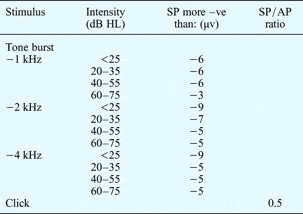
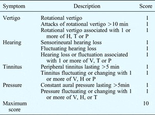
The use of tone burst stimuli that generate a summating potential lasting as long as the stimulus has been shown to significantly increase the sensitivity of the test for diagnosing hydrops.Reference Gibson5 Normal reference ranges have been publishedReference Gibson and Höhmann6 (Table I). However, in the absence of a post-mortem such reference ranges inevitably rely on a symptom classification system or scoreReference Gibson and Arenberg7 (see Table II) to indicate the likelihood of hydrops being present.
Ecochg criteria for hydrops

The diagnostic level was chosen as the nearest whole figure to the level that provided a false positive diagnosis.Reference Gibson and Höhmann6 A Gibson scoreReference Gibson and Arenberg7 of >7/10 indicates the likely presence of hydrops (Table II). HL = hearing level; SP = summating potential; AP = action potential
Gibson 10-point score for ménière's disease clinical diagnosisReference Gibson and Arenberg7

Score based on patient's answer to the question ‘Have you ever had…?’, for each symptom.
Min = minutes; H = hearing; T = tinnitus; P = pressure; V = vertigo
Until recently, delineation of the bony borders of the otic capsule has only been possible using computed tomography (CT) scanning. Clinical imaging of the internal structures has been difficult, so most inner ear anatomy and pathology has been elucidated from temporal bone histology. In guinea pigs, it was shown that magnetic resonance imaging (MRI) with long scanning times and high intravenous doses of gadolinium could produce a three-dimensional model of the cochlea, differentiating perilymph from endolymph,Reference Koizuka, Seo, Murakami, Seo and Kato8 and could show experimentally induced hydrops.Reference Niyazov, Andrews, Strelioff, Sinha and Luftkin9
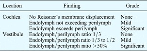
Although Reissner's membrane was visualised in a human ear in 1995,Reference Koizuka, Seo, Kubo, Matsunaga, Murakami and Seo10 inner ear imaging was limited by the special resolution of 1.5 Tesla scanners. In 2005, a 1.5 Tesla scanner was used to demonstrate that intratympanically administered gadolinium could enter the inner ear (presumably through the round window membrane). In 2007, Nakashima et al. Reference Nakashima, Naganawa, Sugiura, Teranishi, Sone and Hayashi11 used intratympanic gadolinium to produce clear images of endolymphatic hydrops in a Ménière's disease ear, utilising a 3 Tesla scanner. Newer scanners have improved image sequencing and made identifying ultrastructural detail consistently possible.Reference Carfrae, Holtzman, Eames, Parnes and Lupinetti12–Reference Hornibrook, Coates, Goh and Bird15 The dose and timing of gadolinium administration and the necessary safety parameters have all been well established, and a hydrops grading system proposed (Table III).Reference Nakashima, Naganawa, Pyykko, Gibson, Sone and Nakata16
Endolymphatic hydrops: mri grading systemReference Nakashima, Naganawa, Pyykko, Gibson, Sone and Nakata16

MRI = magnetic resonance imaging.
In our department, with 16 years' experience with transtympanic tone burst electrocochleography, we undertook a small pilot study of inner ear MRI in three patients who had already undergone electrocochleography.
Materials and methods
Three patients underwent transtympanic electrocochleography with tone burst and click stimuli. After full pure tone audiography, an insulated transtympanic needle was passed through the tympanic membrane to lie in the round window niche. Both ears were tested, using an Amplaid mk 15 diagnostic system (Amplaid, Milan, Italy). The criteria for hydrops are outlined in Table I.Reference Gibson and Höhmann6 Gadolinium (Multihance® 1.6 ml in 10 ml saline) was introduced into the chosen middle ear and replenished four to five times over 45 minutes (to a total of 1–1.5 ml), with the patient lying supine, with the neck extended and towards the opposite side.
Magnetic resonance imaging was conducted 24 hours later using a 3 Tesla magnet General Electric HDX scanner (General Electric Medical Systems, Milwaukee, Wisconson, U.S.A.). Two inversion recovery sequences were obtained with inversion times of T1 1000 (endolymph) and 2500 (perilymph), in addition to three-dimensional FIESTA imaging of the inner ear and internal auditory canals.
Patient histories and results
Patient one
An 80-year-old male had a five-month history of vertigo attacks. He had been admitted to hospital on one previous occasion as a result, and two attacks had been witnessed in his home by one of the authors (JH). The patient had suffered a total of seven attacks, which began with no warning, involved vomiting and lasted for about 4 hours. There were no associated aural symptoms or headache. He had been aware of some hearing loss in his right ear for a year. Using American Academy of Otolaryngology – Head and Neck Surgery criteria, he did not fulfil the criteria for probable or definite Ménière's disease, and his Gibson score was 4/10.
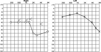
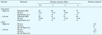
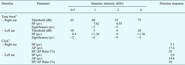
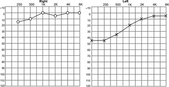
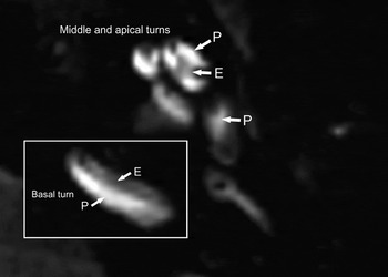
On pure tone audiometry, there was a bilateral high tone sensorineural hearing loss (SNHL), with an additional low tone loss in the right ear. The electrocochleography data, audiogram and inner ear MRI scan for the right ear are shown in Table IV and Figures 1 and 2. Tone burst electrocochleography gave no indication of hydrops in either ear. Magnetic resonance imaging of the right inner ear (the ear most likely to have hydrops if Ménière's disease was present) showed no evidence of hydrops.
Pure tone audiogram for patient one.

Magnetic resonance imaging of the right inner ear of patient one. The perilymph sequence in this ear demonstrated the absence of hydrops. P = perilymph; E = endolymph

Patient one: electrocochleography results

Parameters in both ears were normal. *100 dB; †90 dB. SP = summating potential; AP = action potential
Patient two
A 46-year-old male had a two-year history of vertigo attacks accompanied by fluctuating hearing loss, non-fluctuating tinnitus and fluctuating aural fullness in the right ear. He fulfilled the American Academy of Otolaryngology – Head and Neck Surgery criteria for definite Ménière's disease, and his Gibson score was 9/10.
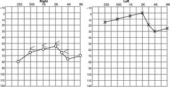
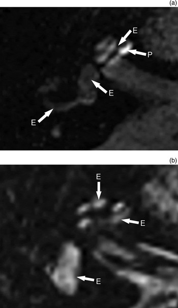
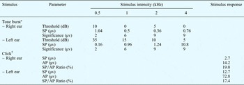
On pure tone audiometry, there was a ‘flat’ sensorineural loss at about 60 dB in the right ear (Figure 3). The electrocochleography data and MRI scan for the right ear are shown in Table V and Figure 4. Tone burst electrocochleography summating potentials at 1 and 2 kHz strongly implied the presence of hydrops. Magnetic resonance imaging showed hydrops in the cochlea, vestibule and semicircular canal.
Pure tone audiogram for patient two.

Magnetic resonance imaging of the right inner ear of patient two. (a) Perilymph sequence showing significant enlargement (33–50%) of the endolymphatic compartment of the cochlea; in the vestibule and semicircular canal, endolymphatic hydrops (>50%) have replaced almost all perilymph (a). (b) Endolymph sequence showing enlargement of the endolymphatic compartment in the cochlea and endolymph filling the vestibule. P = perilymph; E = endolymph

Patient two: electrocochleography results
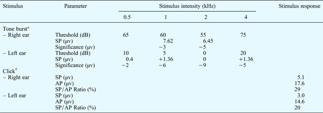
Parameters were normal in the left ear but strongly suggestive of hydrops in the right ear (see right ear tone burst SP values). *100 dB; †90 dB. SP = summating potential; AP = action potential
Patient three
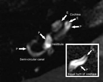
A 42-year-old female presented with a four-month history of humming tinnitus in the left ear and a ‘blocked’ feeling. There was low tone hearing loss in the left ear. She had not had vertigo. The electrocochleography data, audiogram and MRI scan for the left ear are shown in Table VI and Figures 5 and 6. Tone burst electrocochleography at 4 kHz implied hydrops. Magnetic resonance imaging showed hydrops in the cochlea only, sparing the basal turn.
Pure tone audiogram for patient three.

Magnetic resonance imaging (perilymph sequence) of the left inner ear of patient three, showing cochlear hydrops only, sparing the basal turn. P = perilymph; E = endolymph

Patient three: electrocochleography results

Left ear SP result at 4 kHz suggests the presence of hydrops. *100 dB; †90 dB. SP = summating potential; AP = action potential
Discussion
In this small pilot study, MRI of the inner ear showed endolymphatic hydrops in two patients in whom its presence had been predicted by tone burst electrocochleography. Patient two had ‘classical’ Ménière's disease with hydrops in the cochlea, vestibule and semicircular canals. Patient three had hydrops in the upper cochlea, but had never had vertigo. Her presentation with a relatively sudden low tone hearing loss is a common one (i.e. sudden idiopathic SNHL) but this could also be a harbinger of Ménière's disease.
In patient one, tone burst electrocochleography indicated no hydrops and imaging showed none. On strict application of the current criteria (i.e. American Academy of Otolaryngology – Head and Neck Surgery criteria and a Gibson Score of 4/10), a diagnosis of Ménière's disease should not be made. However, the patient's low tone hearing loss (which had been advancing) could result in such a diagnosis made in error.
A review of the literature on treatment outcomes for Ménière's disease found errors in 50 per cent of cases in which the American Academy of Otolaryngology – Head and Neck Surgery guidelines were used.Reference Thorp, Shehab, Bance and Tutka17 As these guidelines offer only a symptom-based definition, and do not refer to in vivo testing, it is therefore likely that diagnostic errors are inevitable.
In humans, intratympanically administered gadolinium enhances cochlear perilymph within 4 hoursReference Carfrae, Holtzman, Eames, Parnes and Lupinetti12 and reaches the vestibule and semicircular canals by 24 hours.Reference Zou, Pyykko, Bjelke, Dastidar and Toppila18 In animals, an eightfold dilution of gadolinium had no discernible effects on the stria vascularis.Reference Kakigi, Nishimura, Takeda, Okada, Murata and Ogawa19 There are no reports of intratympanic gadolinium administration causing hearing loss or tinnitus. Thus far, it appears to be a simple and safe technique.
In summary, MRI of the inner ear is an exciting new technique. In addition to its use in Ménière's disease, it has been used to study idiopathic sudden SNHL and fluctuating hearing loss without vertigo.Reference Nakashima, Naganawa, Katayama, Teranishi, Nakata and Sugiura14 It should contribute to our understanding of these inner ear disorders. In Ménière's disease, it could be the in vivo ‘proof’ of hydrops, and the investigation by which electrophysiological tests are validated. Magnetic resonance imaging of the inner ear could contribute to a more precise clinical diagnosis of Ménière's disease than is currently possible, in particular to distinguish it from other causes of recurrent vertigo. To achieve this, we plan to undertake rigorous testing on patients with recurrent vertigo, sudden hearing loss with and without vertigo, and unilateral tinnitus.