Cardiac point-of-care ultrasound is a promising tool for rapid and focused cardiac evaluation and is increasingly used in various contexts. Reference Yamada, Ito and Fujiwara1–Reference Flores, Su, Moher, Adler and Riley3 The paediatric cardiac ICU is a logical environment for deployment of cardiac point-of-care ultrasound; however, the use of cardiac point-of-care ultrasound in this setting has not been well-evaluated. Furthermore, data on quality characteristics are limited. In this study, we evaluated 1) quality of cardiac point-of-care ultrasound studies obtained in our paediatric cardiac ICU, and 2) compared performing provider qualitative interpretation of cardiac point-of-care ultrasound images with that of a cardiologist with expertise in echocardiography.
During implementation of cardiac point-of-care ultrasound in our paediatric cardiac ICU, cardiac intensivists performed cardiac point-of-care ultrasound in qualifying patients as part of the physical exam. Patients were excluded if they were supported with extracorporeal membrane oxygenation, if the chest was open, or if there was need for escalation in support. Patients with single and biventricular anatomies were included. Patients were selected with a preference for smaller (< 5 kg) patients to optimise image quality and on whom formal echocardiography had not been completed within 24 hours of the encounter. All cardiac intensivists performing cardiac point-of-care ultrasound had completed a categorical paediatric cardiology fellowship, which includes training in performance of echocardiograms. A Philips Lumify S4-1 Phased Array transducer was used for imaging. Prior to the initial encounter, participating physicians were oriented to the device and the imaging protocol. Additional training was not required. Four imaging planes were targeted for each encounter. Providers performing the exam recorded an interpretation of findings at the time of completion. A single, blinded reader with expertise in echocardiography independently completed a modified Rapid Assessment of Competency in Echocardiography score for each study to assess quality and made independent assessments of ventricular systolic function, pleural effusions, and pericardial effusions. Reference Millington, Arntfield and Hewak4 The Rapid Assessment of Competency in Echocardiography score was modified by adding a category for single ventricle systolic function and removing a category for inferior vena cava assessment. Sensitivity and specificity for clinically meaningful dichotomised findings defined a priori were calculated for each interpretation category using the expert reader’s interpretation as the gold standard. This study was approved by our centre’s Institutional Review Board.
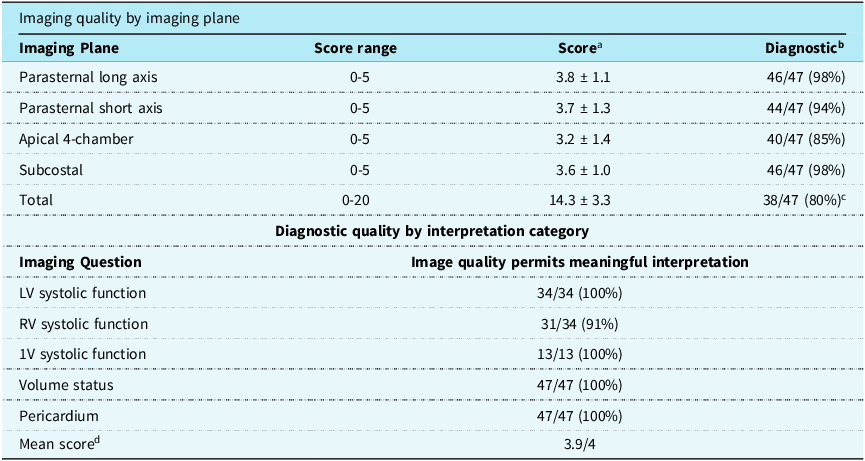
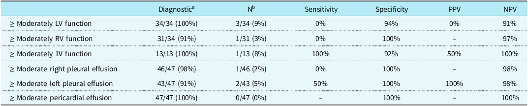
Nine cardiac intensivists completed 47 studies. The median patient age was 37 (Interquartile Range 14, 123.5) days; a total of 13/47 (28%) patients had single ventricle anatomy. Diagnostic imaging was completed in most patients by imaging plane and interpretation category (Table 1, Supplemental Figure and Video). In patients with single ventricular anatomies, imaging to evaluate systolic function was diagnostic in 13/13 (100%) of patients. The mean modified Rapid Assessment of Competency in Echocardiography score was 14.3 ± 3.3 out of 20. Specificities and negative predictive values for dichotomised findings of moderate or greater effusions or moderately or severely reduced ventricular systolic function were high, although sensitivity was limited in some cases in the setting of overall low frequency of positive findings (Table 2).
Modified Rapid Assessment of Competency in Echocardiography (RACE) scale for determination of imaging quality

a Image quality using the validated RACE scale is scored along a continuum as follows: 0 = Not obtained; 1 = Image quality too poor to permit meaningful interpretation; 3 = suboptimal image quality, but basic image interpretation possible; 5 = Good image quality, meaningful interpretation easy. bNumber of patients with diagnostic imaging (ratings of 2-5) for each imaging plane cNumber of patients among entire group with diagnostic imaging in all imaging planes. dEach patient with single ventricle anatomy for whom imaging was felt to be diagnostic received a score of 2; all other categories were scored as 1. Maximal scale score = 4. Data are presented as mean ± standard deviation or n (%). LV = left ventricle; RV = right ventricle; 1V = single ventricle.
Sensitivity, specificity, positive predictive value, and negative predictive value for Cardiac POCUS studies using expert interpretation as gold standard

a Images felt to be of satisfactory diagnostic quality for rendering interpretation of specified finding. bNumber of patients with abnormal finding according to expert interpretation. Data are presented as n (%). LV = left ventricle; RV = right ventricle; 1V = single ventricle; PPV = positive predictive value; NPV = negative predictive value.
These data show that diagnostic imaging with satisfactory image quality can be obtained in a high proportion of patients undergoing cardiac point-of-care ultrasound in the paediatric cardiac ICU, including patients with single ventricle anatomy. These results are encouraging, given the many devices, instruments, and dressings that are often present in patients and present challenging imaging windows. While diagnostic imaging from all imaging planes was obtained in only 80% of patients, the portfolio of images for each patient was satisfactory to provide meaningful commentary on targeted clinical questions such as ventricular systolic function in a very high percentage of patients. This finding supports the use of multiple imaging planes for cardiac point-of-care ultrasound exam in paediatric patients, where images from one imaging plane may be limited, another plane may provide information to answer clinical questions.
We chose to compare cardiac intensivist image interpretation with blinded interpretation of images by a cardiologist with expertise in cardiovascular imaging. Given the limited number of patients, we dichotomised data using clinically meaningful cutoffs. There was high specificity and negative predictive value among interpretation categories suggesting utility at excluding clinically important findings evaluated by cardiac point-of-care ultrasound. Notably, there were few positive findings.
In this setting, sensitivity was low in some categories. For example, there were three cases where left ventricular systolic dysfunction was interpreted as at least moderately reduced by the independent reviewer but not by the cardiac intensivist. This finding may be attributable to differences in experience in review and interpretation of images. However, the finding is also likely partially attributable to known inherent subjectivity in qualitative assessment of ventricular systolic function. Reference Puchalski, Williams, Askovich, Minich, Mart and Tani5 Furthermore, the cardiac intensivist, having availability of all data at time of exam, may have some bias in interpretation influenced by their assessment of the entire clinical picture. Finally, although there were specific imaging targets for the encounter, the use of cardiac point-of-care ultrasound in this setting was not problem-oriented as is traditionally recommended. It is possible that in looking at a multitude of cardiac structures, less focus is given to a single target, such as left ventricular function, and accuracy and reliability diminish as a result. Our findings suggest the challenges of accurately assessing depressed ventricular function and the risk of discrepant interpretations exist even amongst frequent users with extensive training and experience. Factors contributing to differences in assessment and interventions that increase agreement between the cardiac intensivist and the imaging expert should be the focus of future investigations and it is critical to maintain strong collaboration between clinicians with expertise in intensive care and cardiovascular imaging when developing a paradigm for clinical use of cardiac point-of-care ultrasound. Reference Lu, Riley and Conlon6
This study has several limitations, including the exclusion of unstable patients, the focus on the neonatal population, and the inherent differences in image quality between a handheld device and traditional echocardiographic machines, which would be considered the true gold standard and might impact physician interpretation of cardiac performance. Despite these limitations, this report highlights the potential benefits and considerations of an innovative practice on a uniquely fragile population that may serve as a stimulus for future investigation ideally in the form of a randomised prospective investigation.
In conclusion, cardiac point-of-care ultrasound can be successfully performed in a paediatric cardiac ICU by cardiac intensivists with good imaging quality and a high rate of diagnostic imaging. Even in the presence of adequate imaging quality, however, differences in interpretation of cardiac performance may exist between experienced physicians. Our findings offer a platform for continued quality improvement and ongoing efforts to integrate cardiac point-of-care ultrasound into routine clinical care in a high-impact clinical environment.
Financial support
This work was supported by a grant from the Children’s Healthcare of Atlanta 1998 Society.
Competing interests
None.