INTRODUCTION
Chitin is an important component in the shells of crustaceans, the exoskeletons of insects, and the cell walls of fungi. It is a polymer that consists of N-acetylglucosamine monomers (GlcNAc) linked by β-1,4-glucosidic bonds. The annual production of chitin is estimated to be at least 1011 tons in the aquatic biosphere alone, and it is the second largest biomass after cellulose (Haki & Rakshit, Reference Haki and Rakshit2003). Chitosan is the totally or partially de-acetylated derivative of chitin, which is produced at industrial scale through the alkaline deacetylation of chitin (GAF, Reference Gaf1992). Polymers containing more than 50% (Miller, Reference Miller1959) of acetylates are considered chitin, whereas those with less than 50% are called chitosan. Chitosan has various applications in the field of medicine (Enriquez de Salamanca et al., Reference Enriquez de Salamanca, Diebold, Calonge, Garcia-Vazquez, Callejo, Vila and Alonso2006), agriculture (El Hadrami et al., Reference El Hadrami, Adam, El Hadrami and Daayf2010), dietetics (Vargas & Gonzalez-Martinez, Reference Vargas and Gonzalez-Martinez2010), environmental protection (Chen JK, Reference Chen, Wang, Liou, Shen and Liu2011) and several other fields (Friedman & Juneja, Reference Friedman and Juneja2010; Friedman et al., Reference Friedman, Phan, Schairer, Champer, Qin, Pirouz, Blecher-Paz, Oren, Liu, Modlin and Kim2012). Studies on chitin and chitosan have focused on their conversion to oligosaccharides, which possess versatile functional properties (Aam et al., Reference Aam, Heggest, Norberg, Sørlie, Vårum and Eijsink2010) including antitumour (M Yabuki, Reference Yabuki, Uchiyama, Suzuki, Ando and Fujii1988), antimicrobial, anti-osteoporotic (Jung et al., Reference Jung, Moon and Kim2006) and immuno-enhancing properties (Tokoro et al., Reference Tokoro, Suzuki, Matsumoto, Mikami, Suzuki and Suzuki1988).
Chitosanases, which are found in a number of organisms, can be classified into six glycoside hydrolase families, namely, GH-5, GH-7, GH-8, GH-46, GH-75 and GH-80 (CAZy. http://www.cazy.org). Among these families, GH-46, especially that from Bacillus circulans (MH-K1) and Streptomyces sp. (CsnN174), has been studied extensively in terms of its catalytic features, enzymatic mechanisms, and protein structures (Saito et al., Reference Saito, Kita, Higuchi, Nagata, Ando and Miki1999; Tremblay et al., Reference Tremblay, Yamaguchi, Fukamizo and Brzezinski2001).
Renibacterium salmoninarum is a gram-positive bacterium that causes disease in young salmonid fish. The infection is commonly known as bacterial kidney disease (BKD) and it occurs worldwide where salmonid fish are found (Smith, Reference Smith1964; Wiens & Kaattari, Reference Wiens, Kaattari, Woo and Bruno1999). However, despite considerable effort and years of research, the process of infection by Renibacterium salmoninarum remains poorly understood. It is the main cause of BKD in susceptible fish. Once accumulated on the surface of the fish, the strain secretes a number of soluble proteins that destroy the gills and skin (Flaño, Reference Flaño, Kattan, Razquin and Villena1996; A.J. Evenden, Reference Evenden, Grayson, Gilpin and Munn1993), and then migrate into the organs. The scales outside the gills and skin contain large amounts of chitin and chitosan, thus, the chitosanase we described in this paper might have a decisive effect in bacterial infection.
MATERIALS AND METHODS
Materials
All chemicals were of analytical or higher grade unless otherwise stated. Chitosan (≥90% deacetylated) was purchased from Qingdao MdBio, Inc. (China). Colloidal chitosan was prepared as previously described (Yabuki et al., Reference Yabuki, Uchiyama, Suzuki, Ando and Fujii1988). Simply, 1 g of chitosan was dissolved in 100 ml of 0.1 M HCl and incubated overnight at room temperature. The medium was then adjusted to a pH of 6.0 with 1 M NaOH. Chitosan dimers and trimers, which were used as standards, were prepared and identified via nuclear magnetic resonance and gas chromatography/mass spectrometry.
Screening of chitosanase producing strains
Small crustaceans, mainly crabs with a diameter of less than 1 cm, caught from the coast of QingDao, China were ground in seawater and filtered through a sterile gauze to remove any debris. The supernatant was then plated on agar plates containing 0.5% colloidal chitosan. Colonies with surrounding clear zones were harvested and maintained in a liquid medium containing 0.5% chitosan for one to two days.
Sequencing of the 16srDNA gene of strain QD1
The chromosomal DNA of QD1 chitosanlytic bacterial cells was purified using the Wizard Genomic DNA Purification Kit (Qiagen). The 16S rDNA gene was amplified by polymerase chain reaction (PCR) with eubacterial primers 27f and 1492r. The amplified 1.5 kb PCR product was sequenced and blasted at http://blast.ncbi.nlm.nih.gov/Blast.cgi to obtain closely matched species (Xing et al., Reference Xing, Liu and Yu2013).
Purification of chitosanase from culture of QD1
The Renibacterium sp. QD1 strain was cultured at 30°C for 36 h with shaking in a medium containing 0.5% colloidal chitosan as inducer to harvest the extracellular chitosanase. Ammonium sulphate was added to the supernatant until it reached 60% saturation, and the sediment was removed by centrifugation. The supernatants were collected and loaded onto a phenyl sepharose HP chromatography column and eluted with phosphate buffer (20 mM, pH7.0). Fractions with high chitosanase activity were pooled and dialysed at 4°C against phosphate buffer for three times then used for further experimentation.
Protein concentration was measured using the BCA protein assay kit with bovine serum albumin as a standard (Miller, Reference Miller1959).
Sodium dodecyl sulphate–polyacrylamide gel electrophoresis (SDS-PAGE) was carried out to determine the purity and molecular weight of the purified chitosanase.
Enzyme activity assay
Chitosanase activity was measured with colloidal chitosan as the substrate. The reaction was initiated by adding 100 µl of purified enzyme solution to 900 µl of 0.5% colloidal chitosan dissolved in 0.2 M sodium acetate buffer (pH 5.6). The mixture was incubated at 55°C for 10 min and then terminated by adding 750 µl of DNS reagent (Miller, Reference Miller1959). After boiling at 100°C for 10 min, the mixture was centrifuged, and the amount of the produced reducing sugar was determined by measuring the A520 absorbance. One unit of chitosanase was defined as the amount of enzyme that liberated reducing sugars corresponding to 1 µM of glucosamine-hydrochloride per minute.
Characterization of purified Csn-A
The pH dependence of Csn-A was evaluated in the range of pH 4–8 in different buffers, including NaAc (pH 4.0–5.8, 0.2 M), Na2HPO4–KH2PO4 (pH 4.9–6.2, 1/15 M) and Na2HPO4–NaH2PO4 buffers (pH 5.7–8.0, 0.2 M). Temperature dependence was assessed over the range of 20–70°C for 1 h. pH stability was determined by incubating the enzyme at 4°C for 24 h in NaAc (pH 3.6–5.8, 0.2 M), Na2HPO4–KH2PO4 (pH 4.9–6.2, 1/15 M), Na2HPO4–citric acid (pH 5.4–8.0), sodium acetate (pH 5.0–5.4, 0.2 M), Na2HPO4–NaH2PO4 (pH 5.7–8.0, 0.2 M), glycine–NaOH (pH 8.6–10.6, 50 mM) and Na2HPO4–NaOH buffers (pH 10.9–11.6). After incubation, residual activity was plotted against the pH value. Temperature stability was determined over the range 0–70°C in a phosphate buffer (pH 7.0) for 1 h.
The effects of metal salts and inhibitors on activity were determined by adding each salt into the standard assay mixtures at a final concentration of 1 mM and measuring the relative activities. The enzyme mixture without adding any metal ion or inhibitor was considered as control.
Hydrolysis product analysis
Thin layer chromatography (TLC) was performed for hydrolysis product analysis. Suitably diluted Csn-A was incubated with colloidal chitosan at 37°C, pH 5.6, over a 24 h reaction period. The enzyme was added every 2 h until the sizes of the products no longer changed. The extent of substrate depolymerization was monitored by removing an aliquot of each sample for analysis by TLC (Yoon et al., Reference Yoon, Kim, Kim, Hong, Shin and Cho2001).
Amino acid sequence determination and cloning of the chitosanase gene
Automatic Edman degradations and N-terminal amino acid analysis were performed at Xinongsheng Biotechnology Co., Ltd (Beijing, China). Homology searches for amino-acid sequences on public databases were performed using the NCBI BLASTP program. Primers chitoF (5′-CATATGAAACTGTCTTGCATAAGAC-3′) and chitoR (5′-AAGCTTCTTGATCTCGTACGGGTCTC-3′) used for csn-A cloning were designed based on the known chitosanase of Renibacterium sp. ATCC 33209.
RESULTS
Fermentation conditions and screening of strains with higher activity for colloidal chitosan
As described above, strains from the homogenate of crustaceans were placed on a chitosan-selective agar plate and colonies with clear haloes were chosen for further analysis. Among the screened isolates, strain QD1, which produced the largest clearing zone on the chitosan plate, show good signs of chitosanase production. Isolate QD1 is a gram-positive strain that formed a colourless colony on a screening plate within 2 d (Figure S1). Based on the 16srRNA sequence, QD1 was assessed to be Renibacterium sp., and is thus named Renibacterium sp. QD1. Optimal fermentation conditions, which include a temperature of 30°C (Figure S2 and S3) and pH of 6.0, as well as medium composition are shown in Table S1.
Purification of chitosanases
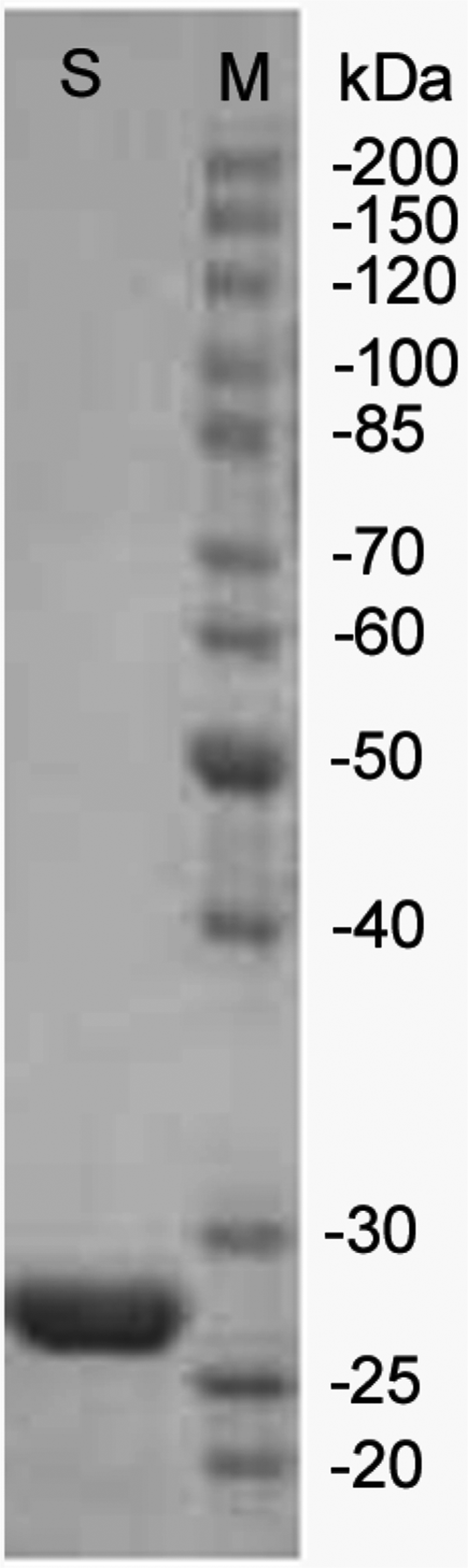
The Renibacterium sp. QD1 strain was cultured at 30°C for 36 h with shaking in a medium containing 0.5% colloidal chitosan as inducer to harvest the extracellular chitosanase. Ammonium sulphate was added to the supernatant until it reached 60% saturation, and the sediment was removed by centrifugation. The supernatant were collected and placed in a phenyl sepharose HP chromatography column. Fractions with high chitosanase activity were pooled and used in the SDS-PAGE analysis. After the purification step, a single band was observed with a molecular weight of about 26.1 kDa based on the SDS-PAGE results (Figure 1).
SDS-PAGE analysis of purified Csn-A. SDS-PAGE was performed with 10% polyacrylamide gel and stained with Coomassie Brilliant Blue R-250. S, purified chitosanase; M, standard molecular weight markers.

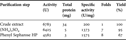
A summary of the purification process is presented in Table 1. The enzyme was purified eight-fold, with an overall yield of 67% and a specific activity of 1575 U/mg.
Purification process of the chitosanase from Renibacterium sp. QD1. Chitosanase was purified using the following procedure: all purification steps were performed at 4°C, and each fraction was assayed for activity. Step 1: bacterial cells were pelleted out from the culture medium by centrifugation (12,000 g, 10 min). Anhydrous ammonium sulphate was added to the supernatant to 60% saturation, and the sediment was removed by centrifugation (12,000 g, 10 min). Step 2: the supernatant was loaded to a Phenyl Sepharose HP column (1.0 × 20 cm) previously equilibrated with 60% saturation of ammonium sulphate. The column was washed with 36% saturation of ammonium sulphate at a flow rate of 1.5 ml/min. The eluent was dialysed with deionized water.
General properties of purified chitosanase
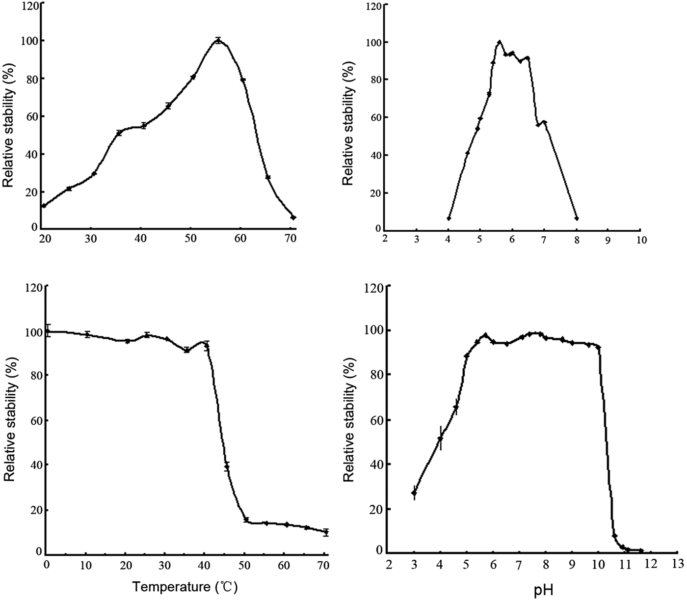
The temperature profile was studied in a temperature range of 20–70°C. Figure 2A, C shows that the enzyme displayed an optimum temperature activity at 55°C, but its stability declined sharply above 40°C.
Effect of pH and temperature on enzyme activities: (A) effect of temperature on enzyme activities; (B) effect of pH on enzyme activities; (C) temperature stability of the enzyme. Temperature dependence was assessed over the range of 20–70°C for 1 h; (D) pH stability of the enzyme. pH stability of chitosanase was assayed from the residual activity after incubating at 4°C in buffers of various pH (3–11.6) for 24 h. Enzyme activities were measured in standard assay conditions described above. The y-axis represents the relative activity. Values were means of three replications ±standard deviation.
Figure 2B, D illustrates the influence of pH on chitosanase activity and stability. The optimal pH was 5.3–6.0 and was very stable at pH 5–10, where more than 90% of its original activity was maintained after incubation at a pH range of 5–10 for 24 h.
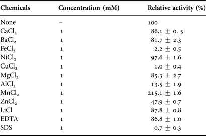
Enzyme assay was performed at the optimal pH and temperature with the tested compounds at a final concentration of 1 mM to determine the effects of metal ions on enzyme activity. The relative activity was calculated with respect to the control sample where the reaction was carried out in the absence of any additive. Table 2 shows that with the addition of 1 mM Fe3+, Cu2+ and SDS, the enzyme retained no less chitosanlytic activity. Remarkable inhibition was found for Al3+ and Zn2+, which resulted in over 80% and 50% loss of enzyme activity, respectively. Ca2+, Ba2+, Mg2+ and EDTA had a smaller inhibition effect. Metal ion Mn2+ was a strong activator of CsnA, and its activity more than doubled.
Effects of chemicals on the activity of Csn-A. Chitosanase activity was assayed after adding metal ions and compounds, such as Ca2+, Ba2+, Fe3+, Ni2+, Cu2+, Mg2+, Al3+, Mn2+, Zn2+, Li+, EDTA and SDS to a final concentration of 1 mM. Relative activity was expressed as the percentage ratio of the purified chitosanase with metals and compounds to those without metals.
Hydrolysis product analysis
The enzyme was hydrolytically active on chitosan with a de-acetyl degree of ≥70%, and could only hydrolyse acidolysed colloidal chitosan instead of insoluble powder. TLC analysis results showed that the final hydrolysis product was chito-biose and -triose (Figure 3). The time course analysis results of the colloidal chitosan show that this enzyme hydrolysed chitosan to polymeric glucosamines at the early stage and subsequently hydrolysed them to form accumulated oligomerized saccharides (Figure 4). Degradation speed rapidly decreased when the polymer had less than five glucosamine residues, which indicates that the enzyme was more active towards polymeric glucosamines with a degree of ≥5. Taken together, the release mode and the rapid decrease in the viscosity of the products suggest that the chitosanase was active in an endolytic manner (Heggset et al., Reference Heggset, Dybvik, Hoell, Norberg, Sørlie, Eijsink and Vårum2010).
Analysis of enzymatic hydrolysates by thin layer chromatography. The reaction was done at 37°C for 12 h: (A) substrate only; (B) reaction products from the enzyme by using 90% deacetylated chitiosan as the substrate; M, marker, a mixture of dimer (GlcN)2 and trimer (GlcN)3.
Time course analysis of enzymatic hydrolysates by thin layer chromatography. The reaction was done at 37°C and the reaction system includes 100 uL of the crude enzyme (about 2 U) and 900 ul of the substrate supplemented with the enzyme when the mixture was incubated for 12 h and sampled at different times (min). M, marker, a mixture of dimer (GlcN)2 and trimer (GlcN)3.
Cloning of the chitosanase gene
The N-terminal sequence of the first ten amino acid residues of purified chitosanase was Ser-Thr-Gly-Asp-Leu-Ser-Ala-Pro-Ala-Lys. This N-terminal sequence shared 100% identity with part of a putative chitosanase of Renibacterium sp. ATCC 33209, whose genome had been previously sequenced. We designed two primers based on the genome sequence of ATCC 33209 to amplify the chitosanase gene of QD1. The results indicate that the full-length gene, named csn-A, consists of 855 nucleotides and encodes an open reading frame of 284 amino acid residues. The encoded chitosanase, Csn-A, was 30 amino acids short at the N-terminal side compared with the chitosanase of ATCC33209, whereas the remaining portion shared 98% identity with the chitosanase of ATCC33209 Csn-A was classified into the GH46 family based on the amino acid sequencing.
DISCUSSION
In this study, we discovered an abundantly expressed extracellular chitosanase with high activity, which is produced by a gram-positive bacterium Renibacterium sp. QD1. Such bacteria are commonly found in salmonid fish, while, strain QD1 was isolated from small crabs with higher frequency. This foundation might provide a new transmission path or host of Renibacterium salmoninarum.
The purified chitosanase enzyme, Csn-A, from Renibacterium sp. QD1 displayed a broad pH stability of 5.0–10.0 and a higher reaction temperature of 55°C . The enzyme activity was greatly inhibited by Fe3+, Cu2+, Al3+, Zn2+ and SDS, but less affected by Ca2+ and Mg2+. Chelating reagent EDTA also has a small effect on enzyme activity, indicating the independent activity of divalent ions. Mn2+ was the only activator for Csn-A and the chitolytic activity more than doubled. The activation effect of Mn2+ was also observed in chitosanase of Penicillium sp. D-1 (Zhu et al., Reference Zhu, Tan, Zhu, Liao, Zhang and Wu2012), but the opposite was observed in Pseudomonas sp. TKU015 (Wang et al., Reference Wang, Peng, Liang and Liu2008) and Serratia sp. TKU016 (Wang et al., Reference Wang, Chang and Liang2010). The purified chitosanase efficiently catalysed an endo-type cleavage reaction in the chitosan polymer and mainly produced chito-biose and -triose as the final product.
Sequence analysis results indicate that Csn-A had nearly 90% identity with the putative chitosanase of ATCC33209 and 67% identity with the well-characterized chitosanase from Streptomyces sp. N174 (Lacombe-Harvey et al., Reference Lacombe-Harvey, Fukamizo, Gagnon, Ghinet, Dennhart, Letzel and Brzezinski2009). Csn-A was classified as GH46 based on the amino acid sequence homology. Chitosanases belonging to this family are among the most studied of all chitosanases. Compared with the known chitosanase of ATCC33209, Csn-A has 30 amino acids deletion at the N-terminal, while this deletion did not result in the loss of enzyme activity, indicating that the N-terminal might not be the catalytic core.
The characteristics of Renibacterium salmoninarum, particularly its selective nature and slow growth (Embley, Reference Embley, Goodfellow and Austin1982; Ordal & Earp, Reference Ordal and Earp1956), together with its relatively unaggressive nature, have undoubtedly made it a difficult organism to study (Evenden, Reference Evenden, Grayson, Gilpin and Munn1993). However, strain Renibacterium sp. QD1 achieved an OD600 of 2–3 after 40 h fermentation and there was no need for L-cysteine, which was shown to be indispensable for Renibacterium salmoninarum cultivation (Evelyn, Reference Evelyn1977; Austin, Reference Austin, Embley and Goodfellow1983; Austin & Rayment (Reference Austin and Rayment1985); Daly, Reference Daly and Stevenson1985). The optimal temperature for QD1 growth is 30°C, it is slow at 0–20°C and does not occur at 37°C.
FINANCIAL SUPPORT
This work was supported by the National Natural Science Foundation of China (No. 31070712 and No. 81102368), Special Fund for Marine Scientific Research in the Public Interest (No. 201105027 and No. 201005024), National High Technology Research and Development Program (863 programme) (No. 2011AA09070304) and National Program on Key Basic Research Project (973 project) (No. 2011CB200906).
Supplementary materials and methods
The supplementary material refered to in this paper can be found online at journals.cambridge.org/mbi.