Introduction
Obesity is recognised as an important driver of non-communicable diseases.Reference Poirier, Giles and Bray1 The Developmental Origins of Health and Disease (DOHaD) paradigm and the application of a lifecourse epidemiology approach have shown the importance of fetal development for obesity in later life.Reference Hanson and Gluckman2 Therefore, understanding intergenerational factors and determinants, particularly maternal health prior to and during pregnancy, are key to the promotion of optimal fetal growth and birth outcomes. However, the role of individual maternal factors has typically been investigated separately, hindering a holistic understanding of the complex interactions between maternal social factors, biological factors, fetal development, and birth outcomes.
Several maternal metabolic health characteristics have been associated with fetal development and birth outcomes. Women who are overweight or obese before conception are more likely to develop gestational diabetes mellitus (GDM) during pregnancy, and both obesity and GDM increase the risks for fetal defects and congenital anomalies, newborn macrosomia, neonatal hypoglycaemia, and/or stillbirth.Reference Poston, Caleyachetty and Cnattingius3 Furthermore, during pregnancy, infectious diseases (e.g. HIV/AIDS) as well as maternal undernutrition have been associated with higher risks of low birthweight, preterm birth, and small for gestational age.Reference Wedi, Kirtley, Hopewell, Corrigan, Kennedy and Hemelaar4–Reference Rahman, Abe and Rahman7 In addition to maternal biological factors, studies mainly conducted in high-income countries have shed light on the role of maternal sociodemographic, socio-economic, and psychosocial contexts on fetal development and birth outcomes.Reference Kim and Saada8,Reference Aizer and Currie9 Further, in four low-to-middle-income countries, a pooled analysis of birth cohort data found that social factors (including, but not limited to lower maternal and paternal schooling, lower income and social class, inadequate toilet and water facilities) were associated with infant linear growth delay through their effect on birthweight.Reference Richter, Orkin and Roman10 In addition, maternal stress, as well as behavioural factors such as alcohol or tobacco consumption before and during pregnancy, has been shown to impact negatively on fetal development and birth outcomes.Reference Accortt, Cheadle and Dunkel Schetter11–Reference Abraham, Alramadhan and Iniguez13 While studies have generally focused on factors associated with birth outcomes as a surrogate marker of fetal development, less information is available concerning maternal factors associated with fetal linear growth and adiposity accumulation. However, fetal growth measures are key to identify specific periods during which fetal growth is influenced by maternal environmental factors. Recently, fetal growth standards were developed allowing comparisons across different settings.Reference Ioannou, Talbot and Ohuma14
In low- and middle-income countries, ongoing economic development, rapid urbanization, and nutrition transition have been associated with a unique epidemiologic situation.Reference Black, Victora and Walker15 In sub-Saharan Africa, an estimated 12% of newborns are preterm,Reference Chawanpaiboon, Vogel and Moller16 13% are low birthweight, and 25.5% are small for gestational age.Reference Lee, Katz and Blencowe17 In South Africa, in addition to HIV affecting 30% of pregnant women,18 the nutrition transition is far advanced with 64% of women of reproductive ages affected by overweight or obesity, while 22% of the same age group are affected by anaemia.Reference Shisana, Labadarios and Rehle19 The combined consequences of infectious diseases, undernutrition, obesity, and cardiovascular diseases in women at reproductive age on obstetric outcomes have to date had limited attention, and our understanding of the biological and social factors associated with adverse birth outcomes is neither complete nor contextualised.Reference Richter, Orkin and Roman10
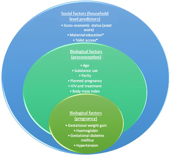
Findings from the Soweto First 1000 Days Study in South Africa, a prospective cohort of pregnant women with repeated ultrasound measures, revealed that 10.2% of women were affected by GDM and 34% by HIV. Exposure to maternal GDM was associated with increased fetal abdominal circumference in utero, and maternal dietary patterns during pregnancy predicted neonatal fat mass. Reference Macaulay, Munthali, Dunger and Norris20,Reference Wrottesley, Ong, Pisa and Norris21 Maternal obesity and weight gain during pregnancy were positively associated with newborn size at birth. In HIV-infected pregnant women, exposure to antiretroviral treatment (ART) (from preconception) was associated with higher newborn adiposity levels.Reference Wrottesley, Ong, Pisa and Norris21 However, these factors have not been considered in combination, nor have social predictors been considered. Using data from the Soweto cohort of pregnant women, this study aimed to understand more fully the social and biological drivers of longitudinal fetal growth parameters using three proposed hierarchical levels of predictors defined in the theoretical framework shown in Fig. 1: linking household, preconception, and pregnancy factors.
Theoretical framework for analysis showing potential social and biological drivers of fetal growth included in the analysis.

Methods
Study setting and participants
The prospective longitudinal pregnancy cohort study (the Soweto First 1000 Days Study; S1000) was conceived and conducted at the South African Medical Research Council/Wits Developmental Pathways for Health Research Unit, at the Chris Hani Baragwanath Academic Hospital in Soweto, Johannesburg, South Africa. Overall, S1000 aimed to understand the complex associations between multiple maternal factors and fetal and infant outcomes within the first1000 days (from conception up until 2 years of age) in an urban African population. A total of 1017 pregnant women were enrolled into the study between 2013 and 2016. Inclusion criteria for S1000 were resident of Soweto or the greater Soweto area, <14 weeks pregnant and no known diagnosis of epilepsy or diabetes at the time of recruitment, 18 years of age or older, and pregnant with a singleton, naturally conceived pregnancy. Data were collected at 6 time points during pregnancy (<14, 14–18, 19–23, 24–28, 29–33, and 34–38 weeks), as well as at delivery. Ethical clearance for the study was obtained from the University of the Witwatersrand’s Human Research Ethics Committee (M120524 and M130309). All study participants provided informed written consent prior to their inclusion in the study.
Data collection
Outcome data: fetal ultrasonography
All participants had a pregnancy dating scan at the first visit using a Philips HD-9 (Philips Ultrasound, Bothell, Washington, USA) ultrasound machine (median(IQR) 12(11–13) weeks). The fetal crown–rump length measurement was used for pregnancy dating in women <14 weeks, with the biparietal diameter, head circumference, and femur length being utilised in more advanced pregnancies (>14 but <20 weeks). Participants were invited for follow-up scans every 5 weeks at the following visits: 14–18, 19–23, 24–28, 29–33, and 34–38 weeks gestation. Gestational age at each visit was calculated using the gestational age determined by the dating scan.Reference Macaulay, Buchmann and Dunger22 Abdominal circumference, biparietal diameter, head circumference, and femur length were recorded at each follow-up scan. This methodology was performed as per the INTERGROWTH-21st study international standards for measuring fetal growth.Reference Papageorghiou, Ohuma and Altman23 All scans underwent external inter-rater reliability quality assessment by colleagues at Oxford University (UK) as per the INTERGROWTH-21st study standards.Reference Sarris, Ioannou and Ohuma24
Longitudinal modelling of fetal growth size and velocity using SITAR analytics
All five serial measurements of femur length, abdominal circumference, biparietal diameter, and head circumference from first to the third trimester of pregnancy were included in the analyses. Fetal growth data were modelled using the Superimposition by Translation and Rotation (SITAR). This shape invariant model with a single fitted curve is particularly useful in that it analyses individual growth patterns and produces three parametersReference Cole, Donaldson and Ben-Shlomo25 – the subject-specific random effects (αi, βi, γi) correspond to the size, tempo, and velocity of growth. This approach simplifies the longitudinal data in variables that can then be inserted into statistical modelling as either exposures or outcomes. Furthermore, SITAR performs well with missing dataReference Cole, Donaldson and Ben-Shlomo25 at any time point. We examined the model fit thoroughly, and for the current analyses, only the size and velocity parameters were obtained. Tempo parameter was excluded to allow the model to reach convergence, as the mean curve for this parameter was close to linear. Given that the growth rate does not change drastically during fetal growth, the growth curves were close to linear making it difficult for the model to distinguish between the tempo (horizontal shift) and size (vertical shift) parameters. Size represents individual variation along the y-axis, giving an absolute deviation of each individual from the sample mean in the units of the measurement. Velocity represents the contraction or expansion of the individual growth curve relative to the mean curve, giving an indication of the rate of change per unit of time. Males and females were modelled together, and the sex variable was included as an interaction term in the model to assess sex differences due to previous analyses on this cohort demonstrating sex differences in fetal growth.Reference Macaulay, Munthali, Dunger and Norris20 Data were modelled using the SITAR version 1.0.10 in R version 3.4.2.
Exposure data: household level
Household-level factors related to the socio-economic environment were collected at the first visit during pregnancy (<14 weeks gestational age) in an interview using questionnaires administered by trained research assistants. Maternal education was defined according to the highest level of education completed (no school or only primary school completed, secondary school completed, or tertiary education). Household socio-economic status (SES) was estimated by scoring each participant according to the number of physical assets possessed out of a possible 11 assets (electricity, radio, television, refrigerator, mobile phone, personal computer, bicycle, motorcycle/scooter, car, agricultural land, and farm animals). This asset index was based on standard items used in the Demographic and Health Surveys household questionnaire (available at: www.measuredhs.com) and has been extensively utilised in this setting.Reference Griffiths, Johnson, Cameron, Pettifor and Norris26,Reference Kagura, Adair, Pisa, Griffiths, Pettifor and Norris27 Toilet access was defined according to whether the households had their own toilet or were sharing a toilet with other households.
Exposure data: preconception level
Individual preconception anthropometry was collected at the first pregnancy visit by trained research assistants. Maternal height was measured to the nearest 1 mm at baseline using a wall-mounted stadiometer (Holtain, UK). A digital scale was used to measure maternal weight to the nearest 0.1 kg at each pregnancy visit. Weight at recruitment (<14 weeks) was used as a proxy for pre-pregnancy weight and, together with height was used to calculate maternal body mass index (BMI) (weight (kg)/height (m2)).Reference Krukowski, West and DiCarlo28 Women self-reported their date of birth at enrolment, from which their age was calculated. Smoking status and alcohol use were assessed as current use of cigarettes and/or tobacco and current alcohol use, respectively. Women reported whether their current pregnancy was planned or unplanned, and parity was defined as the number of previous births at a gestational age of 24 weeks or more – regardless of whether the infant was born alive or was stillborn. This was categorised as no previous births, 1–2 previous births, and ≥3 previous births. Self-reported HIV status was collected at baseline as well as at each subsequent pregnancy visit and confirmed using the results from the participant’s antenatal clinic card. According to South Africa’s national prevention of mother-to-child transmission guidelines, routine HIV counselling and testing are required during pregnancy, and ART is initiated for any HIV-positive woman who is not already on ART. For this reason, all HIV-positive participants were receiving ART during the study and were stratified according to whether they had been initiated on ART prior to pregnancy (pre-pregnancy ART) or during the current pregnancy (antenatal ART).
Exposure data: pregnancy level
At each subsequent pregnancy visit, maternal weight was measured to the nearest 0.1 kg using the same digital scale as the baseline assessment. Gestational weight gain (GWG; kg/week) was calculated as [(weight at final pregnancy visit−weight at recruitment)/weeks of follow-up]. Haemoglobin levels (g/dl) were assessed using a HemoCue at the first pregnancy visit. A 2-h 75 g oral glucose tolerance test was conducted at 24–28 weeks gestation in order to diagnose or rule out GDM. Venous blood samples were taken and assessed on site using the RX Daytona Chemistry Analyzer (Randox, London, UK). GDM was diagnosed using the World Health Organization’s (WHO) 2013 criteria (fasting plasma glucose of 5.1–6.9 mmol/l, or 1-h plasma glucose of ≥10.0 mmol/l or 2-h plasma glucose of 8.5–11.0 mmol/l).29 Maternal blood pressure (mmHg) was measured at the fourth pregnancy visit (24–28 weeks) using an Omron 6 automated machine (Kyoto, Japan). A 5-min seated rest was observed before blood pressure measurements were taken. Seated blood pressure was measured three times on the right side, with a 2-min interval between each measurement. Hypertension was defined as a systolic measure ≥140 and/or a diastolic measure ≥90 using the mean of the second and third readings according to the National Institute for Health and Care Excellence (NICE) guidelines (NG133, 2019).
Statistical analysis
All data were analysed using STATA v13 for Mac. Initially, participant characteristics were summarised and presented using mean (SD) for parametric data and N(%) for non-parametric data. Where exposure level data were missing, multivariate sequential imputation using chained equations (mi impute chained (logit) command) was conducted using maternal age, BMI, parity, gestational age, education, marital status, and SES as auxiliary variables with 10 imputations. Based on the theoretical framework (Fig. 1), predictors at each level of exposure were tested for intra-level correlation, and the variable with the strongest relationship with the outcome was retained. Remaining variables were then included in a series of sex-stratified hierarchical linear models for each fetal growth outcome parameter (sex stratification was based on previously identified sexual dimorphism in this cohort Reference Macaulay, Munthali, Dunger and Norris20 ). That is, for each fetal growth outcome, first, household-level predictors were regressed (model 1). Next, individual preconception-level predictors were regressed (model 2). Third, individual pregnancy-level predictors were regressed (model 3). Last, all three levels of predictors were combined in a final regression (model 4).
Results
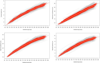
Of the 1017 participants originally included in S1000, 828 participants had outcome data for each of the fetal growth parameters. The sample size and number of measurements for each outcome included in the models of fetal growth are presented in Table 1. SITAR explained 71%, 65%, 69%, and 72% of variance for femur length, abdominal circumference, head circumferences, and biparietal diameter, respectively. Fig. 2 shows the effect of SITAR adjustment (size and velocity) in individual curves. As the curves demonstrate, the mean curve and SITAR-adjusted curves follow closely the pattern of the unadjusted curves, suggesting that SITAR was able to model the data appropriately. The exposure characteristics considered for these participants are shown in Table 2. Most mothers (73%) had at least secondary school level education and a quarter had a high school certificate or further education. There was an equal distribution (33% each) of normal weight, overweight, and obese mothers at the start of pregnancy. Just over half of the pregnancies were unplanned and 52% of pregnancies resulted in male fetuses. Less than 12% of mothers used alcohol and less than 9% smoked prior to pregnancy. GDM was diagnosed in 12% of pregnancies, hypertension in 4% of pregnancies, and 32% of mothers were HIV+.
Variance explained by SITAR for fetal growth measurements

Unadjusted (grey) and SITAR-adjusted (red) individual plots and mean plot (white) to demonstrate the fitting of the SITAR model for raw data.

Participant characteristics at each heirachical level (n = 828)

SES, socio-economic status; BMI, body mass index; GWG, gestational weight gain; GDM, gestational diabetes mellitus; ART, antiretroviral treatment.
Household level
When considering the results from the final regression models (Table 3), SES was not associated with any fetal growth parameters in males or females. No other household-level predictors were included in the regression models due to collinearity.
Results from the final regression model for each fetal growth parameters stratified by fetal sex

***p < 0.01, **p < 0.05.
Preconception level
At maternal baseline level, the final regression models (Table 3) showed that higher parity was negatively associated with most male fetal growth parameters, including biparietal diameter size (β = −0.15, CI = −0.24; −0.06) and velocity (β = −0.03, CI = −0.05; −0.01), head circumference size (β = −0.31, CI = −0.57; −0.04), and femur length size (β = −0.08, CI = −0.15; −0.01) and velocity(β = −0.02, CI = −0.03; −0.01); but positively with biparietal diameter velocity (β = 0.02, CI = 0.00; 0.034) in females. Baseline BMI was positively associated with all fetal growth outcomes in males, and with abdominal circumference size and velocity, head circumference size, and femur length size and velocity in females; however, these beta coefficients were all relatively small (β = <0.10) meaning that one unit increase in maternal BMI (kg/m2) would result in less than 0.1 unit shift in growth rate in comparison to the sample. Having a planned pregnancy was associated with lower abdominal circumference and slower abdominal growth velocity in males (β = −0.17, CI = −0.37; −0.05, β = −0.02, CI = −0.03; 0.01, respectively), and with shorter femur length and slower femur growth velocity in females (β = −0.06, CI = −0.10; −0.03, β = −0.01, CI = −0.02; −0.00, respectively). Smoking was negatively associated with femur length size in females only (β = −0.09, CI = −0.16; −0.02). Being HIV+ with ARTs initiated prenatally was negatively associated with head circumference size (β = −0.32, CI = −0.57; −0.07) and velocity (β = −0.02, CI = −0.04; −0.00) in females only.
Pregnancy level
At the individual pregnancy level, the final regressions (Table 3) showed that having GDM was positively associated with abdominal circumference size and velocity (β = 0.33, CI = 0.09; 0.57, β = 0.02, CI = 0.00; 0.04, respectively) and with biparietal diameter size and velocity (β=0.10, CI = 0.03; 0.17, β = 0.02, CI = 0.00; 0.04, respectively) in males. Conversely, having GDM was not associated with any outcomes in females. GWG was positively associated with abdominal circumference size (β = 0.62, CI = 0.24; 1.00, β = 0.79, CI = 0.34; 1.24) and velocity (β = 0.05, CI = 0.02; 0.08, β = 0.07, CI = 0.03; 0.10) in males and females, respectively, and with biparietal dimeter size and velocity (β = 0.15, CI = 0.03; 0.27 and β = 0.03, CI = 0.00; 0.06) and head circumference size and velocity (β = 0.43, CI = 0.07; 0.79 and β = 0.03, CI = 0.00; 0.05) in females. The relationship between GWG and abdominal circumference size indicated that 1 kg/week increase would result in nearly a full unit increase in size, respectively. Hypertension during pregnancy was negatively associated with abdominal circumference size and velocity in males (β=−0.87, CI = −1.28; −0.46 and β = −0.07, CI = −0.10; −0.03), but not with any female fetal growth parameters. Haemoglobin level was not associated with any fetal growth parameters.
Summative results
Table 4 shows a summary of variables significantly associated with fetal growth outcomes (either size or velocity) by household, preconception, and pregnancy levels. Household-level predictors were not related to fetal growth. Conversely, the individual-level predictors, both preconception and during pregnancy, were related to fetal growth; however, these relationships showed sexual dimorphism. Baseline BMI was the most common predictor of increased fetal growth for both males and females; while GWG affected all growth parameters except for femur length in females. Males appeared to be more sensitive to maternal biological factors (such as blood pressure and hyperglycaemia) than females, while females seem to be sensitive to GWG.
Consolidated results from final regression models

BMI, body mass index; GWG, gestational weight gain; GDM, gestational diabetes mellitus; ART, antiretroviral treatment.
Discussion
This study aimed to quantify the associations between maternal social and biological factors and fetal growth in a sample from Soweto, South Africa. Similar to previous findings, we showed that maternal biological factors were strongly associated with fetal growth. However, social factors did not show significant relationships with fetal growth. These associations differed by fetal sex, showing sexual dimorphism in fetal responsiveness to maternal biology.
The importance of BMI and GWG for predicting infant growth has been well established; Reference Poston, Caleyachetty and Cnattingius3,Reference Barisic, Mandic and Barac30–Reference Ruager-Martin, Hyde and Modi34 however, evidence for associations with fetal growth are less available. Most studies have assessed birthweight as a proxy of fetal growth, yet this limits our understanding of where growth is occurring, and therefore of the longer term implications of these predictors. For example, length gain in fetal life has been associated with decreased infancy length, but with increased childhood BMI and fat mass index, Reference Gishti, Gaillard and Manniesing35 while ultrasound measures of abdominal circumference have been shown to accurately predict macrosomia. Reference Bamberg, Hinkson and Henrich36 In this sample, two-thirds of pregnant, first trimester, women presented as overweight or obese. Baseline BMI was the most consistent predictor of fetal growth across fetal sex and biometry parameters. In addition, GWG was an independent predictor of most growth parameters in females. This highlights the added importance of maintaining healthy behaviours during pregnancy to prevent excess GWG; however, it is evident that maintaining healthy weight gain during pregnancy does not counteract the harmful effect of entering a pregnancy overweight or obese. Therefore, it is essential that researchers and policy-makers develop interventions to combat overweight and obesity prior to conception in order to prevent fetal adiposity and later metabolic disease risk.
Pregnancy is considered as a low-grade inflammatory state. Reference Segovia, Vickers and Reynolds37 Maternal obesity compounds this issue by causing increases in circulating pro-inflammatory cytokines and hormones which are associated with fetal metabolic programming. Reference Segovia, Vickers and Reynolds37 Furthermore, adipose tissue inflammation has also been shown to increase placental inflammation, therefore disrupting function. Reference Segovia, Vickers and Reynolds37 Fetuses exposed to this inflammatory environment are thus predisposed to abnormal development in utero, which can affect birth outcomes, as well as lifelong health and metabolic function. Reference Segovia, Vickers and Reynolds37–Reference Wells, Chomtho and Fewtrell39 Obesity also results in insulin resistance, Reference Segovia, Vickers and Reynolds37 thus increasing risk of GDM, and can increase risk of gestational hypertension, Reference Macdonald-Wallis, Tilling, Fraser, Nelson and Lawlor40 both of which this study and others have shown are independently associated with fetal growth. Reference Macaulay, Munthali, Dunger and Norris20 Prevention of maternal obesity is thus key, and improved understanding of how inflammation mediates the relationship between maternal obesity and fetal growth is warranted.
The presence of hypertension during pregnancy was strongly associated with decreased abdominal circumference in males. Gestational hypertension has regularly been linked to impaired fetal growth, as well as low birthweight, length, and head circumference. Reference Bakker, Steegers, Hofman and Jaddoe41 However, the present study did not corroborate these findings in female fetuses, again highlighting the important differences in sensitivity to the uterine environment according to fetal sex. Male fetuses are known to be more sensitive to the uterine environment than female fetuses, Reference Eriksson, Kajantie, Osmond, Thornburg and Barker42 likely due to having more efficient placentas that are responsive to the womb environment in order to optimise growth rate. Conversely, female fetuses seem to be responsive to pre-existing maternal factors. Reference Eriksson, Kajantie, Osmond, Thornburg and Barker42 The present findings corroborate this, in that male fetuses were more sensitive to hypertension and GDM than female fetuses. Sex differences in the association between GDM and fetal growth have been shown previously in this cohort Reference Macaulay, Munthali, Dunger and Norris20 and are particularly important considering the high prevalence of GDM in South Africa (11% in this cohort). We were also able to determine sex differences in parameters of growth most responsive to predictors. For example, maternal predictors were more often associated with abdominal circumference in males, but with femur length in females. These findings also seem to indicate that maternal biology affects fat storage in males, while in females it affects linear growth. Indeed, sex-specific differences in fetal growth have been shown previously, Reference Broere-Brown, Baan, Schalekamp-Timmermans, Verburg, Jaddoe and Steegers43 in that from the second trimester onwards, males have larger abdominal circumference than females, while females have larger femur length.
A significant portion of the variance in the fetal growth characteristics could be explained by the model. However, we only adjusted for the size and velocity parameters and omitted the tempo parameter due to the near-linear shape of the curve. It is possible that this contributed to the lower than expected variance explained by the model. While Soweto is one of South Africa’s highly transitioned urban townships and is thus at least indicative of the rapid transition underway in both urban and rural South Africa; Soweto may not be indicative of all of South Africa – therefore limiting the external validity of the study. Further, there is limitation in using BMI at presentation as a proxy for pre-pregnancy BMI; however, women presented early in their first trimester (<14 weeks) and were thus unlikely to have gained much weight since conception. Reference Wrottesley, Ong, Pisa and Norris21,Reference Krukowski, West and DiCarlo28 While we did assess use of toxic substances such as alcohol and smoking at presentation (considered as a preconception variable), these were not retested during pregnancy and consumption may have changed during this time. Lastly, we did not assess how complex lifestyle factors, such as physical activity, diet, and depression, may modify these associations. These lifestyle factors should be included in future studies in order to better understand complex and related predictors of fetal growth. There is also a limitation introduced through the separation of exposure variables into hierarchical levels, which may not be so clearly distinct in reality and are likely interrelated. However, we based this framework and the hierarchical levels on existing literature and in consultation with experts in maternal and child health. Future studies should aim to better understand the interplay between a wider set of maternal biological factors and how these interactions may effect fetal growth.
In conclusion, this study is the first to examine biological and social predictors of fetal growth in a South African population using SITAR growth modelling. Sex differences in the responsiveness of fetal biometry to biological exposures were evident; however, both male and female growth were largely predicted by maternal BMI at the start of pregnancy; and social exposures did not seem to affect fetal growth. These findings are important for understanding contextual origins of infant growth and body composition trajectories, and how to intervene in resource-constrained settings such as South Africa.
Acknowledgements
None.
Financial support
GHR Project:17/63/154 – The NIHR Global Health Research Group on leveraging improved nutrition preconception, during pregnancy and postpartum in sub-Saharan Africa through novel intervention models, Southampton 1000 DaysPlus Global Nutrition, at the University of Southampton.
Conflicts of interest
None.
Ethical standards
The authors assert that all procedures contributing to this work comply with the ethical standards of the Human Research Ethics Committee of the University of the Witwatersrand and with the Helsinki Declaration of 1975, as revised in 2008, and have been approved by the Human Research Ethics Committee of the University of the Witwatersrand.