Introduction
Bicuspid aortic valves progressively develop aortic valve stenosis (12–27%) and/or aortic valve insufficiency (13–30%), usually requiring intervention in adult life. Reference Masri, Svensson, Griffin and Desai1 A small subset of patients with congenital aortic valve dysfunction may require intervention at a younger age. While children with congenital aortic valve stenosis may be palliated by balloon aortic valvuloplasty, they eventually require surgical intervention. Reference Boe, Zampi and Kennedy2 The various surgical options for the management of aortic valve dysfunction include the aortic valve repair, Ross procedure (replacement of aortic valve with pulmonary autograft), or aortic valve replacement with either a mechanical or bioprosthetic valve. The need for surgical intervention for aortic valve dysfunction is often guided by the presence of severe stenosis or insufficiency and clinical symptoms, decreased left ventricular systolic function in aortic stenosis, or severe left ventricular dilation in aortic insufficiency. Reference Otto, Nishimura and Bonow3 The feasibility of mechanical aortic valve replacement in the paediatric population is often limited by the size of the patient and the availability of appropriately sized valves. Nevertheless, prosthetic aortic valve replacement is preferred in young adults due to its longevity compared to bioprosthetic valves. Reference Brennan, Edwards and Zhao4,Reference Schlein, Simon, Wollenek, Base, Laufer and Zimpfer5 The bioprosthetic aortic valve develops lipid infiltration, inflammation, and mineralisation over time and degrades early compared to prosthetic valves. Reference Salaun, Mahjoub and Girerd6 However, prosthetic valves lack growth and eventually develop patient prosthesis size mismatch. Reference Bilkhu, Jahangiri and Otto7 Global longitudinal strain measured by speckle tracking echocardiography is a more sensitive marker of ventricular systolic function compared to traditional methods such as ejection fraction and fractional shortening. Reference Amzulescu, De Craene and Langet8,Reference Adamo, Perry, Novak, Makan, Lindman and Mann9 It is also highly reproducible, with a variability ranging between 5 and 10%, which is much lower than the variability of a left ventricle two-dimensional ejection fraction calculation that ranges between 15 and 20%. Reference Farsalinos, Daraban, Ünlü, Thomas, Badano and Voigt10 Kaler et al used speckle tracking echocardiography in adult patients with aortic stenosis and aortic regurgitation to report subtle changes in myocardial function on longitudinal follow-up. Reference Kaler, Mahla and Mahla11 In their cohort of 90 patients, global longitudinal strain and global circumferential strain were abnormal at baseline in a high proportion of patients with normal left ventricular ejection fraction. The magnitude of recovery of global longitudinal strain and global circumferential strain was significantly higher in patients with normal left ventricular ejection fraction compared to those with abnormal ejection fraction on a 3-month follow-up. A few other adult studies reported similar recovery in post-operative left ventricular ejection fraction and strain after an acute decrease in the immediate post-operative period. Reference Lund, Flø and Jensen12–Reference Iwahashi, Nakatani, Kanzaki, Hasegawa, Abe and Kitakaze14 In addition to changes in function, the left ventricle remodels in the presence of aortic dysfunction. Pressure loading in aortic stenosis causes concentric hypertrophy and an increase in left ventricular mass, whereas volume loading due to aortic insufficiency induces eccentric remodelling and left ventricular dilation. The long-term effect of left ventricular remodelling on left ventricular function after surgery may provide insight for early intervention. There is scant data on the use of speckle tracking echocardiography for the evaluation of changes in myocardial function on post-operative longitudinal follow-up in children with aortic stenosis and aortic insufficiency. Also, there is no longitudinal data on left ventricular remodelling following aortic valve replacement in children. We hypothesised that left ventricular systolic function would continue to progressively deteriorate in young patients following aortic valve replacement. The primary aim of this study was to evaluate left ventricular systolic function by speckle tracking echocardiography in young patients who underwent aortic valve replacement over long-term follow-up. Our secondary aims were to (i) assess changes in fractional shortening and left ventricular ejection fraction and (ii) changes in left ventricular remodelling using left ventricular mass and volumes in patients over long-term following aortic valve replacement.
Methods
This was a single-centre observational study conducted at the Children’s Hospital of Michigan. We included patients who underwent aortic valve replacement for aortic valve dysfunction at our institute over 20 years. Patients with (i) significant mitral valve disease defined as greater than mild stenosis (mean gradient >5 mmHg per ASE 2004 guidelines) and greater than mild regurgitation, (ii) mechanical mitral valve, (iii) single-ventricle physiology, and (iv) poor quality images were excluded. We also excluded patients who did not have a pre-operative echocardiogram available for assessment of left ventricular function. The study was approved by the Institutional Review Board (Wayne State University and Detroit Medical Center), and the requirement for patient consent was waived.
The clinical data including age, gender, CHD diagnosis as well as intraoperative findings such as cardiopulmonary bypass time, cross-clamp time, and size of the prosthetic valve were collected, as cardiopulmonary bypass time, cross-clamp time, and size of the prosthetic valve were collected. The primary indication for surgical referral (aortic stenosis versus regurgitation) was noted from the primary cardiologist and surgeons’ documentation. The primary indication for aortic valve replacement was categorised as (1) aortic stenosis when the mean gradient across the aortic valve was >20 mmHg with no or mild AR, (2) aortic regurgitation when the mean aortic gradient was <20 mmHg and more than moderate regurgitation, and (3) combined aortic stenosis and regurgitation when the mean gradient was >20 mmHg with more than moderate regurgitation. Reference Otto, Nishimura and Bonow3
The echocardiograms were performed using iE 33 (Phillips, Andover, MA) and EpiQ machines and stored in compressed Digital Imaging and Communication in Medical (DICOM) format for offline analysis. Echocardiograms were analysed at four time points: pre-operative (ECHO1) closest to the time of surgery, at discharge (ECHO2), 1-year follow-up (ECHO3), and 5-year follow-up (ECHO4). Echocardiographic data was reviewed by a single observer blinded to clinical data.
Left ventricular dimensions were measured using M-mode at parasternal short-axis view. Left ventricular fractional shortening was calculated by standard method. Reference Lopez, Colan and Frommelt15 Left ventricular mass was calculated from these measurements using the Devereux formula and was indexed to the subject’s height (cm)2.7. Reference Khoury, Mitsnefes, Daniels and Kimball16 For the calculation of left ventricular volumes and ejection fraction by 5/6 area length method, left ventricular area was measured in end-diastole and end-systole in parasternal short-axis view at the level of the papillary muscles, and left ventricular length was measured in end-diastole and end-systole in apical four-chamber view. Left ventricular volume at end-diastole (LVEDV) and end-systole (LVESV) were calculated using the 5/6 area length method from these measurements and indexed to body surface area. Ejection fraction was calculated using (LVEDV-LVESV/LVEDV) × 100. Reference Lu, Ensing, Yu, Thorsson, Donohue and Dorfman17
Vendor-independent software (2D Cardiac Performance Analysis, Tom Tec Imaging Systems, Unterschlessheim, Germany) was used to calculate two-dimensional peak systolic strain in end-systole. The best recorded cardiac cycle loop in the apical four-chamber view was selected, and the left ventricle endocardial border was traced. The software measured peak systolic longitudinal strain of the endocardium and myocardium from these tracings, which were then manually adjusted for best tracking. Similarly, tracing was performed in the parasternal short-axis view at the level of the papillary muscles to obtain peak circumferential strain for the endocardium and myocardium.
For this study, we used an absolute value of <16.3% (p < 0.001) as abnormal four-chamber longitudinal strain based on a study in healthy controls performed at our institution using the same software. Reference Akam-Venkata, Kadiu, Galas, Lipshultz and Aggarwal18 Abnormal circumferential strain was defined as an absolute value <18.3% (p < 0.001) using the same reference.
Continuous variables were expressed as mean (SD) and median (IQR), and categorical variables were expressed as number (%). Categorical variables were analysed using Fisher’s exact test. Repeated-measure ANOVA was used to compare various echocardiogram parameters over time (pre-operative, at discharge, 1-year, and 5-year time points). Post hoc Bonferroni test was applied to compare echo parameters between various time points. Box–Whisker plots were constructed to compare median (IQR) for various echo parameters over four time points. Data were analysed using SPSS version 21 (SPSS, Chicago, IL, USA), and a p-value <0.05 was considered significant.
Results
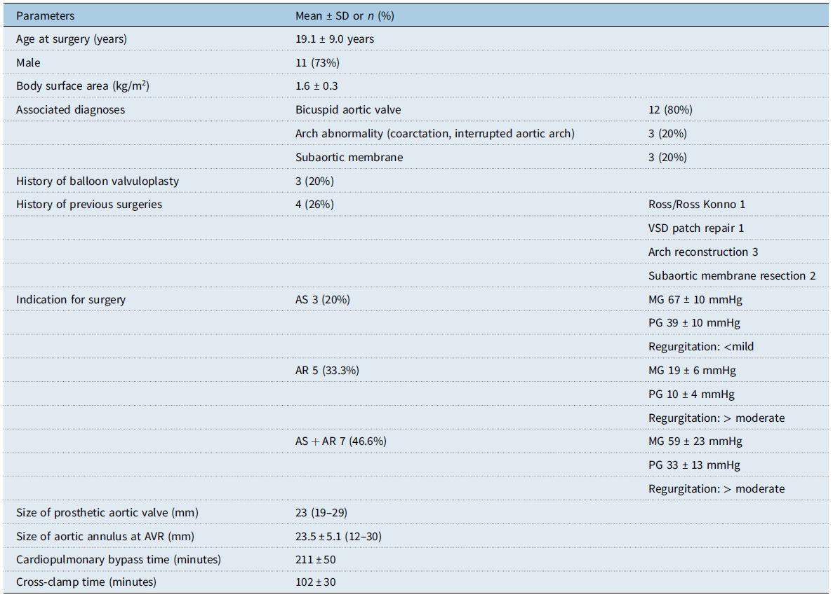
Our cohort included 15 subjects, 11 (73%) of whom were males and had a mean (SD) age of 19.1 (9.0) years at the time of surgery. The patient demographics are described in Table 1. The primary indications for surgery included aortic valve stenosis in three (20%), both aortic stenosis and insufficiency in seven (46.6 %), and aortic insufficiency in five (33.3%) subjects. Bicuspid aortic valve was present in 12 (80%) of the patients. Three patients (20%) had a history of balloon valvuloplasty before aortic valve replacement. The patients who underwent balloon valvuloplasty did not receive any other surgical intervention before aortic valve replacement.
Baseline characteristics of patients

AVR = aortic valve replacement; VSD = ventricular septal defect; AS = aortic stenosis; AR = aortic regurgitation.
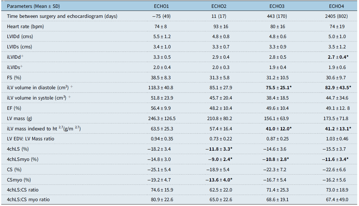
The results of conventional parameters of left ventricular systolic function are described in Table 2. Fractional shortening decreased in the early post-operative period (38.5 ± 8.3% on ECHO 1 versus 31.3 ± 5.8% on ECHO2, p = 0.1) and continued to remain below the baseline at 5-year follow-up (30.6 ± 9.7%, p = 0.1). Similarly, ejection fraction decreased in the early post-operative period (56.4 ± 9.9% versus 48.2 ± 10.4%, p = 0.27) and did not return to baseline by 5 years (49.1 ± 12.8%, p = 0.46) (Fig 1).
Longitudinal assessment of left ventricular systolic function by ejection fraction (EF), peak longitudinal strain (LS), and peak circumferential strain (CS). EF, peak 4chLS, and peak CS decreased in the immediate post-operative period and subsequently improved on 5-year follow-up. However, the peak LS and peak CS at 5-year follow-up remained decreased more than their pre-surgical baseline.

Longitudinal assessment of left ventricular systolic function by conventional echocardiography and speckle tracking echocardiography at four time points

* p < 0.05 compared to ECHO1, +: indexed to body surface area.
LV = left ventricle; LVIDd = left ventricular dimension in diastole; LVIDs = left ventricular dimension in systole; FS = fractional shortening; EF = ejection fraction; EDV = end-diastolic volume; 4chLS = four-chamber longitudinal strain of left ventricular endocardium; CS = circumferential strain of left ventricular endocardium; myo = myocardium.
The results of speckle tracking echocardiography parameters are described in Table 2. Four-chamber longitudinal strain significantly worsened in the immediate post-operative period (−18.2 ± 3.4 versus −11.8 ± 3.3, p = 0.000). Four-chamber longitudinal strain improved over intermediate follow-up but did not return to baseline (−11.8 ± 3.3 versus −14.6 ± 3.6, p = 0.06 on ECHO 3 versus −15.5 ± 3.7, p = 0.28 on ECHO4) (Fig 1). Similarly, circumferential strain worsened in the early post-operative period (−25.1 ± 5.4 versus −18.9 ± 5.4, p = 0.06). Circumferential strain improved over intermediate follow-up but did not return to baseline at 5-year follow-up (−18.9 ± 5.4 versus −22.3 ± 7.2 versus −22.6 ± 6.6, p = 1.00) (Fig. 1).
Abnormal four-chamber longitudinal strain defined as (<−16.3) was present in 20% patients pre-operatively versus 90, 77 and 50% at discharge, 1-year, and 5-year follow-up (Fig. 2). There were 11/15 patients who had normal four-chamber longitudinal strain pre-operatively. Of these patients with normal four-chamber longitudinal strain, 6/11 had decreased four-chamber longitudinal strain post-operatively which persisted at 5-year follow-up. Abnormal circumferential strain was present in 7% patients pre-operatively, versus 42, 8, and 15% patients at discharge, 1-year, and 5-year follow up. The ratio between four-chamber longitudinal strain and circumferential strain was also calculated to account for compensatory changes. If compensatory changes had taken place, the circumferential strain would have increased resulting in decreased four-chamber longitudinal strain: circumferential strain ratio. The four-chamber longitudinal strain: circumferential strain ratio remained unchanged (74.6 ± 15.9 on ECHO 1 versus 73.0 ± 18.9 on ECHO4, p = 1.00), suggesting global dysfunction persisted without any compensatory changes.
Subject-specific line graph showing longitudinal trends in left ventricular systolic function in patients with abnormal peak longitudinal strain (LS) (red) pre-operatively compared to patients with normal peak longitudinal strain (green). The figure shows trends in post-operative LS on long-term follow-up in patients with aortic stenosis (square), aortic insufficiency (triangle), and combined dysfunction (diamond). All patients had a decrease in LS in the immediate post-operative period, which incompletely recovered and remained decreased more than the pre-surgical baseline on 5-year follow-up.

Left ventricular mass indexed to height Reference Boe, Zampi and Kennedy2,Reference Bilkhu, Jahangiri and Otto7 significantly decreased over 5-year period (63.5 ± 25.3 on ECHO1 to 41.2 ± 12.0, p = 0.01 on ECHO3 and 41.2 ± 13.0, p = 0.009 on ECHO4). The indexed left ventricular end-diastolic volume decreased in the immediate post-operative period and continued to remain decreased from the pre-operative volume during 5-year follow-up (118.3 ± 40.8 on ECHO1 versus 85.1 ± 27.9 on ECHO2 versus 75.5 ± 25.1 on ECHO 3 and 82.9 ± 43.5 on ECHO 4, p = 0.08,0.15,0.05). The left ventricular end-diastolic volume: mass ratio decreased acutely in the early post-operative period (0.94 ± 0.35 on ECHO1 versus 0.73 ± 0.22 on ECHO2, p = 0.63) and increased over intermediate follow-up (1.03 ± 0.46 on ECHO4, p = 1.00) (Table 2).
Discussion
To our knowledge, this is the first study in children undergoing aortic valve replacement providing an insight into the trajectory of longitudinal changes of left ventricular systolic function using speckle tracking echocardiography that are not revealed by conventional echocardiographic methods.
Our study demonstrates that in patients with congenital aortic valve dysfunction, a significant number of patients continued to have abnormal left ventricular systolic function following aortic valve replacement. Speckle tracking echocardiography in one-third of our patients had abnormal longitudinal strain and circumferential strain pre-operatively, but it also depicted worsening of left ventricular strain over the 5-year follow-up that did not return to the pre-operative baseline. There was a non-significant decrease in fractional shortening and ejection fraction acutely after surgery, which improved over 5-year follow-up (Fig. 2).
We used speckle tracking echocardiography to assess left ventricular function, which is a novel, more sensitive, and reproducible technique to detect subclinical and clinical left ventricular dysfunction than conventional echocardiographic parameters. It provides a quantitative assessment of myocardial deformation by tracking wall motion frame by frame. Reference Colquitt and Pignatelli19 The long-term data on post-operative strain assessment of left ventricular systolic function following aortic valve replacement in children is limited. In a study by Rost et al, 33 adult patients (68 ± 8 years) with severe aortic stenosis were followed for 6 months after surgical aortic valve replacement. Reference Rost, Korder and Wasmeier20 They reported a non-significant change in four-chamber longitudinal strain ((−17.0 ± 3.9% pre-operatively versus −17.5 ± 4.5%, ns) and circumferential strain (−14.3 ± 4.2% pre-operatively versus −15.3 ± 4.2%, ns) at 1 week post-operatively. Their patients did have a significant improvement in four-chamber longitudinal strain and (−17.0 ± 3.9% versus −19.7 ± 4.5%, p < 0.001) and circumferential strain (−14.3 ± 4.2% versus −18.3 ± 5.5%, P < 0.001) at 6 months follow-up. Kaler et al studied 90 adults with aortic stenosis (mean age 61.6 ± 2.3 years) and aortic regurgitation (59.4 ± 3.4 years) and found four-chamber longitudinal strain improved at 3 months follow-up (−10.9 ± 3.9% at baseline versus −19.4 ± 3.8%, p-value <0.0001) after a small dip at 1st week of follow-up, as did circumferential strain (−17.3 ± 4.5% versus −21.4 ± 3.6%, p-value <0.0001). Reference Kaler, Mahla and Mahla11 Al Rashid et al reported in their cohort of 150 patients (82 ± 4 years old) who underwent transcatheter aortic valve replacement for aortic stenosis, baseline global longitudinal strain improved at 3-month follow-up, and baseline global longitudinal strain correlated with rate of complications ((R = 0.36, p = 0.005). Reference Al-Rashid, Totzeck and Saur13
Our study cohort showed a similar decrease in the acute post-operative period in four-chamber longitudinal strain (−18.2 ± 3.4 versus −11.8 ± 3.3, p = 0.000) and circumferential strain (−25.1 ± 5.4 versus −18.9 ± 5.4, p = 0.06). Although both strain parameters subsequently improved on follow-up, compared to the adult studies, the left ventricular systolic function by strain in our children did not return or surpass their pre-surgical baseline. Reference Kaler, Mahla and Mahla11,Reference Al-Rashid, Totzeck and Saur13,Reference Rost, Korder and Wasmeier20 The gradual development of myocardial fibrosis in aortic dysfunction in adults can result in abnormal global longitudinal strain with normal ejection fraction. Reference Al-Rashid, Totzeck and Saur13,Reference Krayenbuehl, Hess, Monrad, Schneider, Mall and Turina21 Incomplete recovery at 5-year follow-up in our patients may suggest fibrotic changes in the myocardium in utero and in childhood. Reference Krymsky22 Abnormal myoarchitecture and extracellular collagenous matrix exist from the fetal stage in the left ventricle and right ventricle of many CHDs, which, in the presence of patient- and procedure-specific “risk” factors may not lend the left ventricle to favourably remodel to haemodynamic load both before and after surgery, resulting in detrimental ventricular pump function. Reference Krymsky22-Reference Salih, McCarthy and Ho24 Moreover, rapid growth in the paediatric population and early adolescence may result in the development of patient prosthesis mismatch. At 5-year follow-up, the four-chamber longitudinal strain in our patients remained abnormal and below their pre-surgical baseline.
In adults with aortic valve replacement, pre-operative ejection fraction is an independent predictor of mortality after the procedure. In a cohort of 49 children (13.9 ± 3 years) who underwent aortic valve replacement, Tafreshi et al studied predictors of post-operative left ventricular dysfunction at 1-year follow-up. They stated that pre-operative low left ventricular ejection fraction (<60%) and left ventricular dilation >4 standard deviation were significant independent predictors of post-operative left ventricular dysfunction on multivariate analysis. Reference Tafreshi, Shahmohammadi and Davari25 Similar findings were noted in a study by Buddhe et al in 53 children (mean age 11.6 ± 5.9 years at surgery) who underwent aortic valve replacement. Reference Buddhe, Du, Walters, Delius and Pettersen26 Pre-operative left ventricular dysfunction (fractional shortening <28%) was the only significant predictor of persistent left ventricular dysfunction at 6.9 ± 5.6 years follow-up (p < 0.01). They also noted that the left ventricular dimensions normalised in the immediate post-operative period. Their findings corroborated with the adult studies where pre-operative left ventricular ejection fraction is an independent predictor of mortality after the procedure. Dahl et al studied 2017 adult patients with aortic stenosis who underwent surgical aortic valve replacement. They reported that patients with decreased pre-operative left ventricular ejection fraction by Simpson’s biplane had an increased risk of mortality (hazard ratio 0.88 per 10% decrease in ejection fraction). Reference Petersen, Neumann and Naito28 Petersen et al described similar risks in a cohort of 154 patients (mean age 63.5 ± 12.4 years, 71% male) with aortic regurgitation and decreased left ventricular ejection fraction ≤50% who underwent aortic valve surgery. Reference Petersen, Neumann and Naito28 Patients with decreased ejection fraction ≤30% by Simpson’s biplane method at baseline continued to have poor left ventricular function in 40–45% of patients post-operatively when followed up over a period of 4 years.
Our data show no significant difference in the ejection fraction in our patients compared to pre-operatively; however, left ventricular strain by speckle tracking echocardiography remained abnormal. This is comparable to other studies which show strain is a more sensitive marker of subclinical ventricular dysfunction compared to conventional parameters. Stokke et al studied left ventricular strain and ejection fraction in 100 subjects and found that a significant reduction of global longitudinal strain could be compensated by a small increase of circumferential strain, wall thickness, or diameter. Reference Stokke, Hasselberg and Smedsrud29 This could result in a normal ejection fraction despite reduced deformation. We studied the ratio of longitudinal: circumferential strain in our patients and found no significant compensatory changes, suggesting global dysfunction.
Studies suggest reverse cardiac remodelling occurs after aortic valve replacement. Left ventricular end-diastolic dimensions (median z-score (2.0 (1.1 to 3.1) pre-aortic valve replacement versus −0.1 (−0.7 to 1.5) post-aortic valve replacement) and left ventricular mass (median z-score 3.3 (2.0 to 5.3) pre-aortic valve replacement versus 0.4 (−0.7 to 1.9) post-aortic valve replacement) improved over 6 month follow-up in a single-centre paediatric study of 22 patients who underwent transcatheter aortic valve replacement (median age 14.7 years). However, this group of patients had normal ejection fraction and strain pre-operatively and remained normal in the post-operative period. Reference Barfuss, Boucek and McFarland30 Vollema et al have reported a regression in left ventricular dimensions and mass at 10 years post-operatively follow-up in adult patients with aortic valve dysfunction. Reference Vollema, Singh and Prihadi31 In 211 patients (61 ± 14 years old, 61% male), patients with severe aortic stenosis (63%) and aortic regurgitation (37%) underwent left ventricular mass regression independent of the predominant type of aortic valve dysfunction. The authors speculated that the relief from volume overload resulted in a reduction in indexed left ventricular mass in the aortic regurgitation group (154.9 ± 40.0 versus 104.7 ± 28.9 g/m2 at 5 year follow-up), and relief of pressure overload resulted in left ventricular mass regression in the severe aortic stenosis group (127.1 ± 3.0 versus 100.7 ± 28.8 g/m2 at 5-year follow-up). In our study as well, left ventricular dimensions and indexed left ventricular mass significantly decreased over a 5-year follow-up, suggestive of reverse cardiac remodelling. The left ventricular volume: mass ratio did not change significantly.
The age and size of the patient, presence of symptoms, and left ventricular function play an important role in the decision-making process of proceeding with aortic valve replacement. The continued abnormality in strain in our study despite seemingly normal ejection fraction raises a question about the appropriate timing of aortic valve replacement (Fig. 2). Our patients had worse left ventricular hemodynamics post-operatively compared to adults, suggesting that the delay in aortic valve replacement may result in irreversible changes at the cellular level and a long-standing decrease in systolic left ventricular function.
Additionally, as young adults with aortic valve replacement grow older, the mismatch between their native aortic valve area and the effective aortic valve area might increase, resulting in increased afterload for the left ventricle, impeding its complete recovery. Once interstitial fibrosis develops due to chronic pressure or volume overload, the recovery of the left ventricle may be incomplete after aortic valve replacement. Reference Khoury, Mitsnefes, Daniels and Kimball16,Reference Vollema, Singh and Prihadi31 We believe that the incomplete recovery of the myocardium at 5-year follow-up in our patients could be due to a combination of significant pre-surgical left ventricular myocardial changes and chronic afterload from the development of patient prosthesis mismatch with the growth of the patient.
This study has some limitations. This was a single-centre retrospective pilot study, with a small sample size. We were unable to conduct subgroup analysis for patients with associated diagnoses such as bicuspid aortic valve and coarctation of the aorta due to the limited number of patients. Nor could we compare longitudinal function in subgroups of patients with concentric versus eccentric hypertrophy. Some of our patients had abnormal four-chamber longitudinal strain and circumferential strain pre-operatively, but we were unable to stratify their risk of persistent left ventricular dysfunction post-operatively due to small numbers of patients in the two groups. Even though the indication for aortic valve replacement was classified, due to the small sample size no subgroup analysis was obtained. Our study used four-chamber longitudinal strain for the assessment of left ventricular systolic function. Global longitudinal strain utilising 2-chamber, 3-chamber, and four-chamber views may provide a more comprehensive assessment of left ventricular function.
In summary, in this retrospective longitudinal study, we observed that in children who undergo aortic valve replacement for aortic valve dysfunction, pre-operative abnormal strain by speckle tracking echocardiography is indicative of persistently abnormal systolic function post-operatively at 5-year follow up, but fractional shortening and ejection fraction are not. The significance of these subclinical abnormalities needs further investigation with a large sample size, especially in children where the timing of aortic valve replacement is often determined by their clinical symptoms.
Conclusions
Young adults who undergo aortic valve replacement for aortic valve dysfunction have decreased left ventricular systolic function measured by peak longitudinal strain in the immediate post-operative period, which does not return to their pre-operative baseline at 5-year follow-up. Larger multi-centre studies looking at long-term follow-up in young adults who undergo aortic valve replacement are needed to better define these findings.
Acknowledgements
None.
Financial support
This research did not receive any specific grant from funding agencies in the public, commercial, or not-for-profit sectors.
Competing interests
None.