Introduction
Gestational diabetes mellitus (GDM) is one of the most common pregnancy complications, with an incidence of 17.5%.Reference Zhu, Yang and Wei1,Reference Lowe, Scholtens, Sandler and Hayes2 Although GDM is a temporary condition, women with GDM have a 40% increased risk of developing type 2 diabetes within 10–15 years, even if glucose metabolism returns to a normal level after delivery.Reference Damm, Houshmand-Oeregaard, Kelstrup, Lauenborg, Mathiesen and Clausen3,Reference Lauenborg, Hansen and Jensen4 From the developmental origins of health and disease perspective, any disturbance by key events during early life has a profound influence in later life.
The human body contains a large number of microorganisms that are indispensable for sustaining human health when they maintain stable and symbiotic interactions with the host. Unbalanced interactions between microorganisms and human hosts involve the development of numerous metabolic diseases,Reference Cho and Blaser5 such as diabetesReference Karlsson, Tremaroli and Nookaew6,Reference Le Chatelier, Nielsen and Qin7 and obesity.Reference Ley, Turnbaugh, Klein and Gordon8,Reference Backhed, Manchester, Semenkovich and Gordon9 Maternal gut microbiota play an important role in the establishment of the early infant microbiome,Reference Ferretti, Pasolli and Tett10,Reference Jenmalm11 which is associated with the maturation of the immune,Reference Smith, Howitt and Panikov12 nutrient utilization,Reference Nicholson, Holmes and Kinross13 and nervous systems.Reference Robertson, Manges, Finlay and Prendergast14 Therefore, studies on the interaction between gut microbiota and host glucose metabolism may provide new targets for prediction and treatment.
Throughout the whole course of pregnancy, pregnant women undergo considerable metabolic adaptation and hormonal changes,Reference Newbern and Freemark15 in parallel with the shifts in gut microbial compositions and structures.Reference Edwards, Cunningham, Dunlop and Corwin16 One milestone study published in Cell, 2012, reported that the microbial species richness significantly decreased in the third trimester and was characterized by features resembling the bacterial community structure reported in patients with metabolic syndrome.Reference Koren, Goodrich and Cullender17 The authors further verified the influence on adiposity and inflammation through fecal transplantation to germ-free (GF) mice,Reference Koren, Goodrich and Cullender17 suggesting that the shift in the gut microbiota might be associated with metabolic disorders and inflammatory responses during pregnancy and potentially contribute to the pathogeneses of gestational metabolic syndromes.
Previous studies found that GDM was associated with altered gut microbial composition,Reference Wang, Zheng and Shi18 characterized by increased Parabacteroides distasonis and Klebsiella variicola and decreased Bifidobacterium and Eubacterium.Reference Kuang, Lu and Li19 As previous studies were observational studies, the underlying interaction between host glucose metabolism disturbance and gut microbial profiles has yet to be directly evaluated.
Taking advantage of a prospective cohort biobank at the hospital, we conducted a nested case-control study with 45 GDM patients and 45 healthy controls, matched by age and prepregnancy body mass index (BMI) to investigate the association between gut microbial profiles and GDM and verify whether the gut microbiota of GDM donors would cause abnormal glucose metabolism in GF mice.
Materials and methods
Study design and participants
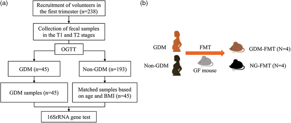
We conducted a nested case-control study at Peking University First Hospital from October 2017 through July 2019. The flow chart is shown in Fig. 1a. Pregnant women were enrolled in their first trimester (T1). The exclusion criteria were existing gastrointestinal diseases, hypertension, type 1 or type 2 diabetes, and history of long-term medication use. Fecal samples and peripheral blood were prospectively collected in T1 and the second trimester (T2), respectively. Clinical information was obtained from medical records and questionnaires (Table S1). In total, 238 participants were enrolled in T1, of whom 45 (18.9%) pregnant women were diagnosed with GDM according to the 75-g oral glucose tolerance test (OGTT) at 24–28 gestational weeks, and 45 non-GDM participants were matched to their GDM counterparts by age and prepregnancy BMI.
Research design flow charts: (a) 90 pregnant women; (b) 6 donors for FMT. FMT, fecal microbiota transfer; GDM, gestational diabetes mellitus; OGTT, oral glucose tolerance test; T1, the first trimester; T2, the second trimester.

To investigate whether the gut microbiota of GDM women could disturb the glucose metabolism in GF mice, six pregnant women were additionally recruited to serve as donors of fresh fecal samples. According to the area under the curve (AUC) calculated from the OGTT results, three GDM donors (AUC > 17) were recruited and matched with three non-GDM donors (AUC < 12) by age and preprenancy BMI. The fecal samples were collected before any treatment (Fig. 1b), and the clinical information was recorded (Table S2).
All participants provided written informed consent, and this study was reviewed and approved by the Institutional Review Board of Peking University First Hospital (V2.0/201504.20). All methods were carried out in accordance with the guidelines.
Screening for gestational diabetes
According to the International Association of Diabetes and Pregnancy Study Group,Reference Metzger20 a universal 75- g, 2-h OGTT was performed during middle pregnancy (24–28 gestational weeks), and a diagnosis of GDM was established if any single threshold value (fasting value, 5.1 mmol/l; 1 h value, 10.0 mmol/l; or 2 h value, 8.5 mmol/l) was met or exceeded.
Sample collection
We provided a labeled sterile stool sampling tube (Sarstedt, Germany, 80.734.311), containing 3–5 ml of RNA storage reagent (Tiangen, China, DP408-02) to all the participants. Fecal samples of all the participants were self-collected at home and transported to the hospital within 3 h. Using a teaspoon, the middle of the fecal core (2–3 g) was placed into the tube within 1 min and immediately stored in a −80 freezer until testing. Blood samples were drawn from the antecubital vein from all the participants. Total blood was centrifuged to isolate plasma with Ficoll-Hypaque density gradient centrifugation (TBD science, Tianjin, China) and then frozen in liquid N2 for further use.
Animal procedures
Animal procedures were approved by the Institutional Animal Care and Use Committee of Peking University First Hospital (J201877). Eight C57/BL6 GF mice (8 weeks old with a body weight 23.44 ± 3.52 g) were purchased from the Institute of Medical Laboratory Animals, Chinese Academy of Medical Sciences. All mice were housed in an isolated sterile environment. Mice were placed into individual cages with a 12 h: 12 h dark–light cycle, a controlled temperature of 21 ± 4 °C and 50–60% humidity, and ad libitum access to food and water. Mice were weighed every 2 days, and food intake was measured.
The fecal samples of the three individuals in the GDM donor group and the three individuals in the non-GDM donor group were respectively mixed, diluted, and packed in an anaerobic incubator (100% N2). The mixture was suspended in 10 ml of PBS buffer and vortexed at room temperature for 5 min. After incubation for an additional 5 min, the supernatant was transferred to an anaerobic tube (500 µl per tube) and immediately stored at −80 °C.Reference Ridaura, Faith and Rey21 The prepared anaerobic tube containing the suspension was placed in a transfer sleeve connected to the sterile isolator, sterilized by ethylene oxide (C2H4O) for 20 min, and then transferred to the sterile isolator.
Eight GF mice were randomized into two groups (GDM-fecal microbiota transplantation (FMT) group, n = 4; non-GDM (NG)-FMT group, n = 4) and inoculated by oral gavage with 200 µl of the fecal mixture from the GDM donor and non-GDM donor groups, respectively. On the first day (D0) and 13 days (D13) after FMT, fresh fecal samples from each mouse were collected with tweezers and placed in a sterile 1.5 ml tube and immediately transferred to the −80 °C freezer. On the same day, mice were fasted for 6 h and then given an intraperitoneal injection of glucose (2 g/kg body weight). Tail blood was collected, blood glucose concentrations were measured with an Accu-Check glucometer (Roche, Germany, H87950), and insulin concentrations were measured with a kit from the American Laboratory Products Company (Salem, NH, 80-INSMSU-E01) before (0 h) and after (1 h) the injection.
Enzyme-linked immunosorbent assay test
The concentrations of TNF-α, IL-6, and IL-8 in plasma were measured with an enzyme-linked immunosorbent assay hypersensitivity kit provided by JingLai Biotechnology Co., Ltd according to the manufacturer’s instructions.
16S rRNA gene sequencing
DNA was extracted with a QIAamp PowerFecal DNA kit (Qiagen, Hilden, Germany) following the manufacturer’s protocols, and the V3–V4 region of the 16S rRNA gene was amplified by polymerase chain reaction (PCR) with the forward primer 5’TCGTCGGCAGCGTCAGATGTGTATAAGAGACAGCCTACGGGGGCGCAG3’ and reverse primer 5’GTCTCGTGGGCTCGGAGATGTGTATAAGAGACAGGACTACGGGTAATCC3’. The PCR products were sequenced on an Illumina HiSeq 2500 platform, and fast length adjustment of short reads (FLASH) was used to merge paired-end reads from the sequencing results.Reference Magoc and Salzberg22 Low-quality reads were filtered by the fastq quality filter in the FASTX Toolkit 0.0.14. The analyzed data retained only high-quality (Q value ≥ 25) reads with a base ratio greater than or equal to 90%, and chimeric reads were removed by USEARCH 64-bit v8.0.1517. The number of reads for each sample was normalized based on the smallest sized sample by random subtraction. Operational taxonomic units (OTUs) were aligned by the UCLUST algorithm with a 97% identity and taxonomically classified using the SILVA 16S rRNA database v128. Alpha and beta diversities were generated in Quantitative Insights into Microbial Ecology.
Statistical analysis
Microbial alpha diversity was evaluated with two indices: the Chao1 index, which is an indicator of species richness,Reference Chao23 and the Shannon index, which is a comprehensive indicator of species richness and homogeneity and is calculated based on the negative sum of each OTU’s proportional abundance multiplied by the log of its proportional abundance.Reference Shannon24,Reference Krebs25 The compositional discrimination of microbial communities between samples was evaluated with Bray−Curtis distance matrices. Clusters were visualized by principal coordinates analysis (PCoA), and statistical comparisons between groups were made using the Adonis method. To identify distinct taxonomic bacterial biomarkers, linear discriminant analysis effect size (LefSE) was used, and the linear discriminant analysis (LDA) log score cutoff value was determined to be 2.Reference Segata, Izard and Waldron26 All the continuous variables were compared between the GDM and non-GDM groups by the nonparametric Mann−Whitney U test. The associations between the relative abundance of taxa and glycemic and inflammatory traits were calculated with the Spearman correlation analyses, and coefficients were computed using R. Image construction was performed in GraphPad Prism 7 and the R software package.Reference Ihaka and Gentleman27 We considered significance at P < 0.05.
Results
The glucose levels and inflammatory factor concentrations were different between the two groups in T2
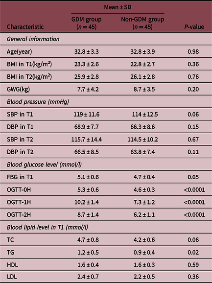
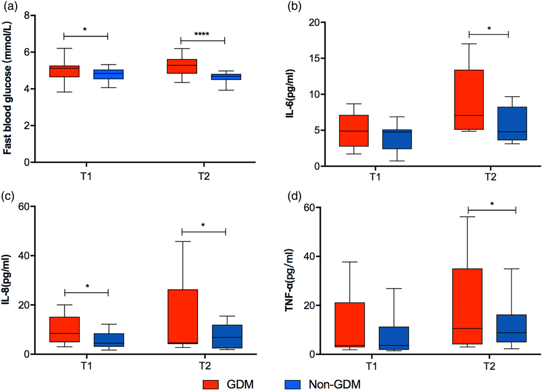
The GDM and control groups were similar in age, BMI, gestational weight gain, and blood pressure (Table 1). Women in the GDM group had a higher level of total triglyceride than women in the non-GDM group during early pregnancy, consistent with previous reports.Reference Enquobahrie, Williams, Qiu and Luthy28,Reference Zheng, Xu and Huang29 No significant difference was observed in the levels of total cholesterol, high-density lipoprotein, or low-density lipoprotein between the two groups. In T1, the fasting blood glucose value in the GDM group was slightly higher than that in the non-GDM group, and this difference was increasingly pronounced in T2 (Fig. 2a). Similar patterns were observed for IL-6, IL-8, and TNF-α between the two groups (Fig. 2b–d), indicating an increasing inflammatory status in GDM women with the progression of pregnancy.
Clinical characteristics of women with and without GDM (n90)

GDM, gestational diabetes mellitus; SD, standard deviation; BMI, body mass index; GWG, gestational weight gain; SBP, systolic blood pressure; DBP, diastolic blood pressure; FBG, fast blood glucose; OGTT, oral glucose tolerance test; T1, first trimester; TC, total cholesterol; TG, triglyceride; HDL, high-density lipoprotein; LDL, low-density lipoprotein.
Comparisons between two groups from T1 to T2: (a) fast blood glucose; (b) the concentration of IL-6; (c) the concentration of IL-8; (d) the concentration of TNF-α. The data are represented as the median and interquartile range in a line graph. GDM, gestational diabetes mellitus; T1, the first trimester; T2, the second trimester. *P < 0.05, ****P < 0.0001.

Comparison of gut microbiota profiles between the GDM and non-GDM groups
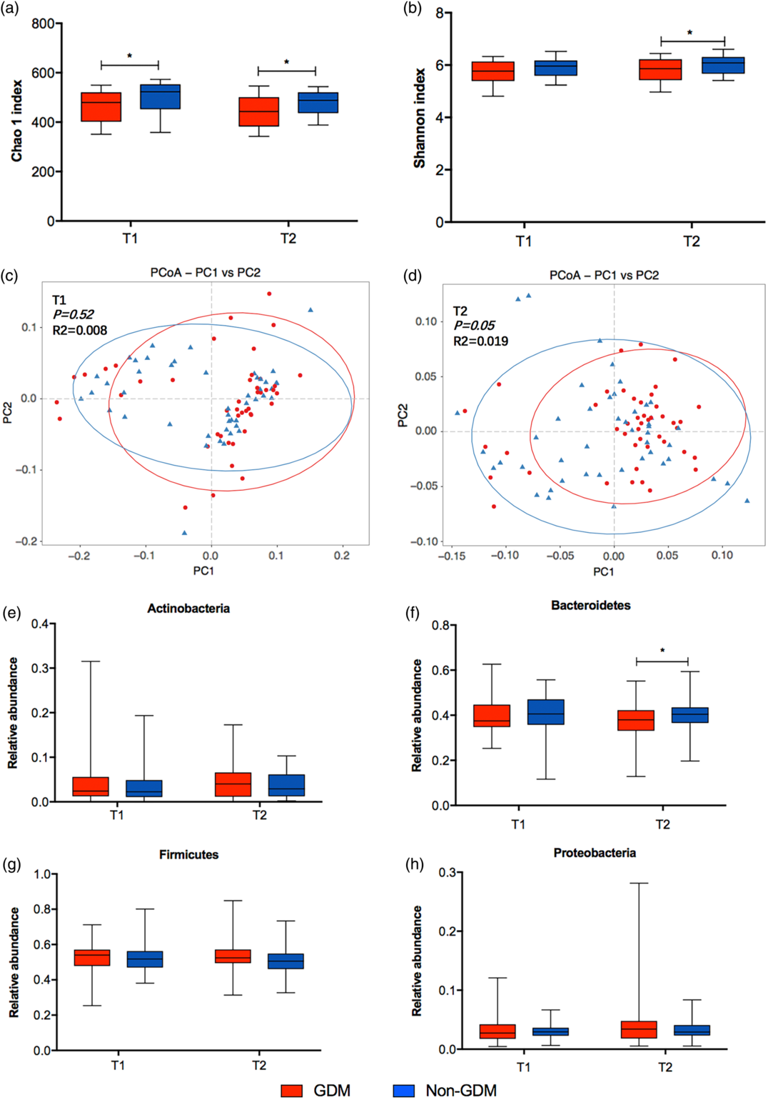
According to 16S rRNA gene sequencing, 72,766 unique OTUs were obtained for further analysis (Table S3). When comparing the alpha diversity of the gut microbiota, we found that the Chao1 index in T1, as well as the Chao1 and Shannon indices in T2, were significantly lower in the GDM group than in the non-GDM group (Fig. 3a and b), suggesting a lower microbial species richness in the GDM group. Moreover, the PCoA results of the gut microbiota between the two groups were similar in T1 (Fig. 3c, Adonis, P = 0.52, R2 = 0.008) but divergent in T2 (Fig. 3d, Adonis, P = 0.05, R2 = 0.019).
Comparisons of gut microbial profiles between two groups from T1 to T2. (a) and (b) alpha diversity in genus level; PCoA analysis in genus level based on Bray−Curtis distance is shown along the first two principal coordinate (PC) axes with Adonis P-value. Individual samples are represented by blue triangles (non-GDM) and red points (non-GDM) at T1 (c) and T2 (d); (e)–(h) the bacteria relative abundance of four bacteria in phylum level. The data are shown as the median with min/max. GDM, gestational diabetes mellitus; T1, the first trimester; T2, the second trimester; PCoA, principal coordinates analysis. *P < 0.05.

The relative abundances of taxa at the phylum level between the two groups were also calculated in T1 and T2. In T2, the relative abundance of Bacteroides was significantly higher in the non-GDM (P = 0.05), while Firmicutes, Actinobacteria, and Proteobacteria were enriched in the GDM group, but the differences did not reach significance (P > 0.05, Fig. 3e–h).
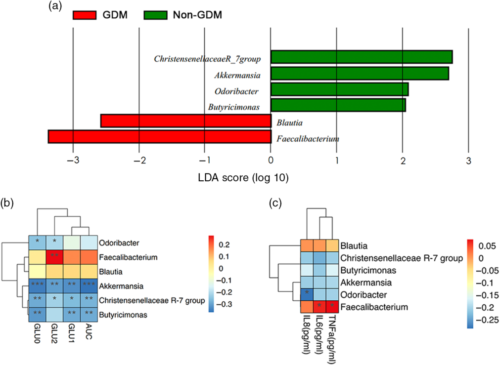
To further evaluate microbial alterations in GDM patients, LDA was used to analyze the scores of significant microbial populations between the GDM and non-GDM groups. According to LefSE analysis, no difference in genara was found between the two groups in T1. In T2, four genera bacteria, namely, Akkermansia, Odoribacter, Butyricimonas, and Christensenellaceae R-7 group, were less abundant in the GDM group than in the non-GDM group, whereas two other genera bacteria, Blautia and Faecalibacterium, were enriched in the GDM group (Fig. 4a).
Relationship between the discriminatory bacteria on the genus level and blood glucose metabolism and inflammatory state. (a) LefSE results in the T2 stage. Enriched taxa of different level from non-GDM group with a positive linear discriminant analysis (LDA) score are shown in green; GDM group with negative LDA score are shown in red (cut off value ≥2); (b) correlative analysis of OGTT values and the relative abundance of discriminatory bacteria; (c) correlative analysis of inflammatory factors concentrations and the relative abundance of discriminatory bacteria. GDM, gestational diabetes mellitus; OGTT, oral glucose tolerance test; AUC, area under curve. *P < 0.05, **P < 0.01, ***P < 0.001.

The associations between gut microbiota and blood glucose metabolism and inflammatory responses in T2
We further explored the correlations between discriminatory bacteria and blood glucose metabolism (OGTT values), as well as inflammatory status (IL-6, IL-8, and TNF-α). The relative abundance of Faecalibacterium was positively correlated with the 2-h blood glucose level, while the Akkermansia, Butyricimonas, Christensenellaceae R-7 group, and Odoribacter were negatively correlated with the OGTT values (Fig. 4b). Additionally, the concentrations of IL-6 and TNF-α were positively correlated with Faecalibacterium and IL-8 was negatively correlated with Odoribacter (Fig. 4c).
Transfer of fecal microbiota to GF mice
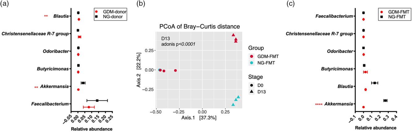
To investigate whether the gut microbiota obtained from a GDM patient could contribute to the glucose metabolic disorders, we conducted a FMT experiment using a GF mouse model. The age, BMI, and gestational weeks of donors were similar between the two groups, while GDM donors had significantly increased OGTT values (Table S2). Among several discriminatory bacteria in the LefSE analysis, the Akkermansia was reduced and Blautia was increased in GDM donors (Fig. 5a, P < 0.01), which was consistent with the previous cohort.
Microbial profiles of donors and GF mice. (a) the comparison of relative abundance of discriminatory bacteria between GDM and non-GDM donors; (b) PCoA analysis based on Bray−Curtis distance is shown along the first two principal coordinate (PC) axes with Adonis P-value. Individual samples are represented by blue points (NG-FMT at D0), red points (GDM-FMT at D0), blue triangles (NG-FMT at D13), and red triangles (GDM-FMT at D13); (c) the comparison of relative abundance of discriminatory bacteria between GDM-FMT and NG-FMT. GDM, gestational diabetes mellitus; NG, Non-GDM; FMT, fecal microbiota transfer; PCoA, principal coordinates analysis. **P < 0.01, ****P < 0.0001.

No difference was found in the mouse gut microbial community structure on D0, while distinct clusters between the two groups were shown on D13 (Fig. 5b, P < 0.0001). Among the several discriminatory bacteria, the relative abundance of Akkermansia was significantly decreased in the GDM-FMT group compared with the NG-FMT group (Fig. 5c, 0.0003 ± 0.0001 vs 0.265 ± 0.020, P < 0.001), while no significant difference was found in the relative abundance of remaining genera between the two groups (Fig. 5c).
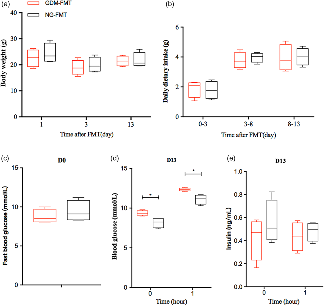
During the whole experimental process (18 days), there was no significant difference in body weight or daily diet intake between the GDM-FMT and NG-FMT mice (Fig. 6a and b), and blood glucose levels were also similar between the two groups at D0 (Fig. 6c). At D13, the blood glucose levels at 0 h (before intraperitoneal injection of glucose solution) and 1 h (after intraperitoneal injection of glucose solution) were significantly increased in the GDM-FMT mice when compared with NG-FMT mice (Fig. 6d). However, at D13, no significant difference was observed in insulin levels in 0 h or 1 h between two groups (Fig. 6e).
GDM microbiota induces hyperglycaemia in GF mice. (a) body weight of mice at 0 day, 3 days, and 13 days after FMT; (b) average diet intake of mice during 0–3 day, 3–8 day, and 8–13 day; (c) fast blood glucose level at 0 day after FMT; (d) fast blood glucose level (0 h) and 1 h-blood glucose level after intraperitoneally injection at 13 days after FMT; (e) fast insulin level (0 h) and 1 h-insulin level after intraperitoneally injection at 13 days after FMT. Data are shown with median with interquartile range. GDM, gestational diabetes mellitus; NG, Non-GDM; FMT, fecal microbiota transfer. *P < 0.05.

Discussion
In this nested case-control study, 45 GDM and 45 non-GDM women were matched by age and prepregnancy BMI, and shifts in the composition and structure of the gut microbiota were found to be associated with GDM status before any treatment. Akkermansia was specifically decreased in GDM women and negatively related to blood glucose level, while the Faecalibacterium was enriched in GDM women and positively related to inflammation factors. The transfer of fecal microbiota to GF mice showed different gut microbiota colonization patterns, and hyperglycemia was induced in GF mice receiving GDM microbiota.
Consistent with recent findings,Reference Kuang, Lu and Li19,Reference Fugmann, Breier and Rottenkolber30,Reference Crusell, Hansen and Nielsen31 we found that the alpha diversity (Chao1 and Shannon indices) in GDM women was significantly reduced, and this shift was observed in the later period of normal pregnancy, which was proven to be the cause of abnormal glucose metabolism and inflammatory responses in mouse models of FMT.Reference Koren, Goodrich and Cullender17 Additionally, we performed comparisons of dynamic microbial compositions and community structures between GDM and non-GDM pregnancies from early to middle gestation. Interestingly, no difference was found in T1, but microbial profiles in the GDM group were different from the non-GDM group in T2, along with a greater inflammatory condition. These distinct change patterns between two groups from T1 to T2 indicated that alterations in the composition and community structure of the gut microbiota contribute to the pathogenesis of GDM.
To further investigate gut microbiota patterns in GDM women, we performed LefSE analysis and found that Faecalibacterium was enriched in the GDM group in the second trimester. This tendency was contrary to a previous study that reported decreased Faecalibacterium in GDM patients in the third trimester.Reference Wang, Zheng and Shi18 GDM is typically diagnosed at approximately 24–28 gestational weeks, and patients may receive exercise-, dietary-, and/or pharmacological-based interventions, which could be the underlying explanation for the discrepant findings.Reference Ponzo, Fedele and Goitre32 Further, Crusell et al. found that different species of Faecalibacterium were both enriched and depleted in GDM women and this alteration was influenced by the adjustment for prepregnancy BMI, suggesting that different strains of Faecalibacterium are involved.Reference Crusell, Hansen and Nielsen31 Faecalibacterium is a butyrate-producer with anti-inflammatory effects and has been shown previously to be reduced in the third trimester of healthy women compared to earlier in pregnancy.Reference Koren, Goodrich and Cullender17 In the present study, the relative abundance of Faecalibacterium was positively correlated with inflammatory status (IL-6 and TNF-α), which was contrary to the previous study.Reference Sokol, Pigneur and Watterlot33 There might be a compensatory mechanism to counterbalance the proinflammatory state. Therefore, further metagenomic investigations are warranted for a functional analysis of Faecalibacterium.
In the current study, Akkermansia, Butyricimonas, and Christensenellaceae R-7 group were less abundant in the GDM group than in the non-GDM group and negatively correlated with the OGTT values. In agreement with previous reports, Christensenellaceae was found to be enriched in individuals with low BMI,Reference Goodrich, Waters and Poole34 while the Butyricimonas, with a function of producing butyrate and which is able to trigger the secretion of gut hormones and improve insulin sensitivity,Reference Canfora, Jocken and Blaak35 was negatively correlated with blood glucose levels.Reference Lee, Lee and Kim36
Another signature genus, Akkermansia, was associated with health-promoting effects.Reference Dao, Everard and Aron-Wisnewsky37,Reference Cani and de Vos38 Consistent with previous studies,Reference Crusell, Hansen and Nielsen31,Reference Cortez, Taddei and Sparvoli39 a decreased relative abundance of Akkermansia was found in GDM patients. Recent studies reported that supplementation with Akkermansia could enhance the bacterium’s ability to improve insulin sensitivity and reduce fat mass in both humansReference Depommier, Everard and Druart40 and mice.Reference Plovier, Everard and Druart41 Additionally, the increased relative abundance of Akkermansia caused by metformin was correlated with improved metabolic profiles,Reference de la Cuesta-Zuluaga, Mueller and Corrales-Agudelo42,Reference Lee and Ko43 and a recent randomized controlled trial revealed that increased Akkermansia was the key to mediating the effects of metformin treatment in newly diagnosed type 2 diabetes patients.Reference Wu, Esteve and Tremaroli44 The mechanism by which Akkermansia improves metabolism might be through reducing proinflammatory bacterial lipopolysaccharides and mediating the intestinal levels of acylglycerols.Reference Everard, Belzer and Geurts45
FMT to GF mice was conducted to determine whether glucose levels were affected by the gut microbiota of GDM patients. To avoid interindividual differences in gut microbiota, three fecal samples from untreated GDM women were mixed before transferring to GF mice. At D0, the microbial community structures were similar between NG-FMT and GDM-FMT mice, but at D13, they had diverged, indicating different gut microbiome colonization patterns between the two groups. Similar to GDM women, the GDM-FMT mice were characterized by reduced Akkermansia, but no difference was found in Faecalibacterium, which might be attributable to incomplete transfer, as in previous reports.Reference Wahlstrom, Kovatcheva-Datchary, Stahlman, Khan, Backhed and Marschall46 The successful transfer of specific species is dependent not only on the composition of donor gut microbiota but also on how well the taxa adapt in the intestinal environment of the recipient. As expected, the GDM microbiota played a causal role in the GF mouse model, inducing higher blood glucose levels than those in the control group.
Strengths and limitations
There were several strengths of this study. First, the gut microbiota shift pattern in GDM women was identified through a nested case-control design. Two confounding factors, maternal age and prepregnancy BMI, were strictly matched. Second, given the potential influence of treatment, which could confound the results, the biological samples were collected at the time of diagnosis, rather than after treatment. Third but most important, this study was not only a simple observation comparing the gut microbial profiles between GDM patients and controls, but also investigated microbiota-induced hyperglycemia in GF mice though FMT. To the best of our knowledge, this is the first time to transfer the gut microbiota of GDM women to GF mice to verify the causal effect of GDM-associated gut microbial changes on glucose metabolic disturbance, providing a new insight into GDM etiology.
Study limitations should be taken into consideration. As a single-center study, the sample size was relatively small; hence, it was difficult to control confounders, including the habits of diet and exercise. The current features of the gut microbiota should be further verified and analyzed in the context of metagenomics and metabolomics. Multiomics could provide further information, including the annotation and function of specific taxa, which might disentangle the key signaling and/or metabolic pathways involved in the pathogenesis of GDM. Last, fecal transfer without microbiological culturomics makes it difficult to target the pathogenic role of a single specific bacterium.
In summary, our study verified the association between GDM and profound shifts in gut microbiota during T2. The specific bacterial patterns in the GDM women were significantly correlated with blood glucose levels and inflammatory states. The fecal microbiota from GDM women transferred to GF mice further verified the causal role of gut microbiota on glucose disturbance in GDM.
Supplementary materials
For supplementary material for this article, please visit https://doi.org/10.1017/S2040174420000768
Acknowledgments
The authors thank all of the participants in the study for their dedication and contribution to the research. Thanks to the technical supports of Chinese Academy of Sciences. Thanks to Danli Yang for modifying the language of this article. The authors acknowledge all participants for their contribution to the scientific research.
Author Contributions
YL was responsible for paper revision, specimen collection, return visits, and clinical data analysis. SQ contributed to the study concept and design, data analysis, manuscript drafting, statistical analysis, and study supervision. YF and YS participated in the enrollment and management of pregnant women and collection of specimens. NL and FL were responsible for the 16S data analysis. XZ contributed to patient follow-up. SW contributed to the clinical data analysis. YW and SS contributed to summarizing the blood glucose management methods for GDM women. SL contributed to designing the questionnaire. WZ contributed to sorting and registering the specimens. YH and YX participated in the 16S rRNA gene-sequencing analysis. BZ was responsible for the integrity and accuracy of the 16S sequencing data and analyses. JM was responsible for the integrity and accuracy of the clinical data and analyses. HY is the guarantor of this work, has full access to all the study data and takes responsibility for the integrity of the data and the accuracy of the data analysis.
Financial Support
This work was supported by the National Natural Science Foundation of China (81671483, 81830044), Beijing Natural Science Foundation (7171011, S150002), National Key Technologies R&D Program (2016YFC1000303), and Key Research Program of the Chinese Academy of Sciences (KFZD-SW-219).
Conflicts of Interest
The authors declare no competing interests.
Public Sharing of Data
These data will be available under BioProject PRJNA515562. Data submission is in progress, and the accession numbers will be provided prior to publication.