Case report
Case 1
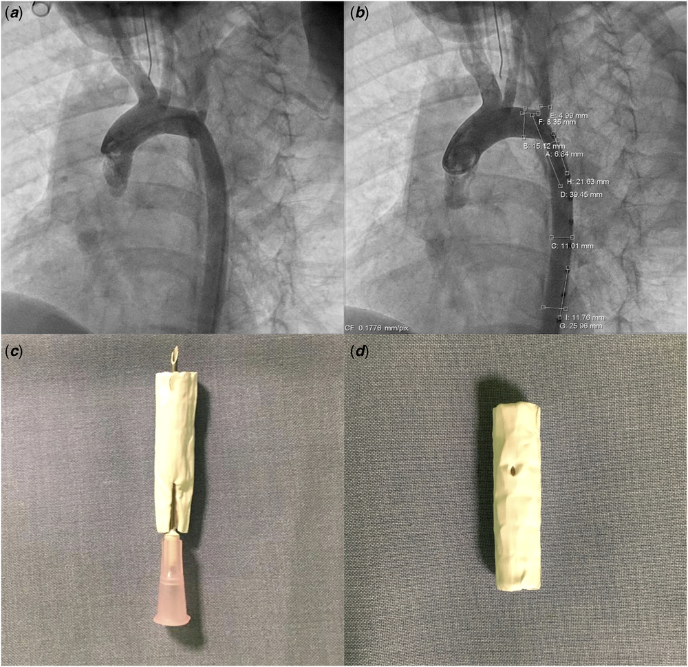
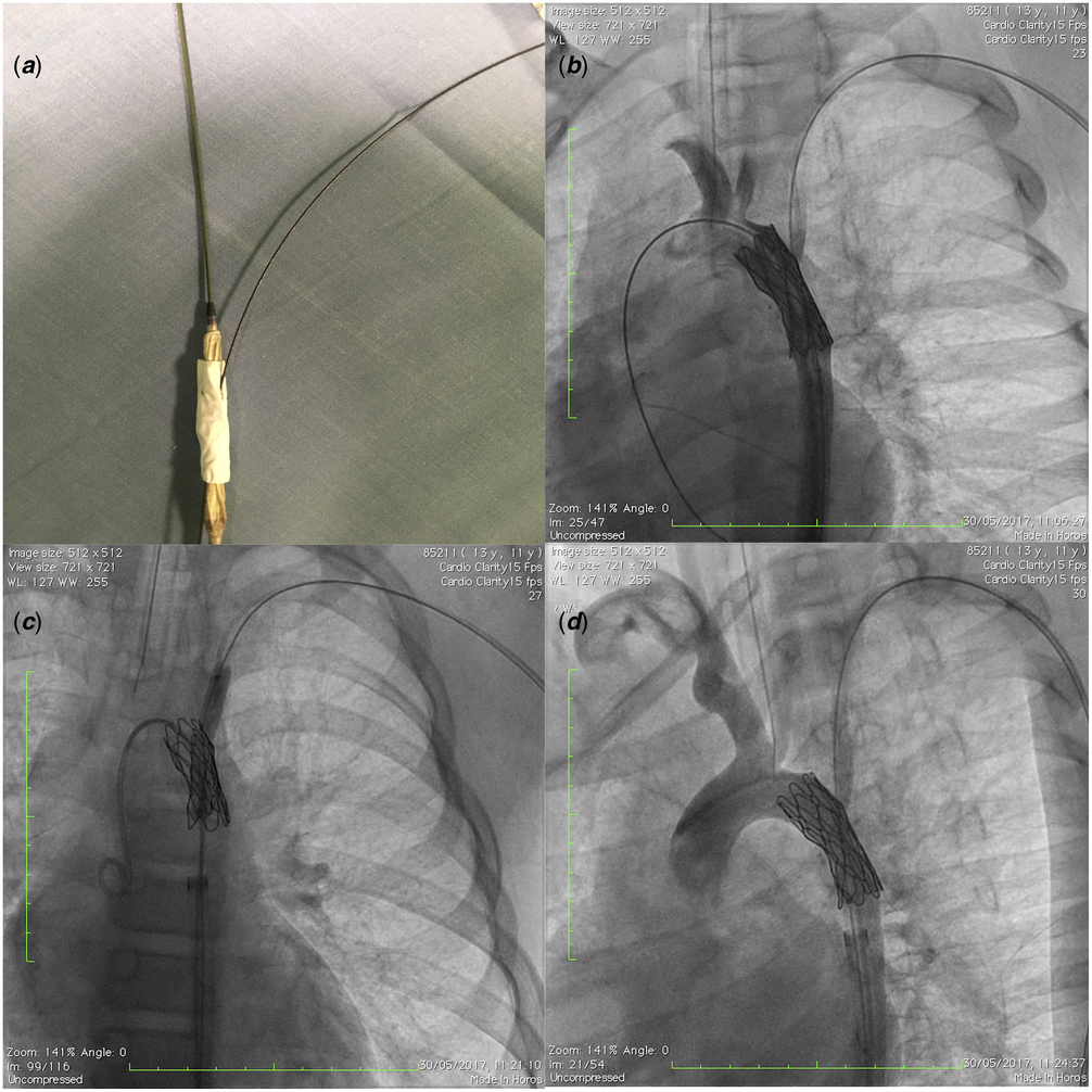
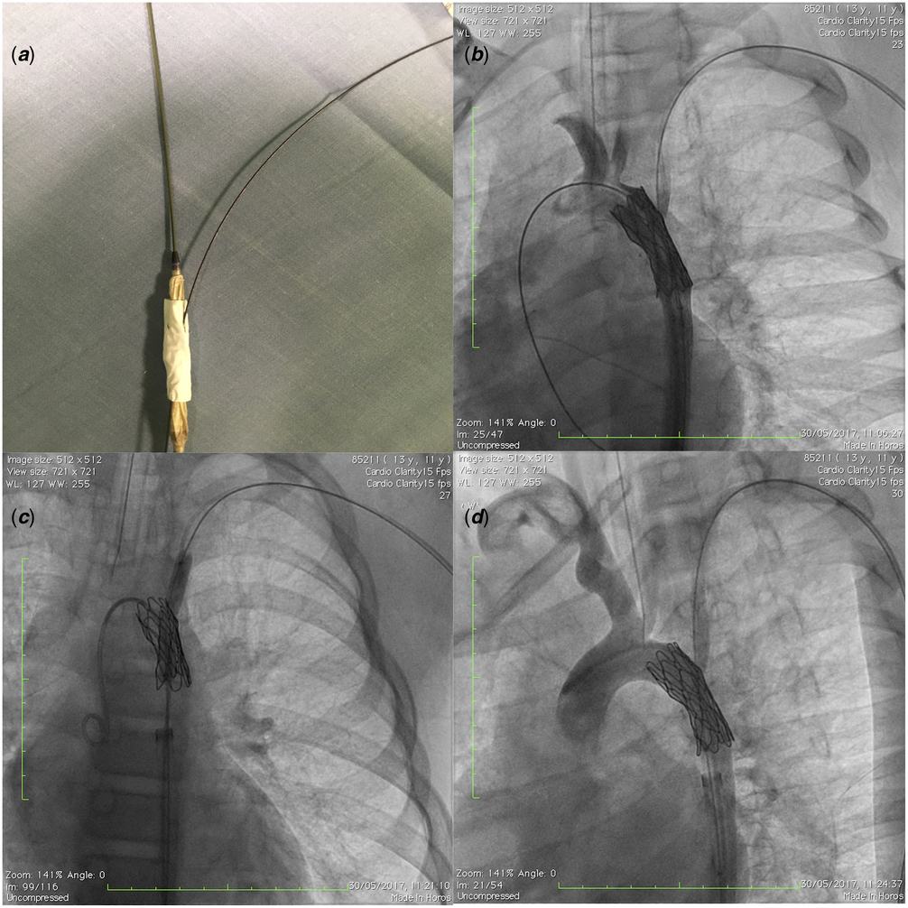
A 12-year-old male affected by Williams syndrome underwent surgical resection with end-to-end anastomosis for aortic arch hypoplasia and native aortic coarctation at 2 months of age. Because of severe recoarctation, the patient underwent a redo surgery at the age of 1 year. When 6 years old, a cardiac catheterisation showed that a recoarctation and a sequential balloon angioplasty (NuMED Tyshak balloon 8 × 20 mm and 10 × 30 mm) were performed lowering the pressure gradient across the aortic arch. During follow-up, the patient remained asymptomatic, but echocardiographic follow-up showed a steady increase of the pressure gradient. At the age of 12 years, a cardiac catheterisation demonstrated a severe recoarctation of the aortic isthmus (minimum diameter 5 mm) involving the origin of the left subclavian artery (Fig 1a) with a pressure gradient of 35 mmHg. As he was deemed a high-risk surgical candidate, a percutaneous catheter intervention was indicated and informed consent was obtained. After induction of general anaesthesia, a 4-Fr arterial catheter was advanced percutaneously in the left radial artery for invasive haemodynamic monitoring. The right femoral artery was accessed after surgical exposure, whereas the right femoral vein was accessed percutaneously and a 6-Fr central venous catheter was advanced. A 34-mm-long NuMED-covered Cheatham-platinum stent was chosen after accurate measurements (Fig 1b) and modified before the implantation to avoid the left subclavian artery occlusion. A tiny fenestration was created at 8 mm of its caudal extremity by a needle 18G (BD Microlance 3; Becton Dickynson, Franklin Lakes, NJ, USA) used to puncture the expanded polytetrafluoroethylene of the stent (Fig 1c-d). Afterwards, the stent was hand crimped on a 12 × 40-mm-long Balloon-in-Balloon delivery catheter (NuMed, Hopkinton, NY, United States of America). A 14-Fr-long sheat Mullins (Medical Cook, Bloomington, IN, United States of America) from the right femoral artery was adopted for the stent delivery using a two-guidewire technique. The stent was advanced over a 0.035-inch Stiff 260-cm Amplatzer J tip exchange guidewire (Boston Scientific, Marlborough, MA, United States of America) in the ascending aorta through the balloon lumen and a 0.035-inch J-Tip Glidewire (Terumo, Tokyo, Japan) placed in the left subclavian artery through the puncture site of the stent (Fig 2a). Multiple angiograms were performed to show a reliable position of the stent and then it was deployed. A following angiogram showed a good flow into the descending aorta and left subclavian artery without findings of dissection (Fig 2b). A balloon Armada 6 × 40 mm (Abbott Vascular, Chicago, IL, United States of America) was advanced over the guidewire in the left subclavian artery, and a further enlargement of the stent fenestration was performed to improve the flow (Fig 2c) according to the artery size. A final angiogram (Fig 2d) showed a correct position of the stent and an adequate perfusion of the descending aorta and subclavian artery. After stent implantation, the gradient across the aortic arch decreased to 3 mmHg.
Left anterior oblique view showing aortic recoarctation: the left subclavian artery was within the area of the coarctation and had a stenosis at its origin ( a ); measurements of the different areas ( b ). Stent fenestration procedure: a needle perforated the covering and passed through the cell of a 34 mm, 8 Zig, Cheatham-Platinum (CP) stent ( c ); a tiny perforation was created ( d ).

Bench simulation of the stent-balloon-guidewires assembly: the balloon lumen was passed over the stiff guidewire and the covered CP stent was crimped on a balloon; an exchange glidewire was passing between the balloon and the stent, then it exited from the pinhole fenestration and was placed in the left subclavian artery ( a ). Left anterior oblique view showing stent implantation within the aortic recoarctation: the stiff guidewire was in the ascending aorta, while the glidewire was passing through the fenestration in the left subclavian artery ( b ); an Armada balloon was advanced over the wire and it was inflated across the fenestration in an anteroposterior view ( c ); improved flow across the left subclavian artery and proper position of the covered CP stent ( d ).
The patient was haemodynamically stable during the procedure and post-procedural course was uneventful. He was continued on beta-blockade, as well as an anti-platelet dose of aspirin. At follow-up 24 months later, the patient was in New York Heart Association (NYHA) class I and assessment of blood pressure revealed increased arterial pressure difference between arms with normal values in the lower extremities. A redo procedure was performed to dilate the stent and it was necessary to accommodate for somatic growth. The flow in the left subclavian artery was normal. Further 6-month follow-up showed no significant problems.
Case 2
A 28-year-old female affected by aortic coarctation underwent surgical resection with end-to-end anastomosis at 4 months of age. Echocardiographic follow-up showed a steady increase of the pressure gradient. When 27 years old, a cardiac catheterisation confirmed a severe recoarctation (minimum diameter 6 mm) involving the origin of the left subclavian artery with a pressure gradient of 45 mmHg. A percutaneous catheter intervention was indicated and informed consent was obtained. The procedural steps were similar to Case 1. A 45-mm-long NuMED-covered Cheatham-platinum stent was chosen and a tiny fenestration was created at 10 mm of its caudal extremity. Afterwards, the stent was hand crimped on a 16 × 40-mm-long Balloon-in-Balloon delivery catheter (NuMed). The stent was implanted as previously described in Case 1. A balloon Armada 8 × 40 mm (Abbott Vascular) was advanced over the guidewire in the left subclavian artery and a further enlargement of the fenestration was performed. Final angiogram showed a correct position of the stent and an adequate perfusion of the descending aorta and subclavian artery. After stent implantation, the gradient across the aortic arch decreased to 2 mmHg. The procedure was uneventful and she was discharged home 2 days later under aspirin. At follow-up 36 months later, the patient was in NYHA class I and assessment revealed no pressure gradient across the aortic arch and good flow in the left subclavian artery.
Discussion
Covered Cheatham-platinum stents have been successfully adopted over the last years, especially in case of complex aortic coarctation anatomy. Reference Tzifa, Ewert and Brzezinska-Rajszys1,Reference Butera, Piazza and Chessa2,Reference Pedra, Fontes and Esteves3 However, the use of a covered stent may be complicated by the occlusion of aortic side branch arteries, and the left subclavian artery is more commonly involved due to the anatomical position. Although it has been suggested that it may be tolerated well, in some cases, it causes a claudication of the left arm that requires a carotid to subclavian graft. Reference Rehders, Petzsch and Ince4,Reference Marcheix, Lamarche and Perrault5
Limited data are available regarding how to deal with the left subclavian artery arising from aortic coarctation in case of covered stents placement. Reference Tsai, Hill and Cheatham6 We dealt with this issue by creating a handmade pinhole in the covered stent before the implantation procedure. Stent delivery was performed by using a two-guidewire-technique as described in case 1. By using the stiffer wire in the ascending aorta, we helped the stent to be directed in the standard position. After stent implantation, the pinhole fenestration was adapted to the left subclavian artery size by performing a balloon angioplasty that increased the artery flow. Our technique does not require stent perforation after its deployment, and the greatest advantage is a significant reduction of the risk of vessel damage.
On the other hand, the procedure requires accurate pre-implantation measurements in multiple angiographic projections. It is mandatory to choose the adequate stent size and perform properly a handmade perforation of the expanded polytetrafluoroethylene at the right position. In addition, several angiograms may be necessary to assess the correct positioning of the stent before its full deployment.
In conclusion, our approach showed its safety and efficacy in two complex cases of severe recoarctation. The main limitation is related to the small number of patients treated and the lack of data about very long-term follow-up.
Acknowledgements
None.
Financial Support
This case report received no specific grant from any funding agency, or from commercial or not-for-profit sectors.
Conflicts of Interest
None.