INTRODUCTION
The modern approach in treatment planning for cervical carcinoma is based on computed tomography (CT) sections and on a three-dimensional (3D) dose distribution. The advantage of 3D image-based brachytherapy planning is the accurate quantification of doses received by various volumes of surrounding critical structures. A gynecologic working group was formed by the Groupe European de Curietherapie and European Society for Therapeutic Radiology and Oncology (GEC-ESTRO) to formulate and describe new terminology regarding 3D image-based treatment planning in cervical cancer brachytherapy.Reference Pötter, Haie-Meder and Van Limbergen 1 , Reference Haie-Meder, Pötter, Van Limbergen, Briot, De Brabandere and Dimopoulos 2 Although conventional treatment planning has generally yielded high tumour control rates, with a low frequency of major complications, image-based planning may allow accurate estimation of doses to the tumour and target volumes and hence lead to improved treatment outcome. As in the case of external beam radiotherapy, 3D planning in brachytherapy offers the advantage of comparing the plans using dose volume histogram (DVH). In addition to this, International Commission on Radiation Units (ICRU 38) 3 recommended point dose such as Point A and B, ICRU reference point for bladder and rectum was also studied.
All the brachytherapy treatment plans were generated traditionally using American Association of Physicists in Medicine (AAPM) Task Group 43 (TG-43) formalism. The TG-43 approach has been widely adopted for high-dose rate (HDR) 192Ir remote after-loading brachytherapy dose planning. The current approach for estimating doses in brachytherapy treatment planning is to superpose pre-calculated dose distributions for single sources in water according to the pattern of the source dwell position and time. The single-source data are derived from one-time source-model specific Monte Carlo (MC) simulations according to specifications outlined in the AAPM TG-43U1 report and are strictly valid only for a homogeneous water phantom of the size and shape used for data derivation. This method is fast and practical in the clinic. However, the influence of tissue and applicator heterogeneity, patient dimensions are all ignored.Reference Beaulieu, Carlsson Tedgren and Carrier 4 Model-based dose-calculation algorithms offer the possibility of departing from water-only geometries by modelling radiation transport in non-water media (tissues, applicators, air-tissue interfaces), resulting in a much more physically accurate reconstruction of the dose distribution actually delivered to the patient.
A 192Ir brachytherapy treatment planning system (TPS) has been made available BRACHYVISIONTM v. 8.8, Varian Medical Systems Inc., Palo Alto, CA, USA that, besides conventional dose calculations based upon the TG-43 protocol,Reference Nath, Anderson, Luxton, Weaver, Williamson and Meigooni 5 – Reference Rivard, Butler and DeWerd 7 incorporates the capability to account for in-homogeneities and the scatter conditions defined by the position among the sources relative to the boundary of a given geometry, hence providing individualised patient dosimetry. This is realised through the implementation of a grid-based Boltzmann equation. A grid-based Boltzmann equation solver was developed specifically to perform accurate and rapid dose calculations for brachytherapy. For brachytherapy, AcurosTM BV solves the linear Boltzmann transport equation for photons on a locally adaptive Cartesian grid. There have been studies in detail of this commercial GBBSReference Mikell and Mourtada 8 – Reference Petrokokkinos, Zourari and Pantelis 10 and its parent code applied to brachytherapy.Reference Gifford, Price, Horton, Wareing and Mourtada 11 , Reference Gifford, Wareing, Failla, Horton, Eifel and Mourtada 12 TPS used was Brachy Vision version 10.0 (Varian Medical Systems) includes both TG-43 and the GBBS AcurosTM BV algorithm (GBBS; AcurosTM BV version 1.4.1 software; Transpire Inc., Gig Harbor, WA, USAReference Mikell and Mourtada 8 , Reference Zourari, Pantelis and Moutsatsos 9 – Reference Vassiliev, Wareing, McGhee, Failla, Salehpour and Mourtada 13 ) which uses the Gammamed plus HDR 192Ir source (Varian Medical Systems) for dose calculation.
MR image-guidance for brachytherapy of carcinoma cervix treatment permits accurate placement of the applicator and analysis of tumour location in relation to surrounding normal tissues.Reference Kapur, Egger, Damato, Schmidt and Viswanathan 14 The combination of MR image-based contouring, and heterogeneity-based dose calculation provides a unique opportunity for precise dose prediction and reporting for gynaecological cancer may improve treatment outcomes. The differences in estimation of dose comparing AcurosTM BV and TG-43 for an interstitial implant of the breast, do not alter the actual treatment as the difference was <5% of clinical target volume (CTV) dose. The AcurosTM BV also showed lower doses to organs at risk (OARs) in that site and these guides us to make minor corrections while reporting.Reference Sinnatamby, Nagarajan, Reddy, Karunanidhi and Singhavajala 15
In this dosimetric study, the comparison was done using 3D MR and CT image-based planning using AcurosTM BV, model-based dose calculation algorithm and classic TG-43 formalism in the carcinoma cervix. In our Institute conventionally, all patients were treated based on the classic TG-43-based calculation. However, for comparison of two planning algorithm, data from the first fraction was used in this study.
AIM
To compare the image-based 3D treatment planning using AcurosTM BV and AAPM TG-43 algorithm for intracavitary brachytherapy of carcinoma cervix.
MATERIAL AND METHODS
Contouring
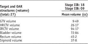
Twenty-seven patients with cancer cervix stage IIB or IIIB with vaginal involvement limited to the upper third of vagina was included in the study. Patients were assessed to be fit for radical treatment. Post implants pelvic CT scans were done with the applicator situated properly for treatment planning. A magnetic resonance imaging (MRI) was done with the applicator situated properly for the first fraction. T2-weighted images were obtained in the para-coronal sections. The MRI images were co-registered with the planning CT using the uterine tandem as the reference and the target volumes were delineated. A 3 mm cut slice thickness for both CT and MRI was used to get 3D image dataset. Single clinical radiation oncologist contoured structures within the patient in this study. Intracavitary brachytherapy (ICBT) treatment to the patients in this study was done with computerised tomography (CT) and MRI compatible applicators. Based on GEC-ESTRO recommendation’s target was delineated, which includes the gross tumour volume (GTV) and two categories of CTV: high-risk clinical target volume (HRCTV) and intermediate risk clinical target volume (IRCTV). Assuming a fixed relation between the applicator and the target, no safety margins were applied, and planning target volume equals CTV were delineated using T2-weighted MR image. The critical normal structures in gynaecological brachytherapy are the hollow visceral organs of the true pelvis: urinary bladder, rectum and sigmoid colon. Pre-imaging preparations performed before every ICBT session, as a large variation in organ position and volume may produce image fusion uncertainties. The pre-empted bladder filled with 100 mL of saline for adequate distension of the bladder during imaging and reproducibility of bladder volume at treatment to stabilise its size and position. Before each application, a urinary catheter was inserted and the catheter balloon inflated with contrast media (7 mL) to localise the bladder neck. Patients were given specific instructions for bowel preparation, to ensure an empty rectum and sigmoid colon before simulation procedure. The bladder, rectum and sigmoid volumes were also delineated. The mean volumes of targets and OAR structures are listed in Table 1. CT and MRI was done for every fraction, and contour was also delineated for every fraction of brachytherapy treatment.
Mean volume of the contoured structure

Abbreviations: OAR, organ at risk; GTV, gross tumour volume; HRCTV, high-risk clinical target volume; IRCTV, intermediate risk clinical target volume.
Applicator
The Ring and Tandem applicator combination 60° set (AL13017000) was designed for use with Varian Gammamed plus iX unit and manufactured from titanium, compatible with both CT and MR imaging system. This applicator features a 32 mm diameter ring made of titanium, which can be used in conjunction with interlocking tandem (titanium) 60 mm length. In addition, included within the set are 5 mm ring build-up acetal cap and a rectal retractor made of titanium and acetal. The titanium has a density of 4·42 g/cm3 and acetal have a density of 1·42 g/cm3. The attenuation of the gamma photon of a 192Ir source with titanium leads to dose variation because of absorption through the titanium metal applicator.
Treatment planning
The treatment planning system includes a library of applicators represented as solid models. The ring CT/MR compatible applicator (AL13017000) selected from the library. The solid model applicator manually registered to the planning CT. During transport; the GBBS substitutes the applicator model and materials for the corresponding CT voxels. GBBS uses CT data outside of the applicators to account for tissue heterogeneity and patient boundaries. The materials available for CT-to-material mapping and the densities that map to them include air, adipose, muscle, cartilage, bone and titanium. In AcurosTM BV, the densities below 0·001 g/cm3 is clamped to 0·001 g/cm3 and densities above 8·0 g/cm3 are clamped to 8·0 g/cm3. To provide enough space for backscatter, the input grid is larger in each dimension than the output grid in the AcurosTM BV. The input grid voxel-size was 0·06×0·06×0·1 with 512×512×196 number-of-voxel were maintained same in both the calculations. AcurosTM BV can report dose-to-water or dose-to-medium. When dose-to-water was selected, the density of water and macroscopical kinetic energy released per unit mass (KERMA) cross section in water for energy group is taken into consideration.Reference Mikell, Klopp and Gonzalez 16 When dose-to-medium was selected, the density of the voxel and macroscopical KERMA cross-section of the voxel mediums for energy group was considered. The dose-to-water-based computation was compared against the dose-to-medium, as the latter can consider of material densities in the CT voxel grid. The account for material heterogeneity and scatter conditions will lead to a dose differences in actual scenario. The dose reduction will lead to a difference in DVH parameters and point doses estimated in this study. The prescription dose was 9 Gy per fraction; a total dose of 18 Gy was delivered in two fractions. For each fraction imaging and planning’s were done. The plan comparison was done with the first fraction in all the cases in this study.
The purpose of this work was to apply the commercial GBBS, AcurosTM BV to estimate doses for cervical cancer patients and compare these doses to standard TG-43 estimates. The DVHs generated by the planning system, were used for recording the volume doses for all these organs. It is to be noted that all the patients who formed study group were treated with TG-43-based calculation using BrachyvisionTM. The treatment plan of this group of patients was evaluated using AcurosTM BV and this treatment planning algorithm was used only for comparison.
Plan Evaluation
Optimisation is based on dose constraints assigned to the target and OAR volumes (location and depth known from clinical assessment). Based on defined dose constraints, the location of active dwell positions and dwell times were modified. The D90 of HRCTV should receive at least the prescribed dose. The minimum dose delivered to 100% and 90% (D90% and D100%) of the volume of interest (GTV, HR-CTV, IR-CTV) were computed. The volume, which were enclosed by 150% or 200% of the prescribed dose (V150%, V200%), was also calculated. The minimum dose in the most irradiated OAR volume close to an applicator such as the bladder, rectum, sigmoid (D0·1cc, D1cc, D2cc, D5cc and D10cc) were calculated. When evaluating the plan, the total dose objectives/constraints for HR-CTV D90 should be ≥85 Gy, IR-CTV D90 should be ≥55 Gy, bladder D2cc≤90 Gy, rectum and sigmoid D2cc≤75 Gy taken into consideration.
Statistical analysis
Normally distributed continuous data were presented as mean with standard deviation. Paired ‘t’ test was used to compare mean difference between paired data. A ‘p’ value of ≤0·05 was considered significant. All data analysis was done using SPSS version 21.
RESULTS
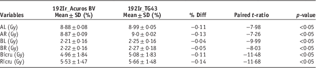
This study intended primarily to compare 3D doses using AcurosTM BV and TG-43 based on GEC-ESTRO recommendations. However, the point doses were also analysed and the differences in the dose between AcurosTM BV and TG-43 formalism, and its significance are shown in Table 2. The Point A dose were compared between AcurosTM BV and TG-43, which indicates 0·11% and 0·13% difference for point A on the left and point A on the right, respectively. ICRU recommended reference point for bladder, and rectum were compared, BICRU point and RICRU point was 0·11% and 0·14% less in comparison with TG-43. The expected point dose on the left and on the right should ideally be symmetrical; however, these two sides values shows a very small difference and are statistically significant. Though the comparison proved statistically significant, but in terms of absolute value, the difference is small, and they are not of any clinical significance.
Comparison between Acuros BV and TG-43 on ICRU 38 recommended points and its significance

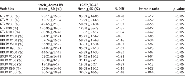
The target volume doses comparison between these algorithms were tabulated in Table 3. The differences in the mean dose to GTV for various volumes are V100% 0·28%, V150% 1·22% and V200% 1·03%; all volumes showed small difference but statistical significant (p<0·05). The dose received by 90% and 100% of volume indicates the reduction in percentage difference of 1·85 and 1·04 compared with TG-43.
Comparison on Acuros BV and TG-43 on GEC-ESTRO recommendation-based target volumes

Abbreviations: GTV, gross tumour volume; HRCTV, high-risk clinical target volume; IRCTV, intermediate risk clinical target volume.
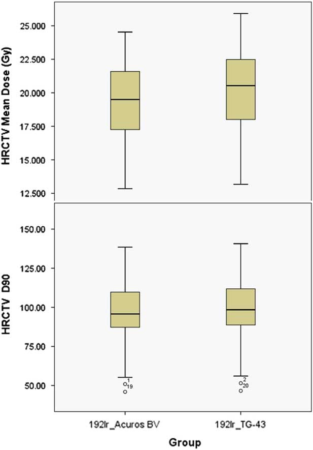
The mean dose of HRCTV D90 using AcurosTM BV was 8·47 Gy (1·63% less compared with TG-43). The mean point ‘A’ dose using AcurosTM BV is 1·04 times the dose to D90 of mean HRCTV. The same difference was observed in comparison with TG-43. The Box and whisker plot comparing the mean doses of HRCTV and HRCTV D90 are shown in Figure 1. The prescribed dose 9 Gy covered 85% of the target (V100%), V150% was 57·7%, V200% was 36·9%, and as compared with TG-43 doses to these volumes were 0·8%, 1·16% and 0·83% less, respectively. D90% and D100% of IRCTV of AcurosTM BV-based plan showed a difference in 1·14% and 1·48% fall against standard formalism. The volume study in IRCTV like V100%, V150% and V200% showed a statistical significant difference compared with TG-43. Of all the target volumes analysed statistically, the maximum dose difference was <2%.
Box and whisker plot comparing AcurosTM BV and TG-43 for HRCTV mean dose and HRCTV D90.

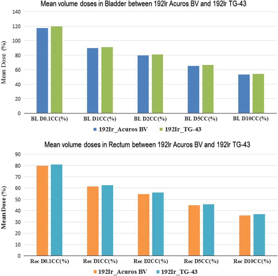
The analyses of mean volume doses of different volumes in bladder and rectum are shown in Figure 2. The mean dose to D2cc of the bladder was 79·77% as against 81·25% using TG-43. The mean dose to D0·1cc of the bladder using AcurosTM BV showed a difference of 2·16% less compared with TG-43. Similarly with D1·0cc, it showed a 1·62% less compared with TG-43. Both indicate statistical significant result (p<0·05). The mean doses to D5cc, D10cc of the bladder were 65·34% and 53·24%, compared with TG-43 it was 1·25% and 1·13% less. The mean minimum and mean maximum dose difference with TG-43 were 0·08% and 0·23%, respectively. A 3% dose difference was seen around the rectal retractor.
Comparison of mean volume doses in different volume in Bladder and Rectum between AcurosTM BV and TG-43.

The mean dose of D0·1cc of the rectum was 0·984 times that of TG-43, and the mean dose to D2cc was 0·980 times that of TG-43. Both D2·0cc and D0·1cc comparison showed a statistical significant difference (p<0·05). Similar differences were observed when doses to sigmoid colon were estimated, and these are listed in Table 4. The overall OARs analysis showed very small differences in the dose, though statistically significant they are clinically insignificant.
Comparison on Sigmoid volumes between AcurosTM BV and TG-43

DISCUSSION
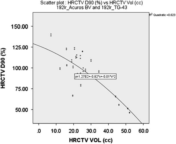
The intention of this study was to use AcurosTM BV (GBBS) algorithm for brachytherapy dose calculation to estimate the total error on the determination of the dose to any given point and volume, which will include, accounting not only for heterogeneity within the patient volumeReference Russell and Ahnesjö 17 but also applicator attenuation. The results from our study showed small dose variations between AcurosTM BV and TG-43 formalism. Of all the parameters statistical analysed the D90 of GTV, HRCTV and IRCTV showed a variation of −1·85%, −1·63% and −1·14%, respectively. The D90 dose to GTV showed a decrease of 1·9%, which agreed with our estimate when accounting for heterogeneity.Reference Hyer, Sheybani, Jacobson and Kim 18 The correlation analysis between HRCTV volume and D90 of HRCTV shown in Figure 3, are statistically significant.
Scatter plot showing the relation between HRCTV volume and the D90 of HRCTV.

Of all the dose volumes studied in OARs, maximum dose difference observed in D0·1cc of the bladder, which showed 2·16%, D0·1cc of the rectum showed 1·22%, sigmoid D0·1cc showed 1·6% difference. As the difference lies within 3%, they proved to have been clinically irrelevant. The ICRU system does not recommend standard points for the sigmoid which is an important OAR and may be a significant contributor for the late toxicity. Image-based planning enables us to optimise dose to the sigmoid colon. Since the contouring of the organ wall is performed, D5·0cc and D10·0cc were also studied and all the OARs like the bladder, rectum. Sigmoid was compared with TG-43 dose calculations, showed a statistically significant results (p<0·05).
The predicted outcome of AcurosTM BV dose calculation in treatment planning of brachytherapy will show a reduction in a dose to the target and OARs. The amount of reductions depends on the heterogeneity associated in planning, which include the material type of applicators and structure. The effect of tissue and applicator heterogeneity for gynecological tandem and ovoid applicator for cervical cancer in HDR treatment showed a dose reduction of up to 10% without normalisation and after normalisation, the doses were seen 6% reduction compared with TG-43-based calculation.Reference Libby, Ter-Antonyan and Schneider 19 In this, study fixed geometry ring applicator with rectal retractor used, and the dose reductions of up to 5% were seen in all parameters analysed. The lesser dose reductions were due to material density of applicator, rectal retraction and geometry of the applicator in position. The dosimetric impact of stainless steel-shielded colpostats in HDR brachytherapy for cervical cancer shows clinically meaningful differences (>5%) from TG-43-based calculation.Reference Mikell, Klopp, Price and Mourtada 20 The mass density of the stainless steel applicator is 8·0 g/cm3, because of which there is a greater reduction in the dose (up to 10%) reported. However, in this study the reductions in the dose were within 5%, which agreed very well with the literature Mikell et al. and Hyer et al.Reference Mikell, Klopp and Gonzalez 16 , Reference Hyer, Sheybani, Jacobson and Kim 18 that a small (<5%) dosimetric impact over TG-43 calculations both in point dose and volume optimised plans.
CONCLUSION
AcurosTM BV dose estimates in HDR brachytherapy found to have only a small dosimetric impact over TG-43-based dose calculations for both conventional points based as well as volume optimised plans. Differences in clinical dosimetric parameters between the AcurosTM BV and TG-43 were <5%, indicating the AcurosTM BV has a minimal impact on clinical parameters. Though all the parameters in this study proved to be statistically significant, the differences are very small, and they are clinically insignificant.
Acknowledgement
None.
Funding
None.
Conflicts of interest
None declared.
Ethical approval
Not required.